Sunday, 31 October 2010

MRCP revision battle 38.2: B cell disorders

B cells are lymphocytes involved in humoral immunity.  Their functions are:
  • production of antibodies (=plasma B cells)
  • memory of antigen (=memory cells, live for a long time and able to respond rapidly if body encounters same antigen again)
  • antigen presenting cells

B cell disorders result in a failure of antibody synthesis, resulting in either hypogammaglobulinaemia or agammaglobulinaemia.  This causes recurrent infections with pyogenic bacteria and fungi.


Examples of B cell disorders include:


1. Common variable immunodeficiency
  • commonest cause of hypogammaglobulinaemia
  • not familial
  • bone marrow shows a normal number of B cells but they fail to mature


2. IgA deficiency
  • commonest isolated Ig in UK
  • increased risk of giardiasis


3. Bruton's agammaglobulinaemia
  • x-linked recessive
  • no circulating B cells
  • usually presents between 3 months and 2 yrs of age


Now on to the T cell disorders......

    MRCP revision battle 38.3: T cell disorders

    T cells are lymphocytes involved in cell-mediated immunity.  T cells mature in the thymus.

    T cell disorders result in increased susceptibility to virus', mycobacteria and fungi.


    Examples of T cell disorders include:


    1. DiGeorge
    • defect in development of thymus and 3rd/4th branchial arches
    • mnemonic CATCH-22:
      • cardiac abnormalities (tetralogy of fallot)
      • abnormal faies
      • thymic aplasmia --> lack of T cells
      • cleft palate
      • hypocalcaemia (due to absent parathyroids)
      • 22 - recessive inheritance on chromosome 22

    2. Nezelof syndrome
    • absent thymus
    • often some B cell involvement

    3. Purine nucleoside phosphorylase deficiency
    • prevents development of T cells


    Onto the final battle of this dull triad - combined B and T cell disorders

    MRCP revision battle 38.4: Combined B and T cell disorders

    So having skated through B cell disorders and T cell disorders its on to combined disorders, the main 3 of which may be recalled as SCID WAS ATAXIC.


    1. Severe Combined Immundeficiency
    • autosomal recessive
    • sometimes due to lack of adenosine deaminase

    2. Wiskott-Aldrich Syndrome
    • x-linked recessive
    • characterised by recurrent infections, eczema and thrombocytopenia
    • associated with an increased risk of maligancy

    3. Ataxic telangiectasia
    • autosomal recessive
    • cerebellar ataxia and telangiectasia
    • increased risk of malignancy
    • low IgE and IgA


    Lets now move back a bit more mainstream with thrombotic thrombocytopenic purpura...

    MRCP revision battle 38.5: Thrombotic thrombocytopenic purpura

    Thrombotic thrombocytopenic purpura is a condition in which large multimers of Von Willebrand's factor clump platelets together, activating the coagulation system, forming fibrin strands and causing microangiopathic haemolytic anaemia (MAHA).


    The 5 key features are:
    • Thrombocytopenia
    • Fever
    • Fluctating CNS signs (visual disturbance, fits, hemiparesis)
    • MAHA
    • Renal failure

    Adult females are most commonly affected


    The cause is often unknown.  Implicated factors include:
    • drugs - clopidogrel, ciclosporin, COC
    • pregnancy
    • HIV
    • SLE

     Treatment is:
    • plasma exchange
    • IV vincristine  (promotes premature release of platelets from marrow)
    • steroids


    Next up... more microangiopathic haemolytic anaemia...

    MRCP revision battle 38.6: Microangiopathic haemolytic anaemia

    Microangiopathic haemolytic anaemia (MAHA) is a nice condition to learn about as you can easily visualise it: it is mechanical disruption of red blood cells in the circulation, so in your mind's eye see the little RBC's being mangled by prosthetic heart valves or sliced by fibrin strands and then imagine them on the other side, reduced in number (anaemia), all with different amounts of haemoglobin in them after being sliced (polychromasia) and lots of partial fragments (schistocytes and helmet cells).


    So to put that above paragraph into a nice list of haematological features of MAHA:
    • anaemia
    • polychromasia
    • helmet cells
    • schistocytes


    Causes of MAHA include:
    • DIC
    • HUS
    • TTP
    • malignant hypertension
    • severe pre-eclampsia
    • prosthetic heart valves
    • septocaemia


    Now onwards to the last MRCP revision battle of the day, sideroblastic anaemia...

    MRCP revision battle 38.7: Sideroblastic anaemia

    Sideroblastic anaemia is a form of anaemia in which the body is not able to incorporate iron successfully in the haem molecule.


    This results in:
    • anaemia
    • sideroblasts in bone marrow
    • rarely haemosiderosis as the body keeps absorbing iron in its futile attempt to correct the anaemia (haemosiderosis may be revised in MRCP revision battle 28.1)

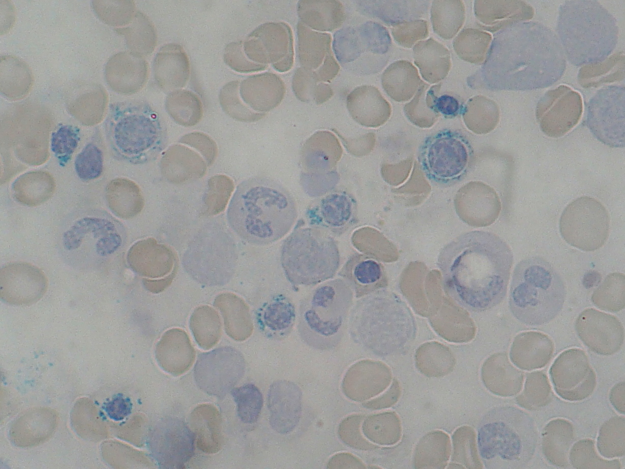
    The image below from Wiki Commons shows ring sideroblasts:


    Causes of sideroblastic anaemia include:
    • congenital causes
      • delta-aminolevulinate synthase deficiency
      • x linked recessive, rare
      • responds to pyridoxine
    • acquired causes
      • myelodysplasias
      • alcohol
      • lead
      • chemotherapy
      • anti-TB meds

    Treatment is to treat the cause, supportive care and pyridoxine.

      Saturday, 30 October 2010

      MRCP revision battle 37.1: TB

      Today has a respiratory feel....


      MRCP revision battle 37.1: TB
      MRCP revision battle 37.2: DVT/PE in pregnancy
      MRCP revision battle 37.3: Asthma
      MRCP revision battle 37.4: Cor pulmonale
      MRCP revision battle 37.5: alpha-1 antitrypsin deficiency
      MRCP revision battle 37.6: Cystic fibrosis
      MRCP revision battle 37.7: HPOA


      MRCP revision battle 37.1: TB


      TB is an infection caused by mycobacterium tuberculosis.  Primary infection is usually in the lungs (ghon focus, usually in the mid or lower zones).  Second favourite site for primary infection = GI, ileocaecal area.


      Incidence =7000/yr in UK


      Spread is by droplet infection. Household contacts have 1 in 4 chance of infection therefore contact tracing is very important and TB is a notifiable disease.


      Primary TB is often symptomless. Although it may cause fever, sweats, anorexia, cough or erthyema nodosum.  If the immune system ‘wins’, the TB is walled off by calcification; however, the TB remains active in ~ 20% calcified areas and may be reactivated when immune system is weakened, eg steroids, diabetes, HIV.


      Post-primary TB makes you ill – fever, malaise, weight loss, anorexia, tiredness, cough, haemoptysis, rarely night sweats.


       It can spread to:
      • brain = meningeal TB
      • spine = Potts fracture
      • blood = miliary
      • skin = lupus vulgaris


      Diagnosis:
      •    CXR
      •    Sputum cultured/stained with Zielh-Nielsen – looking for acid-fast bacilli
      •    Mantoux test of little help in the UK due to BCG

      BCG = bacilli calmette-guerin – decreases risk of TB by 50%


      Treatment: DOTS
      •    2 months rifampicin, isoniazid, pyrazinamide and ethambutol (=RIPE)
      •    then 4 months rifamicin and isoniazid
      •    Pyridoxine throughout to protect against neuropathic effects of isoniazid


      MRCP (and real life) side effects of TB meds to be aware of:

      Rifampicin turns bodily excreations orange
      Isoniazid and rifampicin cause cirrhosis – check LFTs
      Pyrazinamide decreases urea excreation = risk of gout and CI in gout sufferers
      Ethambutol can affect the optic nerve - check colour vision


      On to DVT and PE in pregnancy...

      MRCP revision battle 37.2: DVT/PE in pregnancy

      It is one of those unfortunate situations: pregnancy makes DVT/PE more likely (increases risk around 6x), but the presence of a fetus also makes your preferred investigative tests more risky.


      Risk is more increased if:
      • >35 yrs
      • higher parity
      • raised BMI
      • smoker
      • sickle cell
      • anaemic
      • dehydration
      • not blood group O


      The consensus on management of ?PE in pregnancy seems to be:
      • CXR in all cases
      • compression doppler in all cases - if this shows DVT you can just treat without needing to investigate further


      In terms of the next investigation if you clinically suspect PE but the doppler is normal there is a lack of consensus:
      • CTPA delivers 10-30% less radiation to the fetus but
      • V/Q scan causes less radiation to the maternal breast tissue

      In general CTPA seems to 'win'


      Note that d-dimer is usually 'normal' in the first trimester of pregnancy, rises in the second to third and then returns to baseline at 4-6 weeks post-partum.  However, it is generally not recommended as an investigation in pregnant women.


      Treatment for DVT/PE in pregnancy is LMWH, continued for 6 months (with at least 6 weeks of anticoagulation post-partum)


      For the really keen:


      Next... MRCP revision battle 37.3...

      MRCP revision battle 37.3: Asthma

      Asthma is a condition characterised by periods of dyspnoea, cough and wheeze caused by reversible airways obstruction.


      Asthma diagnosis

      The table below (from the 2009 BTS/SIGN guidelines) lists the factors that increase or decrease the probability of the presentation being asthma:


      If from history/examination you believe there is a:
      • high probability the patient has asthma --> being a trial of treatment
      • intermediate probability of asthma --> perform spirometry:
        • FEV1/FVC <0.7 - trial asthma treatment
        • FEV1/FVC >0.7 - refer to specialist

      After a trial of treatment >400mls improvement in FEV1 or PEFR >15% is significant


      In a peakflow diary, look for diurnal variation >25%


      Treatment of asthma

      Treatment of asthma follows a stepwise approach (BTS/SIGN guidelines):

      1. Mild intermittent asthma
        •  SABA (short-acting inhaled beta 2 agonist) PRN 
        • salamol, salbutamol
      2. Regular preventor therapy
        • corticosteroid 200-800mcg per day
        • beclometasone, fluticasone, budesonide
      3. Initial add-on therapy
        • LABA (long-acting inhaled beta 2 agonist (eg salmeterol))
        • if this doesn't work, stop and trial an oral therapy such as leukotriene receptor antagonist or theophylline
      4. Persistant poor control
        • increase steroid inhaler up to 2000mcg per day
        • add leukotriene receptor antagonist or theophylline
      5. Continuous or frequent oral steroids


      If stable for 3 months, consider stepping down a step.


      Severity of asthma

      In acute asthma attacks, severity may be graded as follows:

      Severe:
      • unable to complete sentences
      • RR>25
      • PEFR<50% predicted or best
      • pulse >110

      Life-threatening:
      • silent chest
      • PEFR <33% predicted or best
      • bradycardia
      • hypotension
      • normal or raised CO2
      • exhaustion


      Management of acute severe asthma

      Think 'O! sip Ma':
      • high flow oxygen
      • 5mg salbutamol neb
      • 500mcg ipratropium neb
      • 30mg prednisolone (or 100mg hydrocortisone IV)
      • magnesium 1.2g and aminophylline if still not improving
      And make sure ITU know if things aren't going well....


      Now for a battle with cor pulmonale...

      MRCP revision battle 37.4: Cor pulmonale


      Cor pulmonale is right heart failure caused by chronic pulmonary hypertension

      Normal pulmonary artery pressure = 15mmHg.
      Pulmonary hypertension = pulmonary artery pressure >25mmHg at rest or >30mmHg on exercise


      Causes of cor pulmonale include:


      • lung disease - of which COPD is by far the commonest
      • pulmonary vascular disease - PE
      •  rib cage deformity


      Presentation is:
      • dyspnoea
      • fatigue
      • syncope


      Signs:
      • raised JVP, a and v waves
      • RV heave
      • loud P2
      • pansystolic murmur - tricuspid regurgitation
      • peripheral oedema
      • hepatomegaly
      • Graham Steell murmur = high-pitched early diastolic murmur, best heard 2nd left intercostal space with pt in full inspiration = pulmonary regurgitation


      Investigations:
      • CXR
      • FBC - look for secondary polycythaemia
      • ECG - R axis deviation, right ventricular hypertrophy


      Management:
      • treat cause
      • treat symptoms
      • ?heart-lung transplant


      Next up: alpha-1 antitrypsin deficiency

      MRCP revision battle 37.5: alpha-1 antitrypsin deficiency

      alpha-1 antitrypsin is a serine protease inhibitor produced in the liver.


      In the lung it prevents neutrophil elastase from breaking down the alveolar wall.
      It is inactivated by cigarette smoke.


      Genetic varients of alpha-1 antitrypsin exist and are labelled based on their electrophoresis mobility, with M (medium), S (slow) and Z (very slow).  MM is 'normal' while 'ZZ' has the worst prognosis with panlobular emphysema.


      Patients with an alpha-1 antitrypsin defiency are likely to develop COPD early (even without smoking) and liver cirrhosis is common.


      Now for a very straightforward battle - cystic fibrosis

      MRCP revision battle 37.6: Cystic fibrosis

      CF is an autosomal recessive genetic disorder which results in a mutation of CFTR, leading to Cl not leaving cells and so more Na and water entering, resulting in thick, sticky secreations.


      The mutation is on chromosome 7.  70% are ΔF5O8


      Incidence = 1/2000.  Carriers = 1/25


      Presentation in neonates is failure to thrive, meconim ileus or rectal prolapse.
      Later presentation tends to be recurrent respiratory infections, cough, wheeze, bronchiectasis.  85% have steatorrhoea.


      Signs include:
      -    cyanosis
      -    clubbing
      -    bilateral course crackles


      Diagnosis is by pilocarpine sweat test.  Na/Cl>60mmol/l, usually Cl>Na.  CXR may show bronchietasis.  PCR can pick up 90% of mutations.  Faecal elastase is a good screening tool for exocrine pancreatic dysfunction.  Spirometry shows obstruction.


      Treatment is MDT, physio, enzyme replacement, heart/lung transplant.


      Complications include  
      • diabetes (1/3) 
      • gallstones
      • cirrhosis
      • OP
      • male infertility (no vas deferens or epididimus)
      • vasculitis
      • nasal polyps (1/3) 
      • HPOA (if you don't know what this is you will by the end of the next battle)
      • infections
        • s.aureus
        • h.influenzae
        • pseudomonas
        • s.pneumoniae
        • burkholderia cepacia (gram negative)
        • aspergillos


      It tends to be the upper lobes of the lungs that are damaged.


      Mean age of death ~30.


      So to the final battle of the day, HPOA...

      MRCP revision battle 37.7: HPOA

      HPOA = hypertrophic pulmonary osteoarthropathy is a paraneoplastic syndrome consisting of periostitis, arthritis and clubbing.


      It is seen in around 50% of patients with mesothelioma.

      Of the 'usual' lung cancers it is most commonly associated with adenocarcinomas and least commonly associated with small cell lung cancer.


      Thats it for today

      Thursday, 28 October 2010

      MRCP revision battle 36.1: Wilson's disease

      Another set of battles written before commencing the joy of a 10 hour shift... and to think we all used to think revising for finals was hard!


      MRCP revision battle 36.1: Wilson's disease
      MRCP revision battle 36.2: Drugs causing impaired glucose tolerance
      MRCP revision battle 36.3: Acromegaly
      MRCP revision battle 36.4: Vitamin C
      MRCP revision battle 36.5: Insulinoma
      MRCP revision battle 36.6: Metabolic syndrome
      MRCP revision battle 36.7: Phaeochromocytoma


      MRCP revision battle 36.1: Wilson's disease


      Wilson's disease is an autosomal recessive condition inherited on chromosome 13.
      Approximately 1 in 400 people carry the gene, giving the disease an incidence of around 1 in 200 000


      Wilson's disease is due to a mutation in the gene coding for a copper transporting ATPase, ATP7B.   This results in the failure of biliary copper excretion, causing raised urinary copper and raised tissue copper with accumulation of copper especially in the liver and basal ganglia.


      Features include:
      • liver disease - hepatitis, cirrhosis
      • CNS  signs - tremor, ataxia, dementia, emotional lability
      • hypoparathyroidism
      • haemolysis
      • Kayser-Fleischer rings - copper deposits in descemet's membrane - pathognomonic
      • blue nails
      • hypermobile joints
      • arthritis


      Diagnosis:
      • low serum caeruloplasmin
      • raised urinary copper

      Treatment: penicillamine - remember this can cause low platelets and white cells so patients should be warned to report bruising/fever/sore throat to their doctor.


      Note that the liver aspects of Wilson's tend to be reversible while the neuro problems have limited reversibility on treating.


      Now on for a short battle about drugs causing impaired insulin tolerance...

      MRCP revision battle 36.2: Drugs causing impaired glucose tolerance

      Drugs associated with impaired glucose tolerance include:
      • thiazide diuretics
      • furosemide
      • steroids
      • tacrolimus
      • ciclosporin
      • IFN alpha
      • nicotinic acid


      I'm afraid I can't think of any clever way to remember this list... if anyone can think of one please email me... marietreasure7 followed by at gmail .com.


      Next up: acromegaly

      MRCP revision battle 36.3: Acromegaly

      Acromegaly is caused by excess growth hormone.  99% of cases are due to a pituitary tumour.  5% of cases are associated with MEN 1 (now might be a good time to re-revise battle 20.1 which covers MEN...)


      For the purposes of revision I'll divide features into 'classical' and 'MRCP smaller print'


      Classical features:
      • soft tissue growth
        • large tongue
        • large hands
        • big ears
        • coarsening facial features
        • widening of gaps between teeth
      • impaired glucose tolerance (40%)/diabetes (20%)
      • raised blood pressure
      • horse voice

      MRCP features:
      • raised phosphate
      • raised triglycerides
      • pseudogout
      • colonic polyps
      • proximal muscle weakness
      • arthralgia


      Diagnosis:
      • failure of GH to decrease (to a lab-specific level) after oral glucose tolerance test
        • false positives to this test may occur in puberty, pregnancy, DM, renal/liver impairment
      • serum IGF-1 is a screening test only


      Treatment:
      • transphenoidal surgery (40-70% cure)
      • ocreotide = somatostatin analogue
      • bromocriptine (<20% effective)
      • pegvisomant = GH receptor antagonist, conclusive data on effectiveness still awaited


      Now for a little vitamin C....

      MRCP revision battle 36.4: Vitamin C

      Vitamin C is an antioxidant.  It is found in many fruits, vegetables and in liver. 


      A deficiency in vitamin C leads to scruvy:
      • gingivitis
      • loose teeth
      • malaise
      • poor wound healing
      • bleeding

      Causes of deficiency include poor diet and pregnancy.


      Now for another short battle, insulinoma...

      MRCP revision battle 36.5: Insulinoma

      Insulinoma is the commonest pancreatic endocrine tumour.

      10% are malignant, 10% are multiple.
      If multiple, 50% are associated with MEN-1.


      Presentation tends to be with the symptoms of hypoglycaemia (revise battle 29.1 if you need to be reminded of these)


      Diagnosis is by supervised prolonged fasting.


      Treatment is surgical excision.


      Onwards for a quick tour of metabolic syndrome...

      MRCP revision battle 36.6: Metabolic syndrome

      Metabolic syndrome (AKA syndrome X) is a combination of disorders that increase a patient's risk of cardiovascular disease/diabetes.


      It is defined as:
      • central obesity (= waist >94cm in europeans) OR BMI >30 
      • PLUS 2 of the following 4:
        • triglycerides >1.7
        • low HDL
        • BP >130/85
        • fasting glucose >5.6 or type 2 DM


      Metabolic syndrome is associated with:
      • raised uric acid levels
      • non-alcoholic fatty liver
      • PCOS


      Now for the final battle of the day, phaeochromocytoma...

      MRCP revision battle 36.7: Phaeochromocytoma

      Phaeochromocytomas are rare, catecholamine (=adrenaline, noradrenaline, dopamine ) producing tumours.


      Presentations are variable and can include:
      • headaches
      • visual disturbances
      • chest tightness
      • palpitations
      • hypertension (70% persistent, 30% episodic)
      • hypotension/postural hypotension is also possible
      • faints
      • abdominal pain

      The triad of headache, sweating and palpitations is 90% predictive.


      Lots of facts to remember about phaeochromocytomas follow the rule of 10%:
      10% are familial (= MEN II, neurofibrosis, Von Hippel Lindau)
      10% are bilateral, 10% are malignant and10% are extraadrenal


      The commonest extraadrenal site is the organ of Zuckerkandl, which is adjacent to the bifurcation of the aorta.


      Diagnosis is by VMA in urine and CT/MRI (show up bright white on T2 weighted MRI)


      Treatment is surgery with pre-op alpha and beta blockade (alpha blocker of choice = phenoxybenzamine)


      Recurrence is possible so these patients should be followed up lifelong.


      Thats all for today, hopefully more tomorrow!