Wednesday, 8 September 2010

MRCP revision battle 5.1: Nephrotic Syndrome

After yesterdays big push today's revision was less epic, mainly because I started reading "The Lovely Bones" by Alice Sebold and I just got too involved.  My motivation to revise also wasn't helped by the first onexamination question I did being 'renal based', with renal medicine being my personal black hole of knowledge... it doesn't matter how many facts I try to throw into it, it still remains just as black.

So, today's battles are:
5.1 nephrotic syndrome
5.2 renal vein thrombosis
5.3 IgA glomerulonephritis
5.4 acute epiglottis


Battle 5.1: Nephrotic syndrome


Nephrotic syndrome is a classic triad of:
  • >3g proteinuria
  • hypoalbuminaemia <25 and
  • oedema
It is also strongly associated with hypercholesterolaemia


80% of the time it is associated with glomerulonephritis, a fiendishly complex set of conditions.


The rest of the time its possible causes are many and varied:
  • diabetes mellitus
  • leprosy/malaria/HBV
  • myeloma/lymphoma
  • amyloidosis
  • SLE
  • gold/penicillamine/NSAIDS/captopril/interferon alpha/heroin
  • pre-eclampsia
  • accelerated hypertension
  • sickle cell disease
  • Alports
  • rarely visico-ureteric reflux
I'm afraid I don't have a clever way/inclination to remember that list, I just hope that by reading it a few times in the exam a faint light of recognition may go on at the appropriate time.


Happily I do have a way of remembering the potential complications of nephrotic syndrome, which is good since sneaky MRCP examiners may be trying to get you to guess nephrotic syndrome from a description of the complications alongside part of the classic triad.

My mnemoric is BOB, I HIT HIM:
  • B12 deficiency
  • Osteomalacia
  • Budd-Chiari
  • Infections
  • Hyperlipidaemia
  • Immunosupression
  • Thrombosis (including renal vein thrombosis)
  • Hyponatraemia
  • Iron deficiency
  • Malnutrition

Treatment for nephrotic syndrome is to restrict salt, treat the cause, prophylactic heparin, treat any hypertension and potentially give furosemide or ACE-i.


On to 5.2... renal vein thrombosis

MRCP revision battle 5.2: Renal vein thrombosis

Renal vein thrombosis can occur due to:
  • nephrotic syndrome inducing a hypercoagulable state
  • invasion of the renal vein by renal cell carcinoma
  • thombophillia

It is often asymptomatic, but equally may present with haematuria, loin pain, palpable mass or a sudden worsening of renal function.


Treatment is with warfarin, anticoagulated to INR 2-3 for 3-6 months.

 Follow me to battle 5.3....

MRCP revision battle 5.3: IgA Glomerulonephritis

As if the glomerulonephropathies aren't tricky enough (hence why series 5 of the battles only discusses one of them) they can also have a series of pseudonyms/ 'nicknames.'  For example, IgA nephropathy also goes by the labels 'Bergers disease' and 'mesangioproliferative glomerulonephritis'.  For simplicity, I'll just refer to it as IgA nephropathy.


IgA nephropathy is the commonest GN in adults, and it classically affects young males after an URTI.  So, in the exam look out for the 25 yr old man who has haematuria following a sore throat.

Symptoms may just be micro/macro haematuria, or match the nephrotic syndrome triad.

Incidence is increased in those from the far east, and those with HLA DQw7 or HLA B35.

Associations include cirrhosis, dermatitis herpetiformis, ank spon, Wiskott-Aldrich syndrome and chronic liver or lung disease.


IgA is positive in 50% of cases and C3 tends to be raised.


25% of cases progress to end stage renal disease.

Frank haematuria is associated with a good prognosis.
Being male, having proteinuria, hypertension, smoking or high lipids are all associated with a poor prognosis.


Phew, thats one of the GN covered... a few more to face at a later date... now lets diversify for the last battle of the day to acute epiglottis.

MRCP revision battle 5.4: Acute Epiglottitis

Since the introduction of the Hib vaccine the potentially life-threatening condition of acute epiglottitis has happily become rare.  Except, of course, in the MRCP exam.

The commonest cause of acute epiglottis was always h.influenzae, but many other pathogens can also cause it.

Presentation tends to be a 'toxic' looking patient with fever and stridor, often with a preceding sore throat.


Treatment is with a 3rd generation cephalosporin.


That's it for today, but if you fancy testing yourself on yesterday's battles click here to go to war 4...

MRCP questions: War 4

As with previous 'wars' after 'battles' these are just a few quick questions to see if your brain cells have retained the information provided in battles 4.1 to 4.5.

Jot down your answers on a piece of paper then compare them to my answers here


Question 1: 
What pattern would COPD produce on spirometry?

Question 2:
With what condition is thermoactinomyces vulgaris associated?

Question 3:
In which zone of the lungs would fibrosis secondary to EAA classically occur?


Question 4:
Can you name 11 causes of lung fibrosis?

Question 5:
Can you name at least 10 causes of clubbing?


answers here

Tuesday, 7 September 2010

MRCP questions: War 3

As with previous 'wars' after 'battles' these are just a few quick questions to see if your brain cells have retained the information provided in battles 3.1 and 3.2.

Jot down your answers then look at my answers here


Question 1: 
Can you now state 10 causes of bilateral hilar lymphadenopathy?


Question 2: 
What are the two main causes of hypercalcaemia?


Question 3:
Name the 2 drugs mentioned that can cause hypercalcaemia


Question 4:
Toxicity of which 2 vitamins can cause hypercalcaemia?


Question 5:
Which 5 cancers are most commonly associated with boney mets?


Question 6:
What effect does vit D have on calcium and phosphate?


Question 7:
What will bloods show in secondary hyperparathyroidism?


Question 8: 
What is the commonest cause of primary hyperparathyroidism?


Question 9:
What is vitamin D made from?


Question 10:
What is the effect of PTH on calcium and phosphate levels?


answers here

MRCP revision battle 4.5: Clubbing

This clinical sign should be on one of those 'you know you're a medic when...' lists, as in "you know you're a medic when clubbing relates to fingers rather than a night out..."

As an undergraduate you probably spent cumulative hours staring purposefully at fingers to convince the examiner you were looking for clubbing, and now at MRCP you need to think about it again..


Firstly, for a definition: clubbing is defined as loss of the obtuse angle between the nail and the dorsum, with thickening of the nail bed and increased curvature of the nail bed in both directions.  There may also be increased fluctuation of the nail bed and the finger can sometimes have a 'drumstick like' appearence.

A picture can be found here


And now, for some causes, and a cry of 'ACE MILC (milk) MCFAB'!

Cardiac causes:
  • atrial myxoma
  • cyanotic congenital heart disease
  • endocarditis
GI causes:
  • malabsorption
  • inflammatory bowel disease
  • lymphoma
  • cirrhosis
Lung causes:
  • mesothioloma
  • CF and cancer
  • fibrosis
  • abscess
  • bronchiectasis

Thats all for today, but if you participated in battle 3 yesterday and fancy testing yourself go on for a brief test here...

MRCP revision battle 4.4: Lung fibrosis

You may need to be able to come up with a list of differentials for lung fibrosis... my way of remembering it is C BAR TAPS (mainly because at some point in the revision process I always get an urge to visit a drinking establishment and 'see' the bar taps, and possibly even purchase something from them...)

So, C BAR TAPS =
  • Cryptogenic fibrosing alveolitis
  • Bleomycin
  • Amiodarone
  • Radiation/rheumatological disease (=RA and SLE)
  • TB
  • Asbestosis
  • Paraquat/pigeon breeders lung
  • Sarcoid/siliosis

And finally for today, battle 4.5...

MRCP revision battle 4.3: Aspergillus

Aspergillus is a group of molds/fungi.  Allegedly they got their name from looking like water does when it falls from an aspergillum, which is an instrument for sprinkling holy water (that fact is unlikely to be necessary in MRCP but its nice for the brain to read bits its not obligated to remember every now and then)


Aspergillus can affect the lungs in 6 main different ways:
  1. It can cause asthma
    • a type 1 hypersensitivity reaction
    • prick test +, precipitans/culture +
  2.   It can cause an aspergilloma
    • = a fungal ball inside a pre-existing cavity
    • usually asymptomatic but may cause haemoptysis, lethargy or weight loss
    • treated by ?surgery ?amphotericin paste under bronchoscopy
    • prick test -, precipitans/culture ++
  3.  It can cause allergic bronchopulmonary aspergillosis
    • type 1 and type 3 hypersensitivity
    • CXR may show bronchietasis, lobar collapse or upper lobe fibrosis, or patchy lesions
    • prick test +, precipitans/culture +
    • also IgE levels are raised
  4. It can cause invasive aspergillosis
    • only occurs in immunocompromised individuals
    • prick test +/-, precipitans/culture +/-
  5. It can cause EAA
    • + serum precipitans to aspergillus clavatus
  6. It can simply colonise.

After that slightly heavy-going section, lets go on to think about BAR TAPS...

MRCP revision battle 4.2: Extrinsic Allergic Alveolitis


This is inflammation of lung tissue due to hypersensitivity to an allergen.  To get more scientific, it is a Gell and Coombs type 3 reaction acutely and a type 4 reaction chronically.


Clinically, an acute reaction occurs 4-6 hours post exposure and manifests as fever, rigors, dry cough, dyspnoea and myalgia.  Auscultation of the lungs might reveal crackles but no wheeze.

Chronically EAA causes dyspnoea, weight loss and can eventually lead to type 1 respiratory failure and cor pulmonale.


There are several different 'flavours' of EAA, each precipitated by something different.  These include:
  • Farmers lung: micropolyspora faeni or thermoactinomyces vulgaris or Saccharopolyspora rectivirgula
  • malt workers lung: aspergillos clavatus
  • mushroom workers lung: thermophilic actinomycetes
or, more modernly:
  • hot tub lung: mycobacterium avium in poorly maintaned hottubs!

A CXR acutely might show mid-zone mottling; chronically upper zone fibrosis might be seen.  It can also be a cause of bilateral hilar lymphadenopathy (remember battle 3.1?)

Spirometry gives a restrictive picture and lavage will show lymphocytes.


Treatment is oxygen, possibly steroids.


Allons-y to battle 4.3!

MRCP revision battle 4.1: lung function tests

After working an excessive number of hours and recovering thanks to chocolate cake (do click here to visit the ultimate chocolate cake recipe, which really is just amazing) I've managed a bit of a revision spurt, mainly through the world of lungs.

So today we have:
battle 4.1: pulmonary function tests
battle 4.2: extrinsic allergic alveolitis
battle 4.3: aspergillosis
battle 4.4: fibrosis
then bringing up the rear battle 4.5, that classic clinical sign clubbing.


Battle 4.1: Pulmonary function tests

Using a spirometer, ask your patient to blow as hard, as fast and as long as possible.  The spirometer will then give you their FEV1 (=forced expiratory volume in 1 second) and FVC (= forced vital capacity.)  These can be plotted, giving graphs that look like this.

In the exam you are far more likely to just get figures and need to be able to recognise them as representing either an obstructive or a restrictive picture.


Obstructive pattern

Obstructive lung function usually gives a very reduced FEV1 and a less severely reduced FVC.  This means that overall the FEV1/FVC ratio is <75%.

Classical causes of an obstructive pattern are COPD and asthma


Restrictive pattern

Restrictive lung function causes a reduced FVC, and FEV1/FVC remains either the same or may increase.

Causes include LONE K: lung fibrosis, obesity, neuromuscular problems, effusion, kyphoscoliosis.


That was short and sweet... onwards to battle 4.2...

Sunday, 5 September 2010

MRCP questions: War 2

As with previous 'wars' after 'battles' these are just a few quick questions to see if your brain cells have retained the information provided in battles 2.1 and 2.2.

Jot down your answers then compare them to mine here


Question 1: 
Name 6 drugs associated with drug-induced lupus


Question 2: 
Name the 3 best tests for monitoring SLE activity.


Question 3:
What is libman-sacks?


Question 4:
Name 3 conditions associated with Jaccouds arthropathy


Question 5:
List as many side effects of amiodarone as you can manage


Click here for the answers

MRCP revision battle 3.2: hypercalcaemia and hyperparathyroidism

Hypercalcaemia

Ah, hypercalcaemia, the classic 'stones, bones, abdominal groans and psychic moans'... and a favourite of MRCP questions.

So what are the causes?
The top 2 to consider are always MALIGNANCY and PRIMARY HYPERPARATHYROIDISM.
Once you've thought of those, think toxic COAT MISST paget...

Malignancy may be hinted at by other blood tests showing low albumin, chloride or an alkalosis.
Think of myeloma, the possiblity of boney mets and of tumours producing PTH-P.
For boney mets remember the brilliant 'bronchus, breast, byroid, brostate and bridney'
For PTH-P the main culprits are squamous cell lung carcinoma, breast and kidney.

Primary hyperparathyroidism is usually due to a single adenoma (85% of the time) and is covered more in the topic of that name below.

So on to the slightly more eoseritc possiblities.... TOXIC COAT MIIST PAGET

TOXIC = vit D or vit A toxicity

COAT = the endocrine causes; essentially any 'growth' hormone excess will increase calcium (GH/oestrogen/thyrotoxicosis/steroids) and COAT is a way to recall the important ones: cushings, acromegaly/addissons, thyrotoxicosis.  Phaechromocytoma can occasionally be a culprit too.

MIIST = the random causes
Milk-alkali syndrome - treat with n.saline and loop diuretics
Iatrogenic - thiazides, lithium
Idiopathic infantile - supravalvular aortic stenosis (think of if 'elfin facies' are mentioned)
Sarcoid
TB/any granulomatosis disease (histoplasmosis, wegeners granulomatosis)

PAGETS - pagets disease of the bone does not normally cause raised calcium but will if the patient has been immobile for a long period.


Having considered the causes, lets take a step back and look at the 2 main 'characters' involved in calcium balance.

Firstly, theres vitamin D.  Vit D is made from cholesterol, meaning that while meat-fans can get some of theirs from food vegetarians are reliant on their body making some (made in the skin with the aide of sunlight, making the UK's weather not ideal...)

Vit D affects calcium by:
- promoting GI absorption of Ca and phos
- promoting renal reabsorption of Ca and phos.

Unsuprisingly, its net effect is therefore to increase both calcium and phosphate.

The second 'character' is PTH.  This affects calcium by:
- promoting renal reabsorption of calcium (and encouraging renal loss of phosphate)
- promoting osteoclast activity --> increasing both Ca and phos
- increasing Vit D hydroxylation - which indirectly increases GI calcium absorption

So, overall its net effect is to increase calcium and decrease phosphate.

Keep these bits in mind and when hypocalcaemia pops up they may come in handy again!


Hyperparathyroidism

PTH is released in response to decreased ionised calcium.  It works in the way described above, causing a net increase in calcium and decrease in phosphate.

Primary hyperparathyroidism causes raised calcium, raised PTH, raised alk phos and low phosphate.
85% of the time it is a solitary adenoma, 15% of the time hyperplasia of all glands and <0.5% of the time a carcinoma.

Rarely  osteitis fibrosa cystica can develop = bone marrow fibrosis and cyst formation due to severe reabsorption.  This results in brown tumours, subperiostial erosions of distal phalanges and pepper-pot skull.

Secondary hyperparathyroidism is the 'good' form - this occurs when calcium is low as a compensatory mechanism, so biochemically the bloods will show low Ca, raised phosphate and raised PTH

Tertiary hyperparathyroidism is back into the 'inappropriate' catagory - it is seen after long term secondary hyperparathyroidism and is due to the parathyroid glands getting 'too big for their boots' and become hyperplasic/acting autonomously.  Biochemically it causes raised calcium and very high PTH.


Phew well thats enough for one day... time for some chocolate!
Unless you're feeling up for testing what you remember from battle 2... if so click here

MRCP revision battle 3.1: Hilar lymphadenopathy

Currently in a stint of antisocial long shifts so struggling to squeeze in revision, but have managed to squash a quick review of 3 topics in, all prompted by one question about a patient with bilateral hilar lymphadenopathy:

1) causes of bilateral hilar lymphadenopathy (sure that one wasn't a suprise)
2) hypercalcaemia (predictable, given topic 1)
3) hyperparathyroidism (an obvious progression from topic 2)


Causes of bilateral hilar lymphadenopathy

I like to try and remember these as a rhyming song:
"cancer, sarcoid and TB, lymphoma HIV.... and the other 5 are 2CHEAP2 to me... oh the joys of bilateral hilar lumphadenopathy..."
CHEAP = 2 Cs (CF, Churg Stauss), EAA, pneumoconiosis, phenytoin (2Ps)


Onwards to battle 3.2...

Saturday, 4 September 2010

MRCP revision battle 2.2: Amiodarone and dronedarone

Amiodarone

Amiodarone is a dirty, dirty drug, and as well has being dirty has a long half life of between 26 and around 126 DAYS.

It has a whole host of side effects to look out for in MRCP questions/real life:
- torsade de pointes
- ataxia
- alveolitis
- pulmonary fibrosis
- corneal microdeposits
- hepatitis
- metallic taste
- photosensitivity
- grey skin
- peripheral neuropathy
- and the classical THYROID PROBLEMS - if hypothyroid, amiodarone can be continued, if hyper, in general it should be stopped.

To try and remember this list I have a little mental cartoon of a man with a grey face, wearing sunglasses (photosensitivity) drinking from a metal can of beer (metallic taste and hepatits) and staggering because of his ataxia and peripheral neuropathy. He also has a goitre (thyroid problems) and is coughing (pulmonary fibrosis/alveolitis).  Due to the amiodarone prolonging his QT he goes into torsades and arrests; he then sees halos and glare (due to either him dying or his corneal microdeposits, depending on my mood)

The onset of amio is in hrs IV or 1-3 weeks when given orally.
As a practical aside remember when giving IV it is given in 5% dextrose and the doses tend to be 300mg over 20mins then 900mg over 24 hrs.


As an MRCP-irrelevant but randomly interesting aside DRONEDARONE has been proposed as a less toxic alternative to amiodarone ... essentially amiodarones brother but without his dirtiness.  Trials such as ADONIS and ATHENA (don't you just love drug companies acronyms?  yet another of my secret fetishes) showed that dronedarone did not cause thyroid problems or fibrosis.  Unfortunately, the ANDROMEDA trial showed it was dangerous in heart failure, and overall while amiodarone is 50-60% successful in treating AF dronedarone is only 30-40% successful.  So, since it costs more and works less, NICE have said no.

MRPC revision battle 2.1: SLE, Libman-Sachs and Jaccouds

After yesterdays achievement of getting through one whole question (which I got wrong) in my allocated hour of revision I approached today with some trepidation.  Happily, in 1.5 hours I got through 5 questions, I got 50% right and yesterday's thoroughness had already started to pay off as one of the questions involved oligoclonal bands in CSF (MS, NS NS, S S, GBS!)

So which topics do I want to recap from today's session?  The main one would definitely be SLE, and the side-dish to go with it for a clashing taste (probably metallic, if its side effects profile is to be believed) is amiodarone.


Revision battle 2.1: SLE
Revision battle 2.2: Amiodarone


Revision battle 2.1: SLE

Defined as a multi-system CTD with small vessel vasculits and non-organ specific antigens

Affects females far more than males (9:1), which I'm sure you all knew already

Is associated wiht HLA B8, DR2 and DR3


Is diagnosed by 4 of the following 11 criteria, which the OHCM helpfully suggests be remembered by A RASH POINTS MD:
  • Arthritis (non-erosive) of  2 or more joints
  • Renal disorder (either persistant >0.5g/l protein in urine or casts)
  • ANA + (occurs in 95% of cases)
  • Serositis - pleuritis or pericarditis
  • Haematological - haemolytic anaemia, leukopenia (WCC<4 on 2 occassions), lymphopenia (<1.5 on 2 occasions) or platelets <100
  • Photosensitivity
  • Oral ulcers
  • Immunological - anti ds DNA, anti SM or antiphospholipid antibody +
  • Neurological disorder
  • Malar rash
  • Discoid rash


3 best tests for monitoring SLE are:
  1. anti DS antibody titre
  2. decreased C3/C4
  3. raised ESR


Classical SLE led into brief foray to drug-induced lupus, which in contrast to SLE is commoner in men than in women.  It tends to be mild and rarely involves renal/neuro systems.  It generally resolves on stopping the culprit drug.

The common culprits are: PC M HIP: procainamide, chlorpromazine, minocycline, hydralazine, isoniazid, phenytoin.

Anti-histone antibodies are often present.


Happily SLE also threw up 2 more eponymous syndromes:

Libman-Sacks endocarditis: non-bacterial endocarditis associated with SLE, commonly affecting the MV.
Jaccouds arthropathy: a reversible deforming arthropathy due to capsular laxity.  As well as being seen in SLE it can feature in rhumatic fever, PD, and hypocomplementemic urticarial vasculitis.


Please click here if you wish to progress to the side-dish of amiodarone, battle 2.2!

Friday, 3 September 2010

MRCP revision battle 1.3: Addisons

The brief review of Addissons disease didn't throw up much that I wasn't already very familar with, and if you're revising for part 2 chances are you're already fairly close buddies with it too.  

However, it was worth looking at for a recap of drugs that can induce Addissons (rifampacin, phenytoin, carbamazepine) and to be reaquinted with the eponymous syndrome Waterhouse Friederchsen...

Waterhouse Friederchsen Syndrome: bilateral adrenal haemorrhage, usually secondary to meningococcal sepsis but can also be due to sepsis caused by strep, staph or h.influenzae.


Should you feel the need for a more comprehensive recap of Addisons disease please click here for wiki's take on it.


MCRP revision battle 1.2: CSF

Analysis of CSF

Questions involving CSF are common, and there are just some facts and lists that you have to 'suck it up' and learn...


Normal values 
  • normal protein 0.2-0.4 g/l
  • red cells 0
  • WCC <5/mm3
  • glucose >2/3 plasma

CSF in meningitis
  • Bacterial: cloudy appearence, high protein, low glucose, polymorphs
  • Viral: clear or cloudy appearence, normal protein, normal glucose, lymphocytes
  • TB: fibrin webs, high protein, low glucose, lymphocytes

Causes of very high protein in CSF (>2g/l)

Remember as BLOCK TB with FUNGAL GBS 
= spinal block, TB meningitis, fungal meningitis and GBS.


Causes of high protein in CSF
  
Remember as ABCDE... and an N
= abscess, bacterial meningitis, cancer, subDural, viral Encephalitis and neurosyphillis.


Causes of Low glucose in CSF

bacterial/fungal/TB meningitis, 20% mumps meningitis, 20% herpes encephalis and occasionally SAH.


Lymphocytosis in CS

Remember as HB HVLLS (a bit like the front of a ship made of pencil lead...)
= 'half bacteria' (ie partially treated bacterial meningitis), HIV, viral meningitis/encephalitis, lymphoma/leukaemia, Lyme disease, SLE.

 
Oligoclonal bands in CSF 

Remember as MS, NS NS, S S, GBS
MS, neurosarcoid, neurosyphillis, SLE, rarely SAH, GBS.


If you're still up for battle 1.3, click here!

MRCP revision battle 1.1: pituitary apoplexy

My approach to revision has always been to attempt a question, then when I fail use it as a 'spring board' to bounce off into a topic that the question has highlighted I'm less familiar with.  What I've always found in the past is that initially each question produces a whole set of 'learning needs', but as revision progresses fewer 'black holes of knowledge' or 'grey holes of partially forgotten information' are revealed.

It was therefore not entirely a surprise that the first time I sat down to revise I only got through one question as it uncovered so many things I needed to read up on!

These were:
- pituitary apoplexy (which in turn prompted revision of the anatomy of the cavernous sinus)
- analysis of CSF fluid
- brief review of Addisons disease

As you can see, there is a certain degree of randomness about the topics generated each day.  To try and make this blog easier for me (and potentially others) to use for revision in the future I've decided to split each day into a series of 'mini-battles', essentially separating the topics into different posts.  So, in the case of today this post is battle 1.1 and will just cover pituitary apoplexy, and then I'll do a post entitled battle 1.2 to cover analysis of CSF fluid and battle 1.3 to cover Addissons disease.

The other benefit to this is that if you want to avoid my waffle you can just skim down the first part of each post headed by 'battle x.1' and then all other battles will be info-dense, waffle light but hopefully still contain a sprinkling of humour!


So, with no further ado onto to the main meat of battle 1.1!


Pituitary apoplexy

Pituitary apoplexy describes a condition caused by sudden haemorrhage into or infarction of the pituitary gland, usually in a patient already known to have a pituitary adenoma.

It should be considered in questions where the patient has meningitis-like symptoms (neck stiffness, headache, vomiting) and also eye signs such as bitemporal superior quadrantanopia or opthalmoplegia.  Low BP may also be a feature.

Pituitary apoplexy is diagnosed by CT/MRI, and treatment is essentially surgical but immediate medical management would involve ABCD, balancing of electrolyte disturbances and a dose of hydrocortisone, since loss of the pituitary is going to cause levels to plummet (and hence the low BP).


I have a guilty love of eponymous syndromes and happily pituitary apoplexy has one associated with it:  Sheehans syndrome.  To highlight any eponymous syndromes snuck into posts I will be writing them in purple...

Sheehans syndrome is pituitary apoplexy after childbirth, usually (but not always) associated with postpartum haemorrhage. 


Having revised pituitary apoplexy, it makes sense to just sneak in a spot of relevant anatomy revision (partly cos I'm a geek, partly because if you understand the anatomy it makes the medicine easier):


Revision of anatomy of cavernous sinus

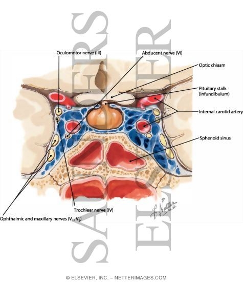
The cavernous sinus around the pituitary gland contains CN III, IV, V1, V2 and VI, also the internal carotid artery.  Note CN II runs just above it.

A nice picture can be found here from the fabulous Netter images collection.


Click here for battle 1.2!

Thursday, 2 September 2010

Preparing for Battle

So the big step of deciding to take exam has been made, meaning its time some procrastinating before actually facing what I've decided to work for entails.  Or, more positively, time for preparing.

Firstly, being a stationary addict, I had to head out to stock up on the essentials.  For me this means highlighters, black biros and Oxford project books - WHSmith sell them at around £3.50 - as they have nice thick paper so you can highlight on both sides to your hearts content.

Next, I gathered a sensible set of books to work from.  I've gone for :

1 ) Essential Revision Notes for MRCP by Kalra, which was utterly invaluable during part 1


 2) Trusty cheese and onion, AKA Oxford Handbook of Clinical Medicine, as combines the virtues of being both comprehensive and concise


3)  Kumar and Clark's Clinical Medicine, for when more depth is needed (hopefully not too often) and finally


4) My First MRCP book which I entirely failed to bond with during part 1 so I'm going to re-attempt using for part 2


So, having prepared my ammunition I joined onexamination.com to see what the battle was likely to be like...