Friday, 17 September 2010

MRCP revision battle 14.1: Schistosomiasis

Its a very random bag today, with our first adventure in the world of infectious diseases, some clotting-based battles in the arena of haematology then a tricyclic overdose to round it all off!


MRCP revision battle 14.1: Schistosomiasis
MRCP revision battle 14.2: The clotting cascade
MRCP revision battle 14.3: Factor V Leiden
MRCP revision battle 14.4: Thrombocytopenia
MRCP revision battle 14.5: ITP
MRCP revision battle 14.6: TCA overdose


MRCP revision battle 14.1: Schistosomiasis

Warning... this is a revision topic likely to make you itch...


Schistosomiasis is a parasitic disease caused by flukes (=trematodes.)   It is generally carried by snail vectors that release cercariae (= parasitic lavae)

The first sign of infection may be 'swimmers itch'.  The MRCP exam may give a history of a patient who has been on holiday to Asia/Africa/South America and has been swimming in a river and is now itchy with a rash.  In part 2 there may also be a picture like the one below:
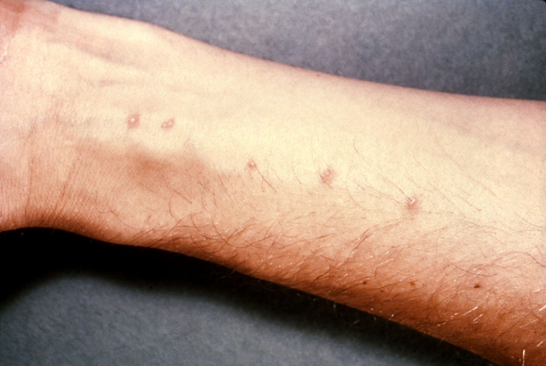


The rash tends of appear within hours of infection and disappear within a week.


The classical presentation of acute schistosomiasis itself occurs roughly 2 weeks after infection with:
  • fever
  • urticaria
  • diarrhoea
  • abdominal pain
  • cough
  • hepatosplenomegaly
This acute version of schistosomiasis is known as Katayama Fever


The treatment is praziquantel.


If schistosomiasis is not recognised and treated it can become a chronic granulomatous disease (granulomas form around the schistosomiasis eggs).  Which parts of the body are affected depends on the type.  For the purposes of MRCP the following associations are worth learning:
  • s.mansoni - liver disease, transverse myelitis
  • s.japonicum - liver disease
  • s.haematobium - urinary tract/bladder disease (haematuria)
    • --> increased risk of squamous cell carcinoma of bladder


The most practical way of diagnosing is by looking for eggs in stool/urine.


It goes without saying that any question with an eosinophillia in the blood results of someone who has been travelling should raise the possibility of this as a diagnosis.


Now on to the less exotic world of haematology and a recap of the clotting cascade!

MRCP revision battle 14.2: The clotting cascade

Before we start tackling clotting disorders a recap of the clotting cascade would be useful.


A schematic of the clotting cascade is:

Its really important to get this in your head as various questions are likely to be indirectly testing your knowledge of it.  I remember the intrinsic pathway into the common pathway as by a little jingle of     " 12,11,9 then 10 (pause) 2, 1 then back again " with the 'back again' bit referring to factor 13 getting in on the action right at the very end to stablise the clot and hence the numbers returning back to being higher.

Also note where VIIIa and Va are involved.


Now look at the same schematic again but with the addition of activated protein C and antithrombin:


So as well as learning the cascade itself make sure the following are etched in your brain:
  • activated protein C inhitbits VIIIa and Va
  • antithrombin inhibits Xa and IIa 
  • protein C is converted to activated protein C by protein S        and finally
  • factors II, VII, IX, X are vitamin-K dependent (method of remembering: '1972')


With all that groundwork covered, lets  move on to battle 14.3 and put a bit of it into practice.

MRCP revision battle 14.3: Factor V Leiden

A point mutation on the gene for clotting factor V results in a varient known as 'factor V Leiden'.
It is inherited in an autosomal dominant fashion.


The result of the mutation is that factor V is insensitive to protein C inactivation, resulting in hypercoagulability.


This leads to increased risk of PE/DVTs and in females increased miscarriage.


Merely being factor V leiden positive doesn't necessitate anticoagulation; warfarin etc would only be considered in response to events rather than prophylactically - indeed, up to 7% of all white europeans/north americans have factor V leiden, making it the commonest inherited thrombophilia.


So having looked at a cause of thrombophillia, lets switch to the opposite and consider low platelets...

MRCP revision battle 14.4: Thrombocytopenia

Thrombocytopenia refers to low numbers of platelets.
Generally the lower limit of normal for platelets is 150 000, with thrombocytopenia often defined as <50 000.


Low platelets increase the risk of bleeding/bruising, commonly manifesting as bleeding gums or nose bleeds.


Causes of thrombocytopenia can be broadly divided into causes that increase platelet destruction and causes that decrease production:
  1. increased platelet destruction (= DISC HH)
    • DIC
    • ITP
    • SLE
    • CLL
    • Heparin
    • Hypersplenism (portal hypertension, leukaemia)
  2. decreased platelet production (=CAB M)
    • Chemo/radiotherapy
    • aplastic anaemia
    • B12/folate deficiency
    • marrow infiltration
Isotretinoin and valproic acid have also been associated with thrombocytopenia.


Treatment is dependent on the cause/the symptoms.


Onwards to the last haematological battle of the day, ITP...

MRCP revision battle 14.5: Idiopathic Thrombocytopenia Purpura

ITP is a condition which results in low numbers of platelets.  Despite its title of 'idiopathic' it is often autoimmune mediated, with IgG autoantibodies against glycoprotein IIb/IIIa/Ib complex.


Acute ITP:
  • affects mainly children
  • boys and girls equally affected
  • often after infection/vaccination
  • tends to be self-limiting over 2 weeks

Chronic ITP:
  • females >males
  • relapsing-remitting


In ITP the bone marrow shows megakaryocytes


Treatment is only needed if the patient is symptomatic or the platelets are <20.
First line = oral prednisolone - 80% respond
If no response in 3 months --> splenectomy
Other options include IV IG or immunosupressive drugs.


One random MRCP type syndrome: Evan's syndrome = ITP +AIHA.


Nearly there, just a quick tricyclic overdose to go...

MRCP revision battle 14.6: TCA overdose

Lets end the day (and the working week) on an overdose.  I've always found it somewhat ironic (and at times in A&E irritating) that a medication which you give to depressed people to treat their depression is so incredibly dangerous in overdose...


TCA in overdose produce symptoms due to their:
  • anticholinergic effects (dry mouth, dilated pupils, tachycardia, constipation, urinary retention, raised intraocular pressure)
  • alpha blockade (hypotension)
  • sodium channel blockade (my personal favourite, cardiac arrhythmias)


On the ECG look out for:
  • long PR
  • wide QRS
  • long QT


In terms of managing these patients you need:
  • ECG and cardiac monitoring
  • consider charcoal if <2 hrs post ingestion
  • bloods for UEs and paracetamol and salicylate (always assume paracetamol taken until proven otherwise)
  • ABG
  • fluids if needed to support BP
  • if acidotic - bicarb
  • dialysis is NOT indicated
  • avoid class Ia/Ic/III antiarrythmics as they all prolong QT.  Also avoid flumazenil (also prolongs QT)
  • observe


Thats all for today folks, and I'm taking tomorrow off.  And possibly the day after too, depending on my mood.  But battles will definitely recommence on Monday.

Thursday, 16 September 2010

MRCP revision battle 13.1: Brugada Syndrome

I'm afraid I'm too tired to even attempt an insightful intro today so lets just go straight to listing the battles:

MRCP revision battle 13.1: Brugada Syndrome
MRCP revision battle 13.2: SVC obstruction
MRCP revision battle 13.3: Causes long PR
MRCP revision battle 13.4: Cauda equina syndrome
MRCP revision battle 13.5: Surviving Sepsis


MRCP revision battle 13.1: Brugada Syndrome


Brugada Syndrome is an autosomal dominant condition that causes abnormal sodium channel proteins, leading to a risk of VF/VT and sudden death.


It is commonest in Asians.

20-40% of cases are associated with a SCN5A mutation.


ECG shows partial/complete RBBB with ST elevation in V1 to V2.  The classical ECG is type 1 below, with the other 2 possible variations.

 Treatment is ICD.


Next up - SVC obstruction...

MRCP revision battle 13.2: SVC obstruction

SVC obstruction can be frightening for the doctor as well as the patient - the first lady I saw with it came in with a lovely slim face and as time went on it became more and more puffy and she became increasingly dyspnoeic.  Once seen it will never be forgotten; I can post this photo:
                                                                     Herbert L. Fred, MD and Hendrik A. van Dijk, Wiki Commons

which demonstrates the type of difference I saw in my patient (normal on right, SVC on left).  My patient however was female and smiled more!


Anyway, lets refocus on points to learn for the MRCP exam:


Features of SVC obstruction:
  • dyspnoea
  • orthopnoea
  • headache 
  • cyanosis
  • cough
  • swollen face/arm
  • engorged veins


There is a rather nice test called Pemberton's Test which involves asking the patient to lift their arms above their head for >1 min and watch for increasing plethora/cyanosis, raised JVP and listen for stridor (that sound every doctor wants to induce in their patient...)  If these things occur it is a positive Pembertons test and suggestive of SVC obstruction.


Causes of SVC obstruction include:
  • lung cancer
  • lymphoma
  • thymus malignancy
  • thrombus around central line


Treatment is surgical, with dexamethasone whilst awaiting this.


Lets return to the small-squared world of the ECG to consider causes of a long PR...

MRCP revision battle 13.3: Long PR interval

The PR interval represents the time taken for the electrical impulse to travel from the sino-atrial node and through the atrioventricular node to the ventricles.

Normal range is 0.12 to 0.2 milliseconds (= 3 to 5 small squares)

A long PR = first degree heart block.


Causes of a long PR ( can be remembered as DRIP SAL, easier to remember if you have a drippy friend called Sally):
  • Drugs (beta blockers, calcium channel blockers, digoxin)
  • Rheumatic fever
  • IHD (anterior MIs in particular can prolong the PR interval)
  • Potassium - either high or low levels
  • Sarcoidosis
  • Aortic root pathology
  • Lyme disease


So that was short and sweet.  Onto another brief battle that should be familiar, cauda equina syndrome...

MRCP revision battle 13.4: Cauda Equina Syndrome

Cauda equina syndrome refers to a set of features caused by compression of the cauda equina.

The cauda equina begins in most people at around L1/L2.


Features of cauda equina syndrome include:
  • low back pain
  • sciatica
  • urinary incontinence/retention
  • faecal incontinence/retention
  • variable motor/sensory loss
  • classically saddle/perianal paraesthesia


Causes of cauda equina are logical when you think about it, you just need to consider what could be there to press on it:
  • commonest = large central disc herniation at L4/L5 or L5/S1
  • tumours
  • trauma
  • epidural abscess (consider this one in MRCP questions that mention a 'post op patient')
  • post op haematoma (another reason to look out for that 'post-op patient')
  • IVC thrombus
  • ankylosing spondylitis
  • sarcoidosis

Treatment is urgent surgery.


So to the final (and juiciest) battle of the day, surviving sepsis...

MRCP revision battle 13.5: Surviving sepsis

There has been a concerted push in recent years to improve the prognosis of patients presenting with sepsis, led by the international 'Surviving Sepsis' campaign.  But what are the key points to pick up for MRCP?


Firstly, a few definitions.


SIRS = systemic inflammatory response = 2 or more of:
  • temperature >38 or <36
  • WCC >12 or <4
  • RR>20 or pCO2 <4.3
  • pulse >90 (note this is lower than you'd probably have expected)


Sepsis is then defined as SIRS plus infection.


Severe sepsis is then defined as sepsis with evidence of organ dysfunction (hypoxia, anuria, raised lactate... so many possible options for the evidenve)

And then septic shock is defined as severe sepsis plus hypotension.


Now we know what sepsis is, what are the key messages from the surviving sepsis campaign?

  1. resuscitate
  • if hypotensive or lactate >4 give fluids
  • aim 
    • CVP 8-12mmHg
    • MAP>65
    • urine output >0.5mls/kg/hr
    • central venous O2 >70% or mixed venous >65%
  • if fluid alone not achieving goals, consider:
    • packed RBC to haematocrit >30% or
    • dobutamine infusion
  1.  =
  2. give antibiotics
  3. keep MAP >65mmHg
    1. norepinephrine 
    2. dopamine
  4. consider hydrocortisone if fluids and vasopressors aren't working
  5. consider recombinant human activated protein c 
    • if apache >25 or multiple organ failure
  6. remember DVT prophylaxis
  7. consider peptic ulceration prophylaxis


As Bugs Bunny would say, thaatts all folks... unless you fancy participating in today's war to check your recall of yesterday's topics...

MRCP questions: War 12

As with previous 'wars' after 'battles' these are just a few quick questions to see if your brain cells have retained the information provided in battles 11.1 to 11.4.

Grab a piece of paper, jot down your answers then compare them to my answers here


Question 1:
Which atria are atrial myxomas found in 75% of the time?


Question 2:
What murmur is associated with aortic dissection?


Question 3:
What % of patients with aortic dissection will have a pulse deficit?


Question 4:
What is the commonest form of aortic dissection?


Question 5:
How are type A dissections managed?


Question 6:
What is the classic cell associated with CLL on blood film?


Question 7:
List 5 features that suggest a poorer prognosis in CLL


Question 8:
What condition does tartrate resistant acid phosphatase positive suggest?


Question 9:
What is Richters syndrome?


Question 10:
How often is the mediastinum wide on a CXR if the patient has aortic dissection?


The answers are here

MRCP revision battle 13


MRCP revision battle 13


Wednesday, 15 September 2010

MRCP revision battle 12.1: Atrial myxoma

Today's battles are a mixed bag; usually they are generated from past questions I've done while today's bunch include some motivated by gaps in clinical knowledge revealed during a recent take.  So the battles are:

MRCP revision battle 12.1: Atrial Myxoma
MRCP revision battle 12.2: CLL
MRCP revision battle 12.3: Hairy cell leukaemia
MRCP revision battle 12.4: Aortic dissection


MRCP revision battle 12.1: Atrial Myxoma

Atrial myxoma are usually benign tumours found in the atria.
75% are in the left atrium.

0.3% of the population are found to have an atrial myxoma at post mortem.
Females >males, 2:1


Atrial myxoma are usually sporadic but an autosomal dominant inheritance also exists.


They are usually benign but as they grow rapidly and embolise they require surgery.


Features of atrial myxoma include:
  • clubbing
  • weight loss
  • fever
  • AF
  • emboli
  • mid diastolic murmur - an 'atrial plop'


Investigations may show:
  • raised wcc
  • low platelets
  • raised ESR in 60%


After that brisk whizz through atrial myxoma, onward to CLL!

MRCP revision battle 12.2: CLL

I keep clerking patients who say they've got 'CLL, doctor' and I nod sagely and scribble it down, while desperately trying to recall any of my med school haematology lectures.  My brief summary for MRCP means that now hopefully my sage nod is slightly more appropriate!!


CLL (=chronic lymphocytci leukaemia) is a monoclonal proliferation of mature lymphocytes.

99% of cases of CLL involve B cells.

CLL is important clinically as it is the commonest cause of lymphocytosis in the elderly.
Males are more often affected than females, 2:1


Presentation:
  • no symptoms in 25%
  • recurrent infections (due to hypogammaglobulinaemia)
  • anaemia (warm AIHA in 10-15%)
  • decreased weight
  • sweats
  • anorexia


Signs includer:
  • enlarged rubbery, non-tender nodes
  • hepatomegaly
  • splenomegaly


Blood tests will show:
  • raised lymphocytes
  • low Hb, neutrophils and platelets
  • smear/smudge cells on blood film


Staging is done by the Rai classification, which can also be used to give the prognosis:
  • Stage 0 - lymphocytosis alone - mean survival >13 yrs
  • Stage 1 - lymphocytosis plus lymphadenopathy - mean survival 8 yrs
  • Stage 2 - lymphocytosis plus spleno/hepato megaly - mean survival 5 yrs
  • Stage 3 - lymphocytosis plus anaemia (defined as Hb <11) - mean survival 2 yrs
  • Stage 4 - lymphocytosis plus platelets <100 - mean survival 1 yr


Indicators of poorer prognosis include:
  • being male
  • age >70
  • lymphocytes >50
  • prolymphocytes >10%
  • doubling rate <12 months
  • raised LDH
  • CD 38 positive
  • ZAP 70 + (if ZAP 70 -ive, mean survival 25 years)


Management of CLL is:
  1. monitoring only if asymptomatic
  2. chlorambucil to decrease lymphocyte count, improve marrow function and decrease node size
  3. fludarabine (a chemo agent, purine analog that works by inhibiting DNA synthesis) if:
    • bulky disease
    • cytopenias due to marrow failure
    • short lymphocyte doubling time


CLL may transform to Richters syndrome.
Richters syndrome is the transformation of CLL to agressive B cell lymphoma.  LDH is raised.


Now onwards to a subtype of CLL, hairy-cell leukaemia.

MRCP revision battle 12.3: Hairy Cell Leukaemia

Hairy cell leukaemia is a subtype of CLL characterised by (suprise, suprise) cells that appear 'hairy' on microscopy.


Hairy cell leukaemia is 4 times more common in men than women.


Features of hairy cell leukaemia include:
  • pancytopenia
  • splenomegaly
  • skin vasculitis in 1/3 cases
  • 'dry tap' 


A key phrase to look out for in MRCP is 'tartrate resistant acid phosphatase positive'


Treatment of hairy cell leukaemia is chemotherapy.  Second line options include alpha interferon and rituximub.


Thats quite enough haematology for one day... lets move on for a 'rippingly' good time with aortic dissection...

MRCP revision battle 12.4: Aortic dissection

Aortic dissection is a scary condition with a mortality rate of 1% per hour.  But before we jump into it, lets begin with a quick recap of some basic anatomy.

The layers of the aorta are, from inside out:
  • tunica intima = epithelial cells
  • tunica media = smooth muscle and elastic fibres
  • tunica adventitia = connective tissue

Aortic dissection usually involves blood collecting in the media.


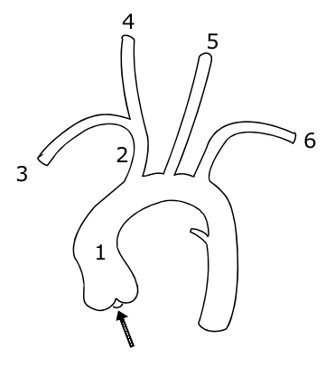
The aorta itself leaves the heart, goes up into an arch and then down again, giving off branches as it goes.  The picture below shows the vessels coming off the aorta:

1 indicates the ascending aorta, 2 the brachiocephalic trunk, 3 the right subclavian artery, 4 the right common carotid, 4 the right common carotid, 5 the left common carotid and 6 the left subclavian artery.

A knowledge of this anatomy aides understanding of the signs and symptoms of aortic dissection.
So what are the features of an aortic dissection?
  • sudden onset of 'tearing' or 'ripping' pain
  • if pain intrascapular, likely a descending dissection
  • MI
  • stroke
  • parathesia of arms
  • hemiparesis
  • many others, depending on the part of the aorta affected

Signs to look out for include:
  • aortic regurgitation - present in 1/3 ascending dissections
  • pulse deficit in an arm - present in 15%
  • BP difference in arms >20mmHg (but note 'normal' individuals may have a BP difference)


Aortic dissections may be classified using either the Stanford or DeBakey systems:
  • Stanford A = ascending dissection - accounts for 2/3 of dissections
    • this covers DeBakey I - ascending and descending aorta involved and
    • DeBakey II - ascending aorta only involved
  • Stanford B = descending aorta only dissected (distal to left subclavian artery) - accounts for 1/3 of dissections
    • = DeBakey III


 Management of aortic dissection:
  • if type A - IV labetalol and urgent surgery
  • if type B - IV labetalol only


Complications associated with type A dissections include:
  • aortic regurgitation
  • inferior MI
  • pericardial effusion
  • carotid dissection
  • loss of pulses


Investigations for dissection:
  • CXR 
    • shows widened mediastinum in 70% of cases
    •  Ring sign - calcification of aorta with wall displaced >5mm
  • CT
  • TOE

Risk factors for dissection include:
  • hypertension (80%)
  • Marfans
  • Noonans
  • Turners
  • trauma
  • pregnancy
  • coarctation
  • congenital bicuspid valve
  • giant cell artertitis
  • cocaine use

If you feel motivated to read more, I'd recommend this webpage: http://www.aorticdissection.com/Aortic%20Diagnosis.htm


A 'war' for yesterday's questions is here


MRCP questions: War 11

As with previous 'wars' after 'battles' these are just a few quick questions to see if your brain cells have retained the information provided in battles 10.1 to 10.6.

Grab a piece of paper, jot down your answers then compare them to my answers here

Question 1
List 4 factors that lead to a worse prognosis in GBS.


Question 2
What percentage of patients with GBS will make a complete or near complete recovery?


Question 3
What regular investigation do patients with GBS require?


Question 4
What is the classic triad of Miller-Fisher syndrome?


Question 5
What antibodies are usually associated with Miller-Fisher syndrome?


Question 6
What is syringomyelia?


Question 7
What are the cardinal symptoms of syrinomyelia?


Question 8
What is the inheritance of Kallmans syndrome?


Question 9
List 5 symptoms/signs of carcinoid syndrome


Question 10
What is the treatment of carcinoid syndrome?


answers here

Tuesday, 14 September 2010

MRCP revision battle 11.1: Guillain Barre Syndrome

When you're working 12 hr days (plus commute) fitting in revision can feel a bit like trying to squeeze that extra towel into the suitcase - you may manage it, but the suitcase is then stretched and prone to snap open at unfortunate times.  If you extend this metaphor to the suitcase being your sanity it explains why junior doctors can often become more emotionally incontinent/irrational as MRCP exams get nearer.  So whats the solution?  Well, in the suitcase scenario you'd add some of those rubber bands round to keep it closed.  In real life, your friends, family and managing to keep some form of hobby/interest going are your rubber bands.

Since I don't want to overpressure my 'rubber bands' I'm going to limit today's battles to

MRCP revision battle 11.1: Guillain Barre Syndrome
MRCP revision battle 11.2: Miller Fisher Syndrome
MRCP revision battle 11.3: Syringomyelia
MRCP revision battle 11.4: Kallmans
MRCP revision battle 11.5: Carcinoid Syndrome


MRCP revision battle 11.1: Guillain Barre Syndrome


Guillain Barre Syndrome is a rare, post-infective demyelinating polyneuropathy.


It tends to occur a few weeks after 'the flu' or a minor illness.  The most commonly associated pathogens are:
  • campylobacter jejuni
  • mycoplasma
  • CMV
  • EBV
  • HIV
  • zoster

Guillain Barre Syndrome is characterised by an ascending, symmetric muscle weakness.  Proximal muscles are more affected than distal ones.  Cranial nerves may also be involved, with CN VII being the most common.


There is usually a 'progressive' phrase for around 4 weeks, after which the patient starts to improve.


Features associated with GBS that appear in MRCP questions include:
  • papilloedema
  • urinary retention
  • arrhythmias

Diagnostically GBS is characterised by:
  • very high protein in CSF
  • decreased nerve velocity on EMG
  • areflexia

The prognosis for patients with GBS is worse if:
  • rapid onset of symptoms
  • older age
  • axomal neuropathoes
  • preceeded by campylobacter infection

Management is:
  • 4 hourly FVC, to check the patient's respiratory muscles aren't affected
  • ventilation if needed
  • plasma exchange
  • IV IG

Overall, 85% of patients make a complete/new complete recovery.
10% will relapse
Less than 5% will die.


Now to to meet a close relative of GBS, Miller Fisher Syndrome.