Friday, 19 November 2010

MRCP revision battle 54.3: Patent ductus arteriosus

The ductus arteriosus is a connection between the pulmonary trunk and the descending aortic arch allowing blood flow in the fetus to avoid the fluid-filled lungs.

It should close shortly after birth, being mostly closed within 48 hrs and fully closed within 3 weeks, leaving the ligamentum arteriosum in its place.


Closure is due to 2 mechanisms:
  1. exposure of infants lungs to oxygen promotes bradykinin production which promotes PDA closure
  2. loss of maternal prostaglandins encourages closure

Closure is more likely to fail (=patent ductus arteriosus) in:
  • premature infants
  • infants exposed to rubella in 1st trimester
  • infants at high altitude

Features of patent ductus arteriosus include:
  • machinery murmur
  • wide, collapsing pusle
  • apical heave

If it does not close the infant may fail to thrive or develop heart failure.
Many cases however will be asymptomatic.
5% of patients with PDA go to develop pulmonary hypertension and Eisenmengers.


Treatment:
  • indomethacin (a prostaglandin antagonist) causes closure in 90%
  • surgical treatment is also an option

In some cases (eg transposition of the great vessels) keeping the ductus arteriosus open may be beneficial to the infant, in which case IV prostaglandin E1 is given.


On to the cardiac axis...

MRCP revision battle 54.4: Cardiac axis

The normal cardiac axis is -30 to +90 degrees.

More negative = left axis deviation
More positive = right axis deviation.


Causes of left axis deviation:
  • LBBB
  • LVH
  • ASD
  • cardiomyopathies
  • aortic stenosis
  • can be normal in:
    • ascites
    • pregnancy
    • obesity

Causes of right axis deviation:
  • RBBB
  • RVH --> lung disease
  • PE
  • can be normal in:
    • infancy
    • thin people


Next: haemolysis

MRCP revision battle 54.5: Haemolysis

Haemolysis (=breakdown of red blood cells) may be intravascular or extravascular.
It causes a microcytic anaemia and raised reticulocyte count.


Intravascular haemolysis

Intravascular haemolysis is characterised by:
  • low haptoglobin (as all used up trying to 'recycle' the broken down red cells)
  • raised free plasma haemoglobin
  • haemoglobuinuria: red-brown urine
  • haemosiderinuria: once haptoglobin binding capacity is surpassed free Hb is filtered by the kidneys and can be detected in the urine in sloughed tubular cells using Prussian blue staining

Causes of intravascular haemolysis include:

Extravascular haemolysis

Extravascular haemolysis may cause splenomegaly.

Causes of extravascular haemolysis include:
  • warm AIHA (remember battle 39.7?)
  • cold haemagglutination disease
  • spherocytosis
  • haemoglobinopathies
  • haemolytic disease of the newborn


Now to the second haem battle of the day...

MRCP revision battle 54.6: Leukoerythroblastic change

Leukoerythroblastic change is the term used to describe nucleated red cells and primative white cells in the peripheral blood.


Causes of leukoertythroblastic change can be split into 2 catagories:

1. Invasion of bone marrow space
  • metastatic carcinoma
  • leukaemia
  • myeloma
  • lymphoma
  • myelofibrosis
  • Gaucher's disease (covered in next battle)
2. Severe illness


Leukoerythroblastic change may be indicated by:
  • high leucocyte alkaline phosphatase
  • Dohle bodies = blue leucocyte inclusions in peripheral cytoplasm of neutrophils
  • left shift of neutrophils = younger neutrophils than normal -->less nuclei


As an aside, Dohle bodies are also seen in burns, infection and trauma.


Now for the final short skirmish, Gaucher's disease...

MRCP revision battle 54.7: Gaucher's disease

Gaucher's disease is the most common of the lysosomal storage disorders.

It is a recessive inherited disorder on chromosome 1 in which lipid accumulates in organs (spleen, liver, brain, kidneys, lungs) and bone marrow due to lack of an enzyme.

Ashkenazi Jews are more affected.


Possible features include:
  • splenomegaly
  • hepatomegaly
  • yellow-brown skin pigmentation
  • seizures
  • low IQ
  • osteoporosis

Treatment can include splenectomy and replacement of the missing enzyme.

Thursday, 18 November 2010

MRCP revision battle 53.1: Genital ulcers

A day of randomness that starts with the delights of genital ulcers and ends in a cat's scratch...


MRCP revision battle 53.1: Genital ulcers
MRCP revision battle 53.2: Vertebral artery dissection
MRCP revision battle 53.3: Intracranial venous thrombosis
MRCP revision battle 53.4: Infective endocarditis
MRCP revision battle 53.5: Upper GI bleeds
MRCP revision battle 53.6: Meckel's diverticulum
MRCP revision battle 53.7: Cat scratch disease


MRCP revision battle 53.1: Genital ulcers


Genital ulcers.  Such a lovely topic.  If you really can't face it there is a summary table at the end.


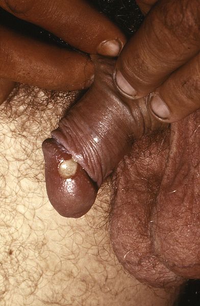
1. Chancroid
  • This is a sexually transmitted infection, most prevalent in third world countries.
  • Chancroid begins as a small lump that then turns into an ulcer.  
  • It is painful
  • The ulcer bleeds easily when rubbed 
  • The ulcer has a greeney-yellow base
  • One third of affected people will develop inguinal lymph node involvement, with half of these developing abscesses after the lymph nodes become so big they break through the skin.
  • Chancroid is caused by the gram negative bacteria haemophillus ducreyi
  • Treatment options are:
    • 1g azithromycin orally or
    • IM ceftriaxone or
    • 7 days erythromycin
Chancroid lesion filled with pus prior to rupture.  From wiki commons, uploaded by Joe Miller


2.  Granuloma inguinale = Donovanosis
  • This is a sexually transmitted infection mainly found in 3rd world countries.
  • Granuloma inguinale begins as a small lump which then bursts into an ulcer/open lesion that continues to spread until treated
  • The ulcer is painless and has a 'beefy red' appearence
  • There is not usually inguinal lymphadenopathy
  • Granuloma inguinale is caused by klebsiella granulomatis
  • Donovan bodies are rod-shaped klebsiella granulomatis found in the cytoplasm of phagocytes in infected individuals.  They stain dark purple with Wright's stain.
  • Treatment is
    • 3 weeks erthyromycin or tetracyline


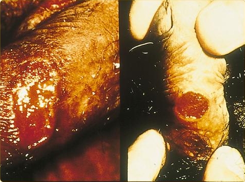
3. Lymphogranuloma venereum
  • This is a sexually transmitted infection
  • It is caused by chlamydia trachomatis (L type)
  • There are several stages of infection:
    • Primary:
      • painless pustule which bursts into a painless ulcer
      • often not noticed by women as may be internal
      • 10% of patients will have accompanying erythema nodosum
    • Secondary
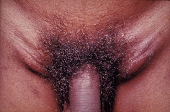
      • tender inguinal lymphadenopathy
    • Tertiary
      • up to 20 yrs later - protocolitis, tenesmus
  • Treatment options:
    • doxycycline or
    • erythromycin

 Image from wiki commons, uploaded by Dr Fred


4. Genital herpes
  • This is a sexually transmitted infection
  • Up to 8 in 10 people who contract it have no symptoms
  • Those who have symptoms tend to develop groups of painful ulcers
  • Primary infection may last up to 3 weeks
  • Subsquent infections tend to be less severe
  • It is highly infective when ulcers are present
  • It is classically caused by HSV 2 but can be caused by HSV 1.
  • Oral aciclovir may be given within the first 5 days of symptoms starting as a 5 day course but there is no cure
  • Subsequent recurrences tend to be less severe.


5. Behcet's disease
  • This is NOT sexually transmitted
  • It is associated with oral ulcers and anterior uveitis
  • See battle 25.2 for more information


Summary of sexually transmitted causes of genital ulcers:


Now for something completely different...

MRCP revision battle 53.2: Vertebral artery dissection

Vertebral artery dissection is an important recognised cause of stroke in patients under 45 years of age.

The vertebral arteries themselves arise from the subclavian and join at the base of the medulla oblongata to form the basilar artery.


Presentation of vertebral artery dissection tends to be:
  • several occipital headache
  • cerebellar signs
  • brainstem signs
    •  CN IX, X, XI and XII:
      • dysarthria
      • dysphagia
    • hiccups
    • loss of sensation to ipilateral face
    • ipsilateral Horner's syndrome in 1/3 of patients
    • 'crossed signs':
      • ipisilateral cranial nerve palsy with contralateral hemiparesis or hemiplegia

Causes include:
  • trauma
  • stretching of neck
    • in MRCP questions look for trips to hairdresser, painting ceiling etc
  • connective tissue disorders

Investigation:
  • CT 
  • 4 vessel angiogram


Treatment:
  • ?neurosurgery
  • anticoagulate if no associated subarachnoid haemorrhage


Now for some intracranial thrombosis...

MRCP revision battle 53.3: Intracranial venous thrombosis

The presentation of intracranial venous thrombosis depends on which venous sinus is affected.

All locations of intracranial venous thrombosis can cause headache.


Isolated sagittal sinus thrombosis (=nearly half of intracranial venous thrombosis) or lateral sinus thrombosis present with:
  • headache
  • vomiting
  • seizures
  • papilloedema
  • potentially focal neurological signs


Cavernous sinus thrombosis can cause:
  • headache
  • oedematous eyelids
  • proptosis
  • painful eye movements/opthalmoplegia
Cavernous sinus thrombosis is often associated with infection spreading from the face or paranasal sinuses.
Remember: cavernous sinus contains CN III, IV, V1, V2 and VI, plus the internal carotid artery.


Sigmoid sinus thrombosis:
  • headache
  • cerebellar signs


Inferior petrosal sinus thrombosis:
  • 5th and 6th nerve palsies

Risk factors for developing intracranial venous thrombosis include:
  • pregnancy
  • head injury
  • recent LP
  • oral contraceptive pill

Investigation is:
  • MRI/CT
    • CT may show 'absent delta sign' which implies a filling defect and therefore a thrombosis
  • ?MRV

Management is by specialists, ?heparin


On to the more familiar topic of infective endocarditis...

MRCP revision battle 53.4: Infective endocarditis

Infective endocarditis is diagnosed by Dukes criteria, which requires:
  • 2 major criteria OR
  • 1 major and 2 minor criteria OR
  • all 5 minor criteria.
Major criteria are:
  • positive blood culture
    • typical organism in 2 separate cultures OR
    • persistently +blood cultures
  • endocardial involvement
    • positive echo
    • new valvular regurgitation
Minor criteria are:
  • fever >38C
  • vascular/immunological signs
  • predisposition (IVDU, valve replacemen)
  • positive blood culture that doesn't mean major criteria
  • positive echo that doesn't meet major criteria

Vascular/immunological signs include:
  • Janeway lesions
  • Oslers nodes
  • splinter haemorrhages
  • Roth spots


40% of cases of endocarditis occur in patients with no previous problems.
30% have had rheumatic heart disease.


Commonest causative organism is streptococcus viridans (alpha haemolytic)
Commonest organism in IVDUs is staphlycoccus aureus.
Commoenst organism within 6 weeks of valve surgery is staph epidermidis.


Usually bicuspid valve affected, except in IVDUs where the tricuspid valve is most commonly affected.


Mortality from strep in 5%, staph around 30%

Poor prognostic factors:
  • s. aureus
  • prosthetic valve
  • culture negative
  • low complement


Treatment:
  • initial blind therapy: fluclox and gent
  • prosthetic valve/penicillin allergy: vancomycin and rifampicin and gent
  • staph: fluclox/vanc and gen
  • strep: benzylpenicillin and gent


Surgery if:
  • abscess
  • recurrent emboli
  • severe valve incompetence
  • cardiac failure


Random MRCP facts:
  • prolongation of PR suggests aortic valve abscess
  • if causative organism is found to be strep bovis look for an associated bowel malignancy.


On to upper GI bleeds...

MRCP revision battle 53.5: Upper GI bleeds

Upper GI bleeds may present with:
  • haematemesis (=vomiting blood)
  • coffee ground vomiting or
  • malaena (=black, tar-like motions)

Commonest causes of upper GI bleeds are:
  • 35% duodenal ulcers
  • 20% gastric ulcers
  • 18% gastric erosins
  • 10% Mallory-Weiss tear
 

Rarer causes of GI bleeds include:
  • variceal haemorrhage
  • aorto-enteric fistula
  • Meckel's diverticulum
  • Peutz-Jeghers syndrome

Risk of rebleeding and mortality from upper GI bleeds is calculated using the Rockall score.
Pre-endoscopy Rockall score is calculated based on:
  • age
    • 0pt <60yrs
    • 1pt 60-79
    • 2pts >80
  • degree of shock
    • 0pt: BP >100 sys and HR <100
    • 1pt: BP >100 sys but HR >100
    • 2pts: BP <100 sys
  • co-morbidities
    • 0pt: none
    • 1pt: heart problems
    • 2pts: liver/renal failure
    • 3pts: mets

Mortality is roughly:
  • 1 in 20 with 2 pts
  • 1 in 10 with 3 points
  • 1 in 4 with 4 points
  • 1 in 2 with 7 points

Post endoscopy mortality is calculated based on the initial score, the diagnosis and the signs of haemorrhage seen.

Below is a summary of the Rockall score from the SIGN guidelines:


Management of upper GI bleeds is:
  • classic ABC resuscitation
  • if cause suspected to be variceal haemorrhage: IV terlipressin 2mg then 2mg/4hr
  • urgent endoscopy
  • surgery if endoscopy unsuccessful
  • if cause is ulcer: omeprazole after endoscopy
  • if cause is variceal haemorrhage: consider transjugular intrahepatic portosystemic shunt to prevent rebleeding (note: nearly 25% of people with a TIPS get hepatic encephalopathy)


Now to Meckel's diverticulum...

MRCP revision battle 53.6: Meckel's diverticulum

Meckel's diverticulum is the vestigial remnant of the vitellointestinal duct.


Its features are often recalled as a series of '2s':
  • found in 2% of the population
  • around 2 inches long
  • at 2 foot from the ileocaecal valve

It may present with:
  • painless rectal bleeding
  • GI obstruction
  • abdominal pain due to faeces trapped inside diverticulum

Investigation is with radionucleotide scan.

Treatment for complications is surgical.


Now to end on an unusual condition - cat scratch disease.

MRCP revision battle 53.7: Cat scratch disease

Cat scratch disease is  usually caused by bartonella henselae (a gram negative rod)

Features:
  • several erythematous, crusted lesions at site of scratch
  • regional lymphadenopathy
  • up to half of patients will feel systemically unwell

If confirmation of diagnosis is required an indirect fluorescent antibody test for bartonella is avaliable.


Treatment is generally supportive and resolution tends to occur within 2 months.

Prevention is keeping cats flea- free!

Wednesday, 17 November 2010

MRCP revision battle 52.1: Zollinger-Ellison Syndrome

Finally I have a topic to put under the 'z' catagory in my A to Z index!

Today we have:

MRCP revision battle 52.1: Zollinger-Ellison Syndrome
MRCP revision battle 52.2: Congential Hyperbilinrubinaemia
MRCP revision battle 52.3: Metformin
MRCP revision battle 52.4: Flecainide
MRCP revision battle 52.5: Arrhythmogenic right ventricular cardiomyopathy
MRCP revision battle 52.6: Pulmonary hypertension and cor pulmonale
MRCP revision battle 52.7: Scromboid poisoning


MRCP revision battle 52.1: Zollinger-Ellison Syndrome


Zollinger-Ellison syndrome is a rare condition in which there are multiple gastric and duodenal ulcers in association with gastrin-secreting adenoma. 

The adenoma is usually pancreatic in origin but may be found in the stomach or duodenum.


50-60% of the adenomas in Zollinger-Ellison syndrome are malignant.
10-30% are associated with MEN-1.
(quick recap: MEN 1 = pancreatic tumours, parathyroid tumours and pituitary tumours)


Presentation:
  • epigastric pain from the ulcers
  • diarrhoea
  • malabsorption/steatorrhoea from inactivation of pancreatic enzymes

Diagnosis is by a raised fasting gastrin level.
Note gastrin levels will also be raised in achlorhydria - this differential can be eliminated by doing a secretin stimulation test which will cause a raised gastrin level in Zollinger-Ellison syndrome but no rise with achlorhydria.


Treatment is with high dose PPI (eg 60mg/day).
Ocreotide (somatostatin analogue) may help with symptom relief.


On to consider some congenital causes of jaundice...

MRCP revision battle 52.2: Congential Hyperbilinrubinaemia

This battle will briefly run through 4 conditions associated with congenital hyperbilinrubinaemia....


1. Gilberts


This is the absolute classic.  Gilberts is inherted in an autosomal recessive fashion and affects 1-2% of the population.


Gilberts is due to low levels of UDP glucuronosyltransferase.
It results in a rise in unconjugated bilirubin.

Gilberts is entirely benign and many sufferers only take on a yellow tinge when they have a concurrent illness.


2. Crigler Najjar

There are 2 types of Crigler Najjar - type 1, which is autosomal recessive, and type 2, which is autosomal dominant.


It is due to there being no UDP glucuronosyltransferase
This results in a catastophic rise in unconjugated bilirubin.


Unless the sufferer has a liver transplant they are likely to die as a baby.


3. Dubin Johnson

Dubin Johnson is an autosomal recessive condition in which there is a mutation in the cMOAT transport protein resulting in a defect of hepatic excretion of bilirubin and a rise in conjugated bilirubin.


This manifests as intermittent jaundice with RUQ pain.


Tests to confirm Dubin Johnson include:
  • coproporphyrin I levels being 3-4x higher than coproporphyrin III- in normal subjects this is reversed
  • normal levels of urine coproporphyrin  but 80% being the I isomer, when normally this would be 25%
  • at postmortem: liver has black pigmentation

Happily Dubin-Johnson is a benign condition


4. Rotor syndrome

Rotor syndrome is an autosomal recessive disorder which is similar to Dubin-Johnson and is also due to a defective mechanism of excretion of conjugated bilirubin.

It is also benign.

Rotor syndrome can be differentiated from Dubin-Johnson as:
  • Dubin-Johnson has normal levels of urinary coproporphyrin while Rotor syndrome has high levels
  • liver in Dubin-Johnson has black pigmentation whereas in Rotor syndrome it is normal


Now on to metformin...

MRCP revision battle 52.3: Metformin

Metformin's mechanism of action is:
  • increasing gluconeogenesis
  • increasing insulin sensitivity and therefore increasing peripheral utilisation of glucose
  • possibly by decreasing GI absorption of carbohydrates

Metformin works only in the presence of endogenous insulin and is therefore only effective if there is residual functioning pancreatic islet cells.


Adverse effects of metformin include:
  • GI upset - to an intolerable degree in up to 20% of subjects
  • decreased B12 absorption - rarely clinically relevant but popular in MRCP questions
  • rarely it may provoke lactic acidosis - usually only if there is also renal impairment.

Contra-indications to metformin include:
  • eGFR <30 
  • IV contrast
  • stop of morning of general anaesthesia
  • pregnancy
  • breast feeding

Now on to a cardiac drug as we enter the cardiology section of today's battles...

MRCP revision battle 52.4: Flecainide

Flecainide is a class Ic antiarrhythmic which works by blocking the sodium channels.

Flecainide causes a wider QRS and longer PR.


Indications for flecainide are:
  • AF
  • SVT with accessory pathway

Its half life is 16 hrs.


Flecainide is contraindicated post MI.
It should be used with caution in those with pacemakers as it can raise the potential needed for stimulation.


Adverse effects of flecainide include:
  • negatively inotropic
  • bradycardic
  • proarrhythmic
  • oral parasthesia
  • visual disturbances
  • rarely: pneumonitis

Next - Arrhythmogenic right ventricular cardiomyopathy

MRCP revision battle 52.5: Arrhythmogenic right ventricular cardiomyopathy

Arrhythmogenic right ventricular cardiomyopathy is the second most common cause of sudden cardiac death in young people after HCM.

It is characterised by fatty and fibrofatty tissue infiltrating predominantly the right ventricle.

It may be inherited in an autosomal dominant fashion with variable penetrance.


Presentation may be:
  • palpitations
  • syncope
  • sudden death

ECG may be normal, or:
  • T wave inversion V1-V3
  • epsilon wave = terminal notch in QRS

The investigation of choice is MRI.


Treatment includes:
  • sotolol
  • ablation
  • ICD


An MRCP-gem to remember is Naxos disease, which is the association of arrhythmogenic right ventricular cardiomyopathy with palmoplantar keratosis and wooley hair.


Now for some pulmonary hypertension...

MRCP revision battle 52.6: Pulmonary hypertension and cor pulmonale

Definition of pulmonary hypertension

Normal pulmonary artery pressure is 12 to 16 mmHg.

Pulmonary hypertension is defined as pulmonary artery pressure:
  • >25mmHg at rest OR
  • >30mmHg on exercising

Note that technically a Swan-Ganz catheter/cardiac catherisation is needed to diagnose pulmonary hypertension; however, cardiac echo is often used.  Echo reports tend to quote systolic pulmonary artery pressure rather than mean - to convert to mean multiply by 0.61 and add 2.


Cor pulmonale is right heart failure caused by pulmonary hypertension.


Features of pulmonary hypertension


Tends to present with progressive shortness of breath.


On examination look for signs of right heart failure:
  • right ventricular heave
  • raised JVP, a waves
  • loud P2
  • pansystolic murmur - tricuspid regurgitation


Types of pulmonary hypertension


Primary - accounts for <1% pulmonary hypertension
10% of cases are familial and inherited in an autosomal dominant fashion


Secondary  - commonest cause is COPD
Other secondary causes of pulmonary hypertension include:
  • IHD
  • MV disease
  • left to right shunts
  • chronic hypoxia


Treatment of pulmonary hypertension
  • If secondary, treat cause
  • Give diuretics
  • anticoagulate
  • vasodilators:
    • calcium channel blockers
    • IV prostaglandins
    • bosentan = endothelial antagonist
    • sildenafil = PDE-5 inhibitor
  • transplant

5 yr survival is less than 50%


On to a fishy battle...

MRCP revision battle 52.7: Scromboid poisoning

Scromboid poisoning is caused by the ingestion of amines, mainly histamines, which are produced by bacterial decarboxylation of histadine in fish meat (mainly tuna, mackeral, sardines, anchoives, marlin)

The commonest cause of scromboid poisoning is ingestion of spoiled fish following improper refridgeration.  Cooking well will not inactivate the hisatmines that have been produced.

Degree of symptoms correlates to amount of fish consumed.

Symptoms include:
  • nausea
  • abdo pain
  • diarrhoea
  • flushing
  • rash
  • headache 
  • palpitations
  • hypo or hypertension
Onset of symptoms tends to be within 10 to 30 mins but may take up to 2 hours
Symptoms tend to settle within 36 hours

Treatment is with antihistamine; corticosteroids are not indicated.


People with asthma or on isoniazid may be more severely affected.

Tuesday, 16 November 2010

MRCP revision battle 51.1: Rheumatic fever

Today includes a triple helping of cardiology with some random topics mixed in...


MRCP revision battle 51.1: Rheumatic fever
MRCP revision battle 51.2: Hypothermia
MRCP revision battle 51.3: Dilated cardiomyopathy
MRCP revision battle 51.4: Coeliac's Disease
MRCP revision battle 51.5: Botulism
MRCP revision battle 51.6: Hypertrophic cardiomyopathy
MRCP revision battle 51.7: Bacterial vaginosis


MRCP revision battle 51.1: Rheumatic fever


Rheumatic fever is a systemic infection caused by group A beta haemolytic strep.


Diagnosis is by the revised Jones criteria which stipulates:
  • evidence of preceeding streptococcl infection AND
  • 2 major criteria OR 1 major and 2 minor criteria

Major criteria are:
  • carditis
  • polyarthritis
  • chorea
  • erythema marginatum
  • subcutaneous nodules

Minor criteria are:
  • fever
  • arthralgia
  • raised ESR/CRP
  • long PR
  • previous rheumatic fever

The cardiac histological marker is the aschoff nodule


Treatment is:
  • bed rest until CRP normal for 2 weeks
  • aspirin
  • penicillin

60% of patients with carditis develop chronic rheumatic heart disease, with the mitral valve most commonly affected.


Rheumatic fever may recur so prophylaxic penicillin should be given until the age of 30 and when dental procedures are being carried out after that.


Now for a cold topic...