Thursday, 4 November 2010

MRCP revision battle 42.2: Mononeuropathies

Mononeuropathies are lesions of individual nerves.  The main causes of mononeuropathies are trauma or entrapment.


If 2 or more nerves are affected it is called mononeuritis multiplex. Causes may be remembered by the mnemonic DRAWS PLC:
  • diabetes
  • rheumatoid
  • amyloid
  • wegeners
  • sarcoid/SLE
  • PAN
  • leprosy/lyme disease
  • carcinomatosis/churg strauss


Important individual mononeuropathies to be familiar with include:


Key nerves of the arm: RUM - radial nerve C5-T1, ulnar never C6-T1 and median nerve C7-T1


Radial Nerve
  • Origin: C5-T1
  • Route: radial groove in humerus then anterior to the lateral epicondyle
  • Sensory supply: lateral 3.5 fingers in back of hand; only small area at base of thumb unique
  • Motor supply: tricipes and extensors of wrist/fingers plus suppinators
  • Classical presentation: 'Saturday night paralysis' - fall asleep with arm across back of chair, compress radial nerve in its groove and bruise it so awake with wrist drop.


Ulnar Nerve
  • Origin: C6-T1
  • Route: posteriomedial aspect of humerus then adjacent to ulna.  Runs superficial to flexor retinaculum into hand.
  • Sensory supply: medial 1.5 fingers
  • Motor supply: interossei, hypothenar eminence, medial 2 lumbicals
  • Classical presentation: claw hand/unable to cross fingers 


Median Nerve
  • Origin: C7-T1
  • Route: medially, goes through carpal tunnel
  • Sensory supply: lateral 3.5 fingers palmar aspect of hand plus nail beds of these fingers
  • Motor supply: pronator, LOAF = 1st and 2nd lumbricals, opponens pollicis, abductor pollicis brevis, flexor pollicis brevis (=thenar enimence)
  • Classical presentation: carpal tunnel syndrome 


Carpal Tunnel Syndrome is so common it deserves its very own battle...

MRCP revision battle 42.3: Carpal tunnel syndrome

Carpal tunnel syndrome is a mononeuropathy of the median nerve at the carpal tunnel = space under the transverse carpal ligament and above the carpal bones.


It is the commonest mononeuropathy.


Classical presentation is pain in hand and arm with paraesthesia in thumb, index and middle fingers.  The pain is often worse at night and is relieved by dangling the hand out of bed and shaking it.


'Bedside tests' include:
  • Phalen's test = maximal wrist flexion ('praying') for 1 minute recreates symptoms
  • Tinel's test = tapping over nerve at wrist recreates symptoms
(I remember which way round these tests go by thinking 't for tapping, t for tinel)


Associations with carpal tunnel syndrome include:
  • diabetes
  • acromegaly
  • hypothyroidism
  • pregnancy
  • RA

 Management may be conservative, spliting, steroid injections or surgery.


Now on to some diabetic neuropathy...

MRCP revision battle 42.4: Diabetic neuropathy

Diabetics may suffer from several different types of neuropathy.


1. Diabetic sensory neuropathy

The classic diabetic neuropathy is sensory neuropathy.  This causes a distal numbness ('glove and stocking') with tingling and pain.  

Treatment:
  • 1st line: TCA
  • 2nd line: gabapentin, pregabalin


2. Diabetic amyotrophy

Diabetic amyotrophy is sometimes known as proximal diabetic neuropathy.  This causes painful wasting of the quadriceps.   It is thought to be caused by occlusion of the vasa nervorum of the proximal lumbar plexus.  There is loss of the knee reflexes.


3. Autonomic neuropathy

If you need a recap refer back to revision battle 32.7


4. Mononeuritis multiplex

Particularly of CN III and VI.


Next up... a bit of nystagmus...

MRCP revision battle 42.5: Nystagmus

Nystagmus is involuntary, jerky eye movements.  

Nystagmus at the extremes of gaze is normal.


Horizontal nystagmus may be due to:
  • vestibular lesions - acutely the nystagmus is away from the affected side
  • cerebellar lesions - tends to be fast nystagmus and towards the affected side
  • MS - suspect if nystagmus is more in the eye which is abducting
  • benign positional vertigo - suspect if nystagmus varies with head position
  • peripheral lesion - suspect if also tinnitus/other signs


Down beat nystagmus is associated with:
  • Arnold Chiari malformations
  • Syringobulbia  (not sure what these are? revise battle 11.3!)


Upbeat nystagmus is associated with:
  • cerebellar vermis lesions
  • organophosphate poisoning


Sticking with eyes we're now going to consider miosis....

MRCP revision battle 42.6: Miosis

Miosis is a small pupil.

Causes of miosis include:

(my personal mnemonic for miosis: AAA pooch)
  • age
  • Argyll Robertson pupil
  • antipsychotics
  • pontine haemorrhage
  • pilocarpine
  • opiods
  • organophosphates
  • cluster headache
  • Horners

On to Pick's disease....

MRCP revision battle 42.7: Picks Disease

Pick's disease is the eponym for frontotemporal dementia.


It presents as loss of inhibition, socially unacceptable behavior, loss of empathy and compulsive behaviour.  There may also be loss of language skills.


Memory and spatial skills remain intact.


Pathophysiologically there are tau-positive inclusions.

 
There is no cure.

Wednesday, 3 November 2010

MRCP revision battle 41.1: Horner's syndrome

Today is another day of completely random battles... with a mnemonic of a addicts hammock and some fevers of unknown cause hopefully each battle should have something of interest in it...


MRCP revision battle 41.1: Horner's syndrome
MRCP revision battle 41.2: Gynaecomastia
MRCP revision battle 41.3: Gingival Hyperplasia
MRCP revision battle 41.4: Leptospirosis
MRCP revision battle 41.5: Opioid and benzo conversions
MRCP revision battle 41.6: Pyrexia of unknown origin
MRCP revision battle 41.7: Fitness to fly


MRCP revision battle 41.1: Horner's syndrome

Horner's syndrome is the triad of:
  • miosis
  • ptosis
  • anhydrosis

 It is caused by an interruption of the sympathetic pupillomotor fibres


Causes may be:
  • central - which causes anhydrosis of the face, arm and trunk
  • pre-ganglionic = second order - which causes anhydrosis of face
  • post-ganglionic = third order - which causes no anhydrosis

A general way to remember the causes is PC Stem:
  • pancoast tumour
  • cervical rib
  • cluster headache
  • carotid artery dissection
  • stroke
  • syringomyelia
  • trauma
  • thyroidectomy
  • encephalitis
  • multiple sclerosis

 An alternative way to try and learn them by their location is:
  • central: STEMS 
    • stroke
    • tumour
    • encephalitis
    • multiple sclerosis
    • syringomyelia
  • pre-ganglionic: PCT
    • pancoast tumour
    • cervical rib
    • trauma
    • thyroidectomy
  • post ganglionic: 2Cs
    • carotid artery dissection
    • cluster headache

Now for some big breasts...

MRCP revision battle 41.2: Gynaecomastia

Gynaecomastia = enlargement of male breast tissue.


Causes can be divided into:


Physiological
  • adolescence
  • increasing age - due to low testosterone

Drugs (a mnemonic to remember drugs causing gynaecomastia is a addicts hammock)
  • antipsychotics
  • ACE inhibitors
  • digioxin
  • diazepam
  • isoniazid
  • cimetidine
  • TCA
  • spironolactone
  • heroin
  • amiodarone
  • metronidazole
  • marijuana
  • omeprazole
  • calcium channel blockers
  • ketoconazole


Pathological
  • klinefelters
  • liver cirrhosis
  • hyperthyroidism


So from enlarged breasts to enlarged gums...

MRCP revision battle 41.3: Gingival Hyperplasia

Gingival hyperplasia is the overgrowth of the ginival (=gum) tissue.


Photos of gingival hyperplasia have appeared in part 2 written exams so a grasp of the main differentials is worthwhile.


Conditions to consider include:
  • poor oral hygiene leading to gingivitis
  • AML
  • vitamin C deficiency
  • pregnancy
  • drug-induced casues:
    • phenytoin
    • ciclosporin
    • calcium channel blockers (nifedipine, verapamil)


Now for a change to a spirochaete...

MRCP revision battle 41.4: Leptospirosis

Leptospirosis, also known as Weil's disease, is a disease caused by infection with the spirochaete leptospira interrogans.


It is classically caught from water contaminated with rat's urine, so suspect it as a diagnosis in those in questions who have recently fallen into rivers/out of rowing boats.


After a 2 to 20 day incubation period the patient develops:
  • fever
  • myalgia
  • cough/haemoptysis
  • headache

The patient then either makes a good recovery or develops:
  • meningitis
  • jaundice
  • renal failure (50%)


Treatment is with penicillin/amoxicillin/doxycycline.


Lets move on to some conversions...

MRCP revision battle 41.5: Opioid and benzo conversions

Occasionally in real-life/MRCP exam you may need to convert between different drugs in the same family, so this revision battle briefly covers 2 popular families: the opioids and the benzos.


Opioids

First, as an aside: remember opiate refers to a substance derived from opium (from the poppy) while opioids are synthetic/semi-synthetic versions of opiates.

  • PO codeine to PO morphine: divide by ten
  • PO tramadol to PO morphine: divide by five
  • PO morphine to PO oxycodone: divide by two
  • PO morphine to S/C morphine: divide by two
  • PO morphine to S/C diamorphine: divide by three
  • PO morphine to S/C oxycodone: divide by three
  • morphine to fentyl: divide by 100


Other misc notes:
  • codeine relies on conversion to an active form by the P450 enzyme group.  Not all patients are able to do this (roughly 10% of the population struggle), and other drugs (such as SSRIs) may interfere with the effectiveness of conversion so the same amount of codeine may have a very variable analgesic effect on different people.
  • doses of all opioids should be reduced in renal failure except fentanyl
  • if asked to prescribe a breakthrough dose this is usually 1/6th of the total 24hr dose.


Benzos (and derivative)


Rough conversions are:

Chlordiazepoxide 15mg = Temazepam 10mg = Diazepam 5mg = Lorazepam 0.5mg


Decrease by 1/8th every 2 weeks.


Now for a little pyrexia of unknown origin...

MRCP revision battle 41.6: Pyrexia of unknown origin

Petersdorf and Beeson defined pyrexia of unknown origin (PUO) as:
  • fever >38.3C on several occasions
  • longer than 3 weeks
  • with no diagnosis reached after 1 week of inpatient investigation

Causes:
  • infections:
    • abscesses
    • TB
    • malaria
    • any other varient...
  •  malignancy
    • lymphoma
    • hypernephroma
    • leukaemia
    • atrial myxoma
  • connective tissue disease
    • RA
    • SLE


Daily spikes: abscess; TB; schistosomiasis
Twice daily spikes: leishmaniasis


On to the final battle of the day - fitness to fly...

MRCP revision battle 41.7: Fitness to fly

When a patient can fly pops up intermittently, particularly in part 1 written.  The following periods after various medical events at which it is considered safe to fly are quoted from the excellent patient.co.uk website and references can be found there.

  • uncomplicated MI: 7 days
  • complicated MI: 4 weeks
  • CABG: 10 days
  • PCI: 5 days
  • pneumothorax: 2 weeks post resolved CXR
  • pregnancy: contraindicated after 36 weeks gestation in singleton pregnancy or 32 weeks gestation if multiple pregnancy
  • laparoscopy/colonoscopy: 24 hrs
  • abdominal surgery: 10 days
  • cast: 24 to 48 hrs
  • CVA: 10 days
  • sickle cell: needs in flight oxygen

Tuesday, 2 November 2010

MRCP revision battle 40.1: Oxygen dissociation curve

After a few days of 'pure' topics its back to the joys of a mixed bag for battle set 40.  Be prepared for a rapid tour through everything from poisoning to mountain sickness and pharmacology!


MRCP revision battle 40.1: Oxygen dissociation curve
MRCP revision battle 40.2: Carbon monoxide poisoning
MRCP revision battle 40.3: Acetylator status
MRCP revision battle 40.4: Generalised lymphadenopathy
MRCP revision battle 40.5: Acute mountain sickness
MRCP revision battle 40.6: Trisomies - Down's, Edward's and Patau's
MRCP revision battle 40.7: Hereditary angioedema


MRCP revision battle 40.1: Oxygen dissociation curve


The oxygen dissociation curve illustrates now easily haemoglobin gives up oxygen at a certain partial pressure of oxygen.


A shift in the curve to the left means lower oxygen delivery (L for left L for lower delivery).  This means the haemoglobin has a higher affinity for the oxygen.

Causes of a shift to the left include:
  • less acidity
  • lower temperature
  • less 2,3 DPG
  • HbF
  • carboxy/methyl haemoglobin


A shift in the curve to the right means higher oxygen delivery, and hence the haemoglobin has a lower affinity for the oxygen.

Causes of a shift to the right include:
  • higher acidity
  • higher temperature
  • higher levels of 2,3 DPG


The Bohr effect states that the curve will move to the right when there is a higher concentration of CO2 (and hence higher acidity).  This explains how the body releases oxygen to the tissues that need it the most.


The Haldane effect states that in the presence of raised oxygen carbon dioxide will bind less well to haemoglobin.


After that chunk of physiology lets go for a bit of poisoning...

MRCP revision battle 40.2: Carbon monoxide poisoning

Carbon monoxide has 200x more affinity for Hb than oxygen
This results in a left shift of the oxygen dissociation curve.


Normal levels of carboxyhaemoglobin are <3% in non-smokers or <10% in smokers.


In terms of carbon monoxide poisoning:
  • 10-30% carboxyhaemoglobin = mild poisoning
  • 30-60% = severe poisoning


Features of carbon monoxide poisoning are vague:
  • 90% have a headache
  • 50% nausea/vomiting
  • 50% vertigo
  • 30% confusion

Patients look pink


Severe CO poisoning can cause:
  • fits
  • coma
  • cardiac arrrest/arrthymias
  • pyrexia
  • muscle necrosis
  • acute renal failure
  • increased reflexes and tone

Treatment is:
  • 100% oxygen
  • hyperbaric oxygen if LOC/pregnant/neuro signs - this decreases the half life from 4 hrs to around 22 minutes.


Next.... some pharmacology with acetylator status

MRCP revision battle 40.3: Acetylator status

Many drugs are metabolised in the liver by acetylation.  However, up to 50% of the UK population is deficient in hepatic N-acetyltransferase, meaning they are likely to accumulate drugs that require acetylation.  These patients are known as slow acetylators.


Drugs affected include (PS HID):
  • procamide
  • sulfasalazine
  • hydrazaline
  • isoniazid
  • dapsone
  • sulphonamides


Now for some generalised lympadenopathy....

MRCP revision battle 40.4: Generalised lymphadenopathy

Causes of generalised lymphadenopathy include:

  • infective
    • glandular fever
    • HIV
    • toxoplasmosis
    • CMV
    • rubella
    • TB
    • roseola infantum
  • neoplastic
    • leukaemia
    • lymphoma
  • drugs
    • phenytoi
    • allopurinol
    • isoniazis
  • rheumatological
    • SLE
    • RA
    • sarcoidosis


After a dull list lets cover the sexier topic of acute mountain sickness....

MRCP revision battle 40.5: Acute mountain sickness, HAPE and HACE

Acute mountain sickness


Acute mountain sickness is usually a self-limiting condition.  

It tends to occur at altitudes higher than 2500m and develops gradually over 6 to 12 hours.

Symptoms are:
  • headache
  • nausea
  • fatigue


Ironically, fitter people are more at risk.

Risk can be decreased by gaining altitude at no more than 500m/day
Acetazolamide (a carbonic anhydrase inhibitor) can also help prevent.


The treatment is to descend.


HAPE = High Altitude Pulmonary oEdema


HAPE presents like pulmonary oedema.


Treatment is:
  • rapid descent
  • nifedipine
  • dexamethasone
  • acetazolamide
  • phosphodiesterase type V inhibitors (=tadalafil)


HACE = High Altitude Cerebral oEdema


This presents as headache, ataxia and papilloedema

Treatment is descent and dexamethasone.


Now for some trisomies....

MRCP revision battle 40.6: Trisomies - Down's, Edward's and Patau's

A trisomy is a genetic abnormality in which there are 3 copies of a chromosome instead of the usual 2.  It is a form of aneuploidy (=when the number of chromosomes is not a multiple of 23).


This battle will look at the 3 commonest trisomies in order of their frequency, which helpful is the same as the order of their names alphabetically.


Down's Syndrome = Trisomy 21

Causes of Down's:
  • non-disjunction: 94%
  • Robertson translocation: 5%
  • mosaicism: 1%

Risk of Downs with age of mother:
  • 30 yrs: 1/1000
  • 35 yrs: 1/350
  • 40 yrs: 1/100
  • 45 yrs: 1/30


The characteristics of patients with Down's are varible but include:
  • microgenia (=small chin)
  • macroglossia
  • epicanthic folds of eyes
  • short neck
  • single palmar crease
  • flat nasal bridge
  • low IQ
  • short stature

Associations with Down's include:
  • duodenal atresia
  • Hirschsprungs disease
  • AV septal defects (40%)
  • ventricular septal defects (30%)
  • atrial septal defects (10%)
  • PDA/tetralogy of fallot
  • Brushfield spots (=white spots on iris)

Later complications include:
  • ALL
  • atlantoaxial instability
  • hypothyroidism


Edwards Syndrome = Trisomy 18


Characterised by:
  • prominent occiput
  • microcephaly
  • overlapping fingers
  • rockerbottom feet
  • cardiac defects

Rare to survive beyond infancy


Patau's Syndrome = Trisomy 13

Characterised by:
  • CNS, renal and cardiac abnormalities
  • low IQ
  • polydactyly
  • rockerbottom feet

Rare to survive beyond infancy.


To the final battle of the day - hereditary angioedema

MRCP revision battle 40.7: Hereditary angioedema

Hereditary angioedema is an autosomal dominant condition in which a decrease in either the amount of C1 inhibitor or the function of C1 inhibitor results in swelling when the body is exposed to certain substances.


Attacks may be preceded by a painful macular rash.  The swelling may be life-threatening.  The image below from wiki commons (uploaded by Dr Heilman) shows a child with hereditary angioedema.


Increased levels of bradykinin play a central role in the pathphysiology and therefore ACE inhibitors (which prevent bradykinin breakdown) can cause angioedema.


Treatment acutely is with IV C1 inhibitor concentration. 
The anabolic steroid danozol may be given prophylactically as it increases liver C1 inhibitor production by an unknown mechanism.

Monday, 1 November 2010

MRCP revision battle 39.1: Hodgkin's Lymphoma

Its the second day of haematology fun!  Lets dive straight in...


MRCP revision battle 39.1: Hodgkin's Lymphoma
MRCP revision battle 39.2: Non-Hodgkin's lymphoma
MRCP revision battle 39.3: Systemic mastocytosis
MRCP revision battle 39.4: Thrombocytosis
MRCP revision battle 39.5: Haemophilia
MRCP revision battle 39.6: The spleen
MRCP revision battle 39.7: Autoimmune haemolytic anaemia


MRCP revision battle 39.1: Hodgkin's Lymphoma


Lymphomas are malignant proliferations of lymphocytes.  These may accumulate in lymph nodes, in organs or in the peripheral blood.


Lymphomas are divided into 2 main types: Hodgkin's, which has characteristic Reed-Sternberg cells, and non-Hodgkin's, which do not have Reed-Sternberg cells.

The slide below (by Dr Ed Uthman) shows a Reed-Sternberg cell in the centre with its characteristic mirror-image nuclei:


Staging of Hodgkin's lymphoma is done by the Ann Arbor system:
  • I = one lymph node group
  • II = 2 areas on one side of the diaphragm
  • III = both sides of the diaphragm
  • IV = extra-nodal tissues (NB the spleen is counted as an 'honorary' node)

Each stage is subdivided into A or B:
  • A = no B symptoms
  • B =  T>38c, night sweats or weight loss >10% in 6 months

Pel Ebstein fever = cyclical fever with long periods of normal temperature - is so rare some believe it to be mythical...


Pruritus or alcohol-induced pain are not B symptoms bit are useful indicators of relapse.


Poor prognostic factors are:
  • B symptoms
  • stage IV
  • Hb <10.5
  • lymphocyte count <8%
  • male

 Histological subtypes of Hodgkin's lymphoma are:
  • nodular sclerosing = most common, good prognosis
  • mixed cellularity - good prognosis
  • lymphocytic predominant = best prognosis
  • lymphocytic depleated = least common, worst prognosis

Bloods will show:
  • neutrophilia
  • anaemia
  • thrombocytosis
  • raised ESR
  • raised LDH - a useful guide to the bulk of the disease

Treatment is with radiotherapy, chemotherapy (ABVD) or both.


Now on to non-Hodgkin's lymphoma...

MRCP revision battle 39.2: Non-Hodgkin's lymphoma

Non-Hodgkin's lymphoma are a hugely diverse group of diseases and this battle is a highly edited set of highlights for MRCP revision.


Low grade 
  • relatively mature cells
  • indolent but usually incurable
  • try local radiotherapy

High grade
  • immature cells
  • rapidly progressive disease but 40% curable
  • treated with CHOP chemo
  • includes Burkitts lymphoma

B cell lymphoma
  • treated with rituximab

Lymphoblastic
  • treated as ALL


Now for a short battle with an esoteric condition...

MRCP revision battle 39.3: Systemic mastocytosis

Systemic mastocytosis is a neoplastic proliferation of mast cells.


Features include:
  • flushing
  • abdominal pain
  • monocytosis on blood film
  • urticaria pigmentosa (Darier's sign)

It is diagnosed by raised serum tryptase and urinary histamine


It is vanishingly rare in real life.


So from something very rare to very common: thrombocytosis

MRCP revision battle 39.4: Thrombocytosis

Thrombocytosis = raised number of platelets.


Primary = essential thrombocythaemia
  • caused by a clonal proliferation of megakaryocytes
  • platelets typically >1000 (x10 to the power 9)
  • abnormal function, risking either thrombosis or bleeding
  • treatment = aspirin.  Hydroxycarbamide is given to lower platelets if pt is >60yrs or they have had a previous thrombosis

Secondary thrombocytosis
  • reactive to
    • bleeding
    • infection
    • trauma
    • thrombosis
    • infarction
    • iron deficiency


Now for some haemophilia....

MRCP revision battle 39.5: Haemophilia

Haemophilia A and Haemophilia B clinically present very similarly, with bleeds into joints and muscles.

Both are inherited in an X-linked recessive way but 1/3 of cases will have no family history.

Both cause raised aPTT.


Haemophilia A = lack of factor VIII
Haemophilia B = lack of factor IX = Christmas disease


Treatment is with desmopressin and tranexamic acid.  Major bleeds may need recombinant factor VIII.  Avoid NSAIDs and IM injections.


On to an organ that has always bewildered me... the spleen

MRCP revision battle 39.6: The spleen

My dubious relationship with the spleen goes waaayyy back to medical school anatomy days when I learnt that any mass of tissue that I had absolutely no idea what it was when placed on a tray in front of me was usually a spleen.


The spleen was equally mysterious to the medical profession in general for many years, as no-one was quite sure as to its function.  It is now known to act as a reservoir for lymphocytes and so is vital in dealing with bacteraemias.


This battle will look at the 2 extremes of spleen: splenomegaly, and hyposplenism/post splenectomy.


Splenomegaly

Massive splenomegaly can occur in:
  • CML
  • myelofibrosis
  • malaria
  • leishmaniasis
  • Gaucher's syndrome (= an inherited lysosomal storage disease)

Moderate splenomegaly can occur in:
  • infections: typhoid, brucella, TB, glandular fever, EBV, schistosomiasis
  • haematological disorders: lymphoma, leukaemia, haemolytic anaemia
  • connective tissue disease: SLE, RA
  • portal hypertension: cirrhosis, CCF


Splenectomy

Splenectomy is performed for a variety of reasons including:
  • trauna
  • ITP
  • warm AIHA

2 weeks prior to splenectomy pneumoccal vaccine and HiB should be given; if the splenectomy is an emergency these should be given ASAP afterwards.
Lifelong penicillin (or erythromycin if penicillin allergic) is then also needed.


Most common serious infections after splenectomy are from encapsulated organisms such as s.pneumoniae, h.influenzae and n.meningitidis.


Blood film after splenectomy shows:
  • Howell-Jolly bodies (=nuclear remnants in RBCs)
  • target cells
  • Pappenheimer bodies (=abnormal iron inside RBCs)

There is low IgM.


As well as hyposplenism being a problem post splenectomy, it can also occur in sickle cell disease, coeliac disease and myeloproliferative diseases


Now onwards to some AIHA...

MRCP revision battle 39.7: Autoimmune haemolytic anaemia

Autoimmune haemolytic anaemia (AIHA) is mediated by autoantibodies and results in mainly extravascular haemolysis.

AIHA will result in a positive direct antiglobulin test (= positive Coombs test)


AIHA is divided depending on the optimal binding temperatures of the autoantibodies to the RBCs.


Warm AIHA = 37C
  • Ig G
  • SLE, lymphoma, CLL, methyl dopa
  • treatment: steroids, immunosupression, splenectomy

Cold AIHA = 4C 
  • IgM
  • lymphoma, mycoplasma, EBV
  • treatment: keep warm, ?chlorambucil

Sunday, 31 October 2010

MRCP revision battle 38.1: Von Willebrands Disease

We're about to enter a 2 day fest of haematology.  Try to get through the dull battles of B and T cell disorders as the topics on the other side of them are slightly more stimulating.  Good luck!


MRCP revision battle 38.1: Von Willebrands Disease
MRCP revision battle 38.2: B cell disorders
MRCP revision battle 38.3: T cell disorders
MRCP revision battle 38.4: Combined B and T cell disorders
MRCP revision battle 38.5: Thrombotic thrombocytopenic purpura
MRCP revision battle 38.6: Microangiopathic haemolytic anaemia
MRCP revision battle 38.7: Sideroblastic anaemia


MRCP revision battle 38.1: Von Willebrands Disease


Von Willebrand's disease is the commonest inherited coagulopathy in the UK.


Von Willebrand's factor is a substance made in epithelial cells which:
  • helps platelets bind to the exposed subendothelium
  • helps platelets bind to each other
  • binds to factor VIII,  helping prevent its destruction in the circulation

There are many types of Von Willebrand's disease.  The commonest 3 are:
  • Type 1: decrease in Von Willebrand's factor 
    • accounts for 80% of Von Willebrand Disease
    • autosomal dominant
  • Type 2: abnormal form of Von Willebrand's factor
    • autosomal dominant
  • Type 3: total lack of Von Willebrand's factor
    • autosomal recessive

Symptoms/signs of Von Willebrand's disease include bruising, menorrhagia, epistaxis and increased bleeding after tooth extraction (a favourite in MRCP exams)


Diagnosis is by:
  • low levels factor VIII
  • low levels VWF antigen
  • deficient ristocetin-induced platelet aggregation

Treatment is:
  • DDVAP (=desmopressin) in mild disease
  • factor VIII concentrate/cryoprecipitate in severe disease
  • avoid NSAIDs.


Next up.... B cell disorders

MRCP revision battle 38.2: B cell disorders

B cells are lymphocytes involved in humoral immunity.  Their functions are:
  • production of antibodies (=plasma B cells)
  • memory of antigen (=memory cells, live for a long time and able to respond rapidly if body encounters same antigen again)
  • antigen presenting cells

B cell disorders result in a failure of antibody synthesis, resulting in either hypogammaglobulinaemia or agammaglobulinaemia.  This causes recurrent infections with pyogenic bacteria and fungi.


Examples of B cell disorders include:


1. Common variable immunodeficiency
  • commonest cause of hypogammaglobulinaemia
  • not familial
  • bone marrow shows a normal number of B cells but they fail to mature


2. IgA deficiency
  • commonest isolated Ig in UK
  • increased risk of giardiasis


3. Bruton's agammaglobulinaemia
  • x-linked recessive
  • no circulating B cells
  • usually presents between 3 months and 2 yrs of age


Now on to the T cell disorders......

    MRCP revision battle 38.3: T cell disorders

    T cells are lymphocytes involved in cell-mediated immunity.  T cells mature in the thymus.

    T cell disorders result in increased susceptibility to virus', mycobacteria and fungi.


    Examples of T cell disorders include:


    1. DiGeorge
    • defect in development of thymus and 3rd/4th branchial arches
    • mnemonic CATCH-22:
      • cardiac abnormalities (tetralogy of fallot)
      • abnormal faies
      • thymic aplasmia --> lack of T cells
      • cleft palate
      • hypocalcaemia (due to absent parathyroids)
      • 22 - recessive inheritance on chromosome 22

    2. Nezelof syndrome
    • absent thymus
    • often some B cell involvement

    3. Purine nucleoside phosphorylase deficiency
    • prevents development of T cells


    Onto the final battle of this dull triad - combined B and T cell disorders

    MRCP revision battle 38.4: Combined B and T cell disorders

    So having skated through B cell disorders and T cell disorders its on to combined disorders, the main 3 of which may be recalled as SCID WAS ATAXIC.


    1. Severe Combined Immundeficiency
    • autosomal recessive
    • sometimes due to lack of adenosine deaminase

    2. Wiskott-Aldrich Syndrome
    • x-linked recessive
    • characterised by recurrent infections, eczema and thrombocytopenia
    • associated with an increased risk of maligancy

    3. Ataxic telangiectasia
    • autosomal recessive
    • cerebellar ataxia and telangiectasia
    • increased risk of malignancy
    • low IgE and IgA


    Lets now move back a bit more mainstream with thrombotic thrombocytopenic purpura...

    MRCP revision battle 38.5: Thrombotic thrombocytopenic purpura

    Thrombotic thrombocytopenic purpura is a condition in which large multimers of Von Willebrand's factor clump platelets together, activating the coagulation system, forming fibrin strands and causing microangiopathic haemolytic anaemia (MAHA).


    The 5 key features are:
    • Thrombocytopenia
    • Fever
    • Fluctating CNS signs (visual disturbance, fits, hemiparesis)
    • MAHA
    • Renal failure

    Adult females are most commonly affected


    The cause is often unknown.  Implicated factors include:
    • drugs - clopidogrel, ciclosporin, COC
    • pregnancy
    • HIV
    • SLE

     Treatment is:
    • plasma exchange
    • IV vincristine  (promotes premature release of platelets from marrow)
    • steroids


    Next up... more microangiopathic haemolytic anaemia...

    MRCP revision battle 38.6: Microangiopathic haemolytic anaemia

    Microangiopathic haemolytic anaemia (MAHA) is a nice condition to learn about as you can easily visualise it: it is mechanical disruption of red blood cells in the circulation, so in your mind's eye see the little RBC's being mangled by prosthetic heart valves or sliced by fibrin strands and then imagine them on the other side, reduced in number (anaemia), all with different amounts of haemoglobin in them after being sliced (polychromasia) and lots of partial fragments (schistocytes and helmet cells).


    So to put that above paragraph into a nice list of haematological features of MAHA:
    • anaemia
    • polychromasia
    • helmet cells
    • schistocytes


    Causes of MAHA include:
    • DIC
    • HUS
    • TTP
    • malignant hypertension
    • severe pre-eclampsia
    • prosthetic heart valves
    • septocaemia


    Now onwards to the last MRCP revision battle of the day, sideroblastic anaemia...

    MRCP revision battle 38.7: Sideroblastic anaemia

    Sideroblastic anaemia is a form of anaemia in which the body is not able to incorporate iron successfully in the haem molecule.


    This results in:
    • anaemia
    • sideroblasts in bone marrow
    • rarely haemosiderosis as the body keeps absorbing iron in its futile attempt to correct the anaemia (haemosiderosis may be revised in MRCP revision battle 28.1)

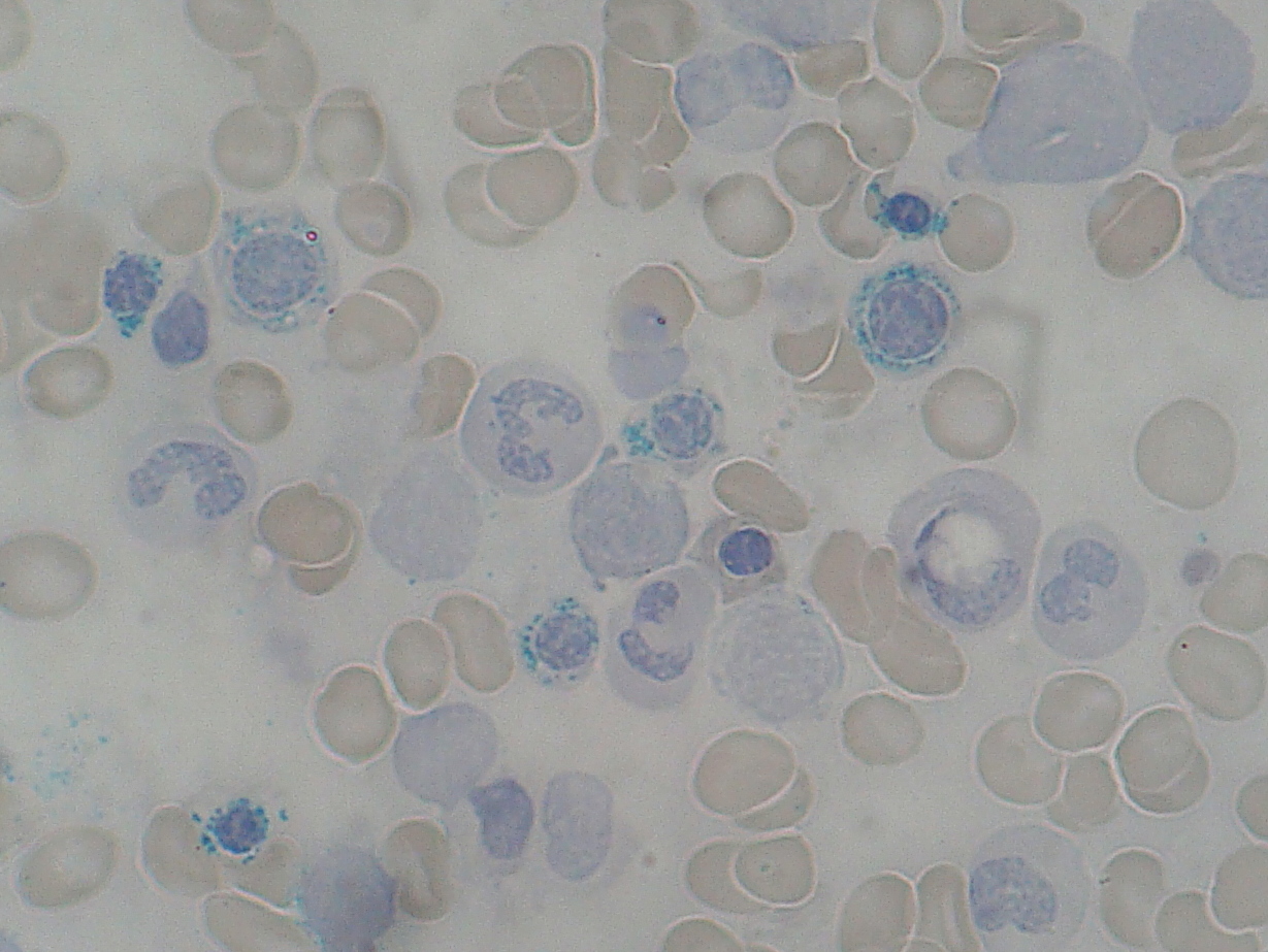
    The image below from Wiki Commons shows ring sideroblasts:


    Causes of sideroblastic anaemia include:
    • congenital causes
      • delta-aminolevulinate synthase deficiency
      • x linked recessive, rare
      • responds to pyridoxine
    • acquired causes
      • myelodysplasias
      • alcohol
      • lead
      • chemotherapy
      • anti-TB meds

    Treatment is to treat the cause, supportive care and pyridoxine.

      Saturday, 30 October 2010

      MRCP revision battle 37.1: TB

      Today has a respiratory feel....


      MRCP revision battle 37.1: TB
      MRCP revision battle 37.2: DVT/PE in pregnancy
      MRCP revision battle 37.3: Asthma
      MRCP revision battle 37.4: Cor pulmonale
      MRCP revision battle 37.5: alpha-1 antitrypsin deficiency
      MRCP revision battle 37.6: Cystic fibrosis
      MRCP revision battle 37.7: HPOA


      MRCP revision battle 37.1: TB


      TB is an infection caused by mycobacterium tuberculosis.  Primary infection is usually in the lungs (ghon focus, usually in the mid or lower zones).  Second favourite site for primary infection = GI, ileocaecal area.


      Incidence =7000/yr in UK


      Spread is by droplet infection. Household contacts have 1 in 4 chance of infection therefore contact tracing is very important and TB is a notifiable disease.


      Primary TB is often symptomless. Although it may cause fever, sweats, anorexia, cough or erthyema nodosum.  If the immune system ‘wins’, the TB is walled off by calcification; however, the TB remains active in ~ 20% calcified areas and may be reactivated when immune system is weakened, eg steroids, diabetes, HIV.


      Post-primary TB makes you ill – fever, malaise, weight loss, anorexia, tiredness, cough, haemoptysis, rarely night sweats.


       It can spread to:
      • brain = meningeal TB
      • spine = Potts fracture
      • blood = miliary
      • skin = lupus vulgaris


      Diagnosis:
      •    CXR
      •    Sputum cultured/stained with Zielh-Nielsen – looking for acid-fast bacilli
      •    Mantoux test of little help in the UK due to BCG

      BCG = bacilli calmette-guerin – decreases risk of TB by 50%


      Treatment: DOTS
      •    2 months rifampicin, isoniazid, pyrazinamide and ethambutol (=RIPE)
      •    then 4 months rifamicin and isoniazid
      •    Pyridoxine throughout to protect against neuropathic effects of isoniazid


      MRCP (and real life) side effects of TB meds to be aware of:

      Rifampicin turns bodily excreations orange
      Isoniazid and rifampicin cause cirrhosis – check LFTs
      Pyrazinamide decreases urea excreation = risk of gout and CI in gout sufferers
      Ethambutol can affect the optic nerve - check colour vision


      On to DVT and PE in pregnancy...

      MRCP revision battle 37.2: DVT/PE in pregnancy

      It is one of those unfortunate situations: pregnancy makes DVT/PE more likely (increases risk around 6x), but the presence of a fetus also makes your preferred investigative tests more risky.


      Risk is more increased if:
      • >35 yrs
      • higher parity
      • raised BMI
      • smoker
      • sickle cell
      • anaemic
      • dehydration
      • not blood group O


      The consensus on management of ?PE in pregnancy seems to be:
      • CXR in all cases
      • compression doppler in all cases - if this shows DVT you can just treat without needing to investigate further


      In terms of the next investigation if you clinically suspect PE but the doppler is normal there is a lack of consensus:
      • CTPA delivers 10-30% less radiation to the fetus but
      • V/Q scan causes less radiation to the maternal breast tissue

      In general CTPA seems to 'win'


      Note that d-dimer is usually 'normal' in the first trimester of pregnancy, rises in the second to third and then returns to baseline at 4-6 weeks post-partum.  However, it is generally not recommended as an investigation in pregnant women.


      Treatment for DVT/PE in pregnancy is LMWH, continued for 6 months (with at least 6 weeks of anticoagulation post-partum)


      For the really keen:


      Next... MRCP revision battle 37.3...

      MRCP revision battle 37.3: Asthma

      Asthma is a condition characterised by periods of dyspnoea, cough and wheeze caused by reversible airways obstruction.


      Asthma diagnosis

      The table below (from the 2009 BTS/SIGN guidelines) lists the factors that increase or decrease the probability of the presentation being asthma:


      If from history/examination you believe there is a:
      • high probability the patient has asthma --> being a trial of treatment
      • intermediate probability of asthma --> perform spirometry:
        • FEV1/FVC <0.7 - trial asthma treatment
        • FEV1/FVC >0.7 - refer to specialist

      After a trial of treatment >400mls improvement in FEV1 or PEFR >15% is significant


      In a peakflow diary, look for diurnal variation >25%


      Treatment of asthma

      Treatment of asthma follows a stepwise approach (BTS/SIGN guidelines):

      1. Mild intermittent asthma
        •  SABA (short-acting inhaled beta 2 agonist) PRN 
        • salamol, salbutamol
      2. Regular preventor therapy
        • corticosteroid 200-800mcg per day
        • beclometasone, fluticasone, budesonide
      3. Initial add-on therapy
        • LABA (long-acting inhaled beta 2 agonist (eg salmeterol))
        • if this doesn't work, stop and trial an oral therapy such as leukotriene receptor antagonist or theophylline
      4. Persistant poor control
        • increase steroid inhaler up to 2000mcg per day
        • add leukotriene receptor antagonist or theophylline
      5. Continuous or frequent oral steroids


      If stable for 3 months, consider stepping down a step.


      Severity of asthma

      In acute asthma attacks, severity may be graded as follows:

      Severe:
      • unable to complete sentences
      • RR>25
      • PEFR<50% predicted or best
      • pulse >110

      Life-threatening:
      • silent chest
      • PEFR <33% predicted or best
      • bradycardia
      • hypotension
      • normal or raised CO2
      • exhaustion


      Management of acute severe asthma

      Think 'O! sip Ma':
      • high flow oxygen
      • 5mg salbutamol neb
      • 500mcg ipratropium neb
      • 30mg prednisolone (or 100mg hydrocortisone IV)
      • magnesium 1.2g and aminophylline if still not improving
      And make sure ITU know if things aren't going well....


      Now for a battle with cor pulmonale...

      MRCP revision battle 37.4: Cor pulmonale


      Cor pulmonale is right heart failure caused by chronic pulmonary hypertension

      Normal pulmonary artery pressure = 15mmHg.
      Pulmonary hypertension = pulmonary artery pressure >25mmHg at rest or >30mmHg on exercise


      Causes of cor pulmonale include:


      • lung disease - of which COPD is by far the commonest
      • pulmonary vascular disease - PE
      •  rib cage deformity


      Presentation is:
      • dyspnoea
      • fatigue
      • syncope


      Signs:
      • raised JVP, a and v waves
      • RV heave
      • loud P2
      • pansystolic murmur - tricuspid regurgitation
      • peripheral oedema
      • hepatomegaly
      • Graham Steell murmur = high-pitched early diastolic murmur, best heard 2nd left intercostal space with pt in full inspiration = pulmonary regurgitation


      Investigations:
      • CXR
      • FBC - look for secondary polycythaemia
      • ECG - R axis deviation, right ventricular hypertrophy


      Management:
      • treat cause
      • treat symptoms
      • ?heart-lung transplant


      Next up: alpha-1 antitrypsin deficiency

      MRCP revision battle 37.5: alpha-1 antitrypsin deficiency

      alpha-1 antitrypsin is a serine protease inhibitor produced in the liver.


      In the lung it prevents neutrophil elastase from breaking down the alveolar wall.
      It is inactivated by cigarette smoke.


      Genetic varients of alpha-1 antitrypsin exist and are labelled based on their electrophoresis mobility, with M (medium), S (slow) and Z (very slow).  MM is 'normal' while 'ZZ' has the worst prognosis with panlobular emphysema.


      Patients with an alpha-1 antitrypsin defiency are likely to develop COPD early (even without smoking) and liver cirrhosis is common.


      Now for a very straightforward battle - cystic fibrosis

      MRCP revision battle 37.6: Cystic fibrosis

      CF is an autosomal recessive genetic disorder which results in a mutation of CFTR, leading to Cl not leaving cells and so more Na and water entering, resulting in thick, sticky secreations.


      The mutation is on chromosome 7.  70% are ΔF5O8


      Incidence = 1/2000.  Carriers = 1/25


      Presentation in neonates is failure to thrive, meconim ileus or rectal prolapse.
      Later presentation tends to be recurrent respiratory infections, cough, wheeze, bronchiectasis.  85% have steatorrhoea.


      Signs include:
      -    cyanosis
      -    clubbing
      -    bilateral course crackles


      Diagnosis is by pilocarpine sweat test.  Na/Cl>60mmol/l, usually Cl>Na.  CXR may show bronchietasis.  PCR can pick up 90% of mutations.  Faecal elastase is a good screening tool for exocrine pancreatic dysfunction.  Spirometry shows obstruction.


      Treatment is MDT, physio, enzyme replacement, heart/lung transplant.


      Complications include  
      • diabetes (1/3) 
      • gallstones
      • cirrhosis
      • OP
      • male infertility (no vas deferens or epididimus)
      • vasculitis
      • nasal polyps (1/3) 
      • HPOA (if you don't know what this is you will by the end of the next battle)
      • infections
        • s.aureus
        • h.influenzae
        • pseudomonas
        • s.pneumoniae
        • burkholderia cepacia (gram negative)
        • aspergillos


      It tends to be the upper lobes of the lungs that are damaged.


      Mean age of death ~30.


      So to the final battle of the day, HPOA...

      MRCP revision battle 37.7: HPOA

      HPOA = hypertrophic pulmonary osteoarthropathy is a paraneoplastic syndrome consisting of periostitis, arthritis and clubbing.


      It is seen in around 50% of patients with mesothelioma.

      Of the 'usual' lung cancers it is most commonly associated with adenocarcinomas and least commonly associated with small cell lung cancer.


      Thats it for today

      Thursday, 28 October 2010

      MRCP revision battle 36.1: Wilson's disease

      Another set of battles written before commencing the joy of a 10 hour shift... and to think we all used to think revising for finals was hard!


      MRCP revision battle 36.1: Wilson's disease
      MRCP revision battle 36.2: Drugs causing impaired glucose tolerance
      MRCP revision battle 36.3: Acromegaly
      MRCP revision battle 36.4: Vitamin C
      MRCP revision battle 36.5: Insulinoma
      MRCP revision battle 36.6: Metabolic syndrome
      MRCP revision battle 36.7: Phaeochromocytoma


      MRCP revision battle 36.1: Wilson's disease


      Wilson's disease is an autosomal recessive condition inherited on chromosome 13.
      Approximately 1 in 400 people carry the gene, giving the disease an incidence of around 1 in 200 000


      Wilson's disease is due to a mutation in the gene coding for a copper transporting ATPase, ATP7B.   This results in the failure of biliary copper excretion, causing raised urinary copper and raised tissue copper with accumulation of copper especially in the liver and basal ganglia.


      Features include:
      • liver disease - hepatitis, cirrhosis
      • CNS  signs - tremor, ataxia, dementia, emotional lability
      • hypoparathyroidism
      • haemolysis
      • Kayser-Fleischer rings - copper deposits in descemet's membrane - pathognomonic
      • blue nails
      • hypermobile joints
      • arthritis


      Diagnosis:
      • low serum caeruloplasmin
      • raised urinary copper

      Treatment: penicillamine - remember this can cause low platelets and white cells so patients should be warned to report bruising/fever/sore throat to their doctor.


      Note that the liver aspects of Wilson's tend to be reversible while the neuro problems have limited reversibility on treating.


      Now on for a short battle about drugs causing impaired insulin tolerance...

      MRCP revision battle 36.2: Drugs causing impaired glucose tolerance

      Drugs associated with impaired glucose tolerance include:
      • thiazide diuretics
      • furosemide
      • steroids
      • tacrolimus
      • ciclosporin
      • IFN alpha
      • nicotinic acid


      I'm afraid I can't think of any clever way to remember this list... if anyone can think of one please email me... marietreasure7 followed by at gmail .com.


      Next up: acromegaly

      MRCP revision battle 36.3: Acromegaly

      Acromegaly is caused by excess growth hormone.  99% of cases are due to a pituitary tumour.  5% of cases are associated with MEN 1 (now might be a good time to re-revise battle 20.1 which covers MEN...)


      For the purposes of revision I'll divide features into 'classical' and 'MRCP smaller print'


      Classical features:
      • soft tissue growth
        • large tongue
        • large hands
        • big ears
        • coarsening facial features
        • widening of gaps between teeth
      • impaired glucose tolerance (40%)/diabetes (20%)
      • raised blood pressure
      • horse voice

      MRCP features:
      • raised phosphate
      • raised triglycerides
      • pseudogout
      • colonic polyps
      • proximal muscle weakness
      • arthralgia


      Diagnosis:
      • failure of GH to decrease (to a lab-specific level) after oral glucose tolerance test
        • false positives to this test may occur in puberty, pregnancy, DM, renal/liver impairment
      • serum IGF-1 is a screening test only


      Treatment:
      • transphenoidal surgery (40-70% cure)
      • ocreotide = somatostatin analogue
      • bromocriptine (<20% effective)
      • pegvisomant = GH receptor antagonist, conclusive data on effectiveness still awaited


      Now for a little vitamin C....

      MRCP revision battle 36.4: Vitamin C

      Vitamin C is an antioxidant.  It is found in many fruits, vegetables and in liver. 


      A deficiency in vitamin C leads to scruvy:
      • gingivitis
      • loose teeth
      • malaise
      • poor wound healing
      • bleeding

      Causes of deficiency include poor diet and pregnancy.


      Now for another short battle, insulinoma...

      MRCP revision battle 36.5: Insulinoma

      Insulinoma is the commonest pancreatic endocrine tumour.

      10% are malignant, 10% are multiple.
      If multiple, 50% are associated with MEN-1.


      Presentation tends to be with the symptoms of hypoglycaemia (revise battle 29.1 if you need to be reminded of these)


      Diagnosis is by supervised prolonged fasting.


      Treatment is surgical excision.


      Onwards for a quick tour of metabolic syndrome...

      MRCP revision battle 36.6: Metabolic syndrome

      Metabolic syndrome (AKA syndrome X) is a combination of disorders that increase a patient's risk of cardiovascular disease/diabetes.


      It is defined as:
      • central obesity (= waist >94cm in europeans) OR BMI >30 
      • PLUS 2 of the following 4:
        • triglycerides >1.7
        • low HDL
        • BP >130/85
        • fasting glucose >5.6 or type 2 DM


      Metabolic syndrome is associated with:
      • raised uric acid levels
      • non-alcoholic fatty liver
      • PCOS


      Now for the final battle of the day, phaeochromocytoma...

      MRCP revision battle 36.7: Phaeochromocytoma

      Phaeochromocytomas are rare, catecholamine (=adrenaline, noradrenaline, dopamine ) producing tumours.


      Presentations are variable and can include:
      • headaches
      • visual disturbances
      • chest tightness
      • palpitations
      • hypertension (70% persistent, 30% episodic)
      • hypotension/postural hypotension is also possible
      • faints
      • abdominal pain

      The triad of headache, sweating and palpitations is 90% predictive.


      Lots of facts to remember about phaeochromocytomas follow the rule of 10%:
      10% are familial (= MEN II, neurofibrosis, Von Hippel Lindau)
      10% are bilateral, 10% are malignant and10% are extraadrenal


      The commonest extraadrenal site is the organ of Zuckerkandl, which is adjacent to the bifurcation of the aorta.


      Diagnosis is by VMA in urine and CT/MRI (show up bright white on T2 weighted MRI)


      Treatment is surgery with pre-op alpha and beta blockade (alpha blocker of choice = phenoxybenzamine)


      Recurrence is possible so these patients should be followed up lifelong.


      Thats all for today, hopefully more tomorrow!

      Tuesday, 26 October 2010

      MRCP revision battle 35.1: Graves' disease

      Today is going to be a bit of a thyroid binge with a little gap to end on...


      MRCP revision battle 35.1: Graves' disease
      MRCP revision battle 35.2: Subacute (de Quervain's) thyroiditis
      MRCP revision battle 35.3: Hashimoto's thyroiditis
      MRCP revision battle 35.4: Drugs causing hypothyoidism
      MRCP revision battle 35.5: Thyroid storm
      MRCP revision battle 35.6: Anion gap


      MRCP revision battle 35.1: Graves' disease


      Graves' disease is an autoimmune condition which causes hyperthyroidism.  Autoantibodies against the thyroid gland are present.  This may be antithyroid peroxidase antibodies or antithyroglobulin antibodies (neither are specific as they may also found in Hashimotos)  or TSH receptor antibody (specific)


      Features of Graves' disease include:
      • eye disease (see revision battle 29.2 to recap which features are Graves' disease specific)
      • pretibial myxoedema - only found in Graves
      • thyroid acropachy = clubbing, painful toe and finger swelling - also only in Graves
      • diffuse thyroid enlargement


      Associated with Graves' disease include:
      • diabetes 
      • vitiligo
      • Addisons


      Treatment options for Graves are:
      1. titrate antithyroid drugs
        • start at 40mg carbimazole
        • continue for 12-18 months
        • less side effects
      2. block and replace
        • start at 40mg carbimazole, levothyroxine once euthyroid
        • treat for 6 to 9 months
        • more side effects
      3. radioiodine


      on to battle 35.2...

        MRCP revision battle 35.2: Subacute (de Quervain's) thyroiditis

        Subacute (de Quervain's) thyroiditis is the label given to a viral infection which causes a painful goitre and potentially transient hyper or hypo thyroidism.


        In addition to a painful goitre the patient may have a fever and dysphagia.


        Classically it occurs following a upper respiratory tract infection but it may occur after any viral illness (mumps, adenovirus) and also post-partum.


        Histology would show multi-nucleated giant cells.
        Radioiodine uptake is typically less than 1% at 24 hrs.


        Treatment is with NSAIDs.  It resolves spontaneously.


        Onwards to Hashimotos...

        MRCP revision battle 35.3: Hashimoto's thyroiditis

        Hashimoto's thyroiditis is an autoimmune condition causing hypothyroidism.


        It is associated with high titres of antithyroid peroxidase antibodies or antithyroglobulin antibodies (neither are specific as they may also found in Graves' disease)


        Patients with Hashimotos have a goitre due to lymphocytic and plasma cell infiltration.  The gotire is firm and non-tender.


        Clinically patients' with Hashimotos may be hypothyroid, euthyroid or occasionally initially even hyperthyroid!


        So lets move away from this autoimmune cause of hypothyroidism to look at drugs causing hypothyroidism...

        MRCP revision battle 35.4: Drugs causing hypothyoidism

        Drugs which can cause hypothyroidism include:
        • lithium
        • interferon
        • amiodarone
        • phenytoin
        • aspirin
        • oestrogens
        • furosemide


         On  to the final bit of thyroid for the day: thyroid storm

        MRCP revision battle 35.5: Thyroid storm

        Thyroid storms may be precipitated by:
        • recent thyroid surgery
        • radioiodine
        • infection
        • MI
        • trauma

        Iatrogenic thyroxine excess doesn't usually cause a storm.


        Features of a thyroid storm include:
        • fever >38.5C
        • tachycardia
        • confusion
        • nausea and vomiting
        • hypertension
        • heart failure

        Treatment:
        • treat precipitating event
        • propranolol
        • antithyroid drugs eg propylthiouracil or carbimazole
        • dexamethasone/hydrocortisone - blocks T4 to T3 
        • Lugol's solution
        • ?digoxin to slow heart.


        Thats the end of the thyroid binge - now to 'mind the gap..'

        MRCP revision battle 35.6: Anion gap

        If you have a patient with metabolic acidosis you need to calculate their anion gap.  This is very simple to do, so long as you can remember the following formula (which unfortunately I never can):

        Anion gap = (Na + K) - (Cl + HCO3)


        A 'normal' anion gap is 10 to 18mmol/l (which can be recalled as roughly the ages you attend secondary school for)


        Causes of a raised anion gap may be recalled as LUKES:
        • lactic acid (shock, infection, hypoxia)
        • urate (renal failure)
        • ketones (diabetes, alcohol)
        • ethylene glycol/methanol
        • salicylate

        A more comprehensive list can be recalled by 'cute dimples' = cyanide, urate, toulene, ethylene glycol, diabetic ketoacidosis, isoniazid, methanol, propylene glycol, lactic acid, salicylates


        Causes of a metabolic acidosis with a normal anion gap can be recalled as FUSEDCARS:
        • fistula (pancreatic)
        • uretogastric conduits
        • saline administration
        • endocrine (hyperparathyroidism)
        • diarrhoea
        • carbonic anhydrase inhibitors (acetazolamide)
        • ammonium chloride
        • renal tubular acidosis
        • spironolactone


        Hopefully thats demystified the anion gap for you.

        Monday, 25 October 2010

        MRCP revision battle 34.1: The first heart sound

        I've just had a whole week off revising.  Very bad from a passing MRCP point of view.  Very good from a remaining sane, keeping friends and staying up-to-date with all the other bits of paper medicine in the UK requires.  Since the exam is now under a month away however I'd better buckle down to it...


        MRCP revision battle 34.1: The first heart sound
        MRCP revision battle 34.2: The second heart sound
        MRCP revision battle 34.3: The third and fourth heart sounds
        MRCP revision battle 34.4: Sodium Valproate
        MRCP revision battle 34.5: Salicylate overdose
        MRCP revision battle 34.6: Haemodialysis in overdose
        MRCP revision battle 34.7: Cyanide poisoning
        MRCP revision battle 34.8: Phentolamine


        MRCP revision battle 34.1: The first heart sound


        The first heart sound is made by the closure of the mitral and tricuspid valves.


        A loud S1 may be due to:
        • mitral stenosis
        • hyperdynamic states
        • short PR


        A soft S1 may be due to:
        • long PR
        • mitral regurgitation


        A    s p l i t  S1 is caused by:
        • RBBB
        • LBBB
        • VT 
        • inspiration
        • Ebsteins abnormality


        A variable S1 is caused by:
        • complete heart block
        • AF


        On to the second heart sound....

          MRCP revision battle 34.2: The second heart sound

          The second heart sound comprises of the closure of the aortic and pulmonary valves.They usually close <0.05secs apart and in alphabetical order, with the aortic valve closing first.


          A loud S2 may be due to:
          • systemic hypertension (=loud A2)
          • pulmonary hypertension (=loud P2)
          • tachycardia


          A soft S2 may be due to aortic stenosis.


          w i d e l y   s p l i t  second heart sound occurs in:
          • RBBB
          • deep inspiration
          • mitral regurgitation
          • pulmonary stenosis


          A reverse split (=paradoxical split) second heart sound occurs in:
          • LBBB
          • severe aortic stenosis
          • right ventricular pacing
          • WPW type 2
          • patent ductus arteriosus


          Onwards for the third and fourth heart sounds...

            MRCP revision battle 34.3: The third and fourth heart sounds

            Third heart sound

            Third heart sound, AKA gallop rhythm, can be perfectly normal in children and young adults.

            It is due to passive filling of the ventricles when the atrioventricular valves open.


            In the over 40s it is pathological and may indicate:
            • mitral regurgitation
            • VSD
            • CCF
            • constrictive pericarditis

            S1          S2  S3

            The sound of the 3rd heart sound may be remembered as 'Kentucky' or 'SLOSH-ing in'


            Fourth heart sound

            A 4th heart sound is caused by atrial contraction against stiff ventricles.

            It occurs in:
            • aortic stenosis
            • HOCM
            • hypertension

            S4  S1     S2

            It can be remembered as 'Tennessee' or  'a STIFF wall'


            I thoroughly recommend this website to listen to them http://www.wilkes.med.ucla.edu/Rubintro.htm


            Now away from cardiology and to a bit of pharmacology - sodium valproate....

            MRCP revision battle 34.4: Sodium valproate

            Sodium valproate is a drug used in epilepsy.


            It is a P450 inhibitor.


            It has the highest risk of birth defects of any of the anti-epileptic drugs.


            Sodium valproate also has an impressive plethora of unfortunate side effects which often appear in MRCP questions:
            • alopecia
            • nausea
            • gynaecomastia
            • weight gain
            • tremor
            • hepatitis
            • pancreatitis
            • tetratrogenic
            • ataxia
            • thrombocytopenia


            As always with long lists of side effects its probable best to try and imprint on your mind a cartoon image of an unfortunate patient who is suffering from all of these side effects.   Personally I have a bald man who has big breasts and a fat, pregnant stomach, who is walking along in an ataxic way, covered in bruises (from his thrombocytopenia and crashing into things with his ataxic gait) while also vomiting and pointing with his trembling fingers to his liver and pancreas.


            On that pretty image, lets move on to battle 33.5...

            MRCP revision battle 34.5: Salicylate overdose

            Patients who have taken a salicylate overdose are potentially more interesting than those who have taken a paracetamol overdose as salicylate poisoning actually has early clinical features you can look out for,  such as:
            • sweating
            • tinnitus
            • dizziness
            • pyrexia
            • hyperventilation


            A blood gas should show a mixed respiratory alkalosis and metabolic acidosis, and potentially a low potassium.


            The sweating and pyrexia are due to uncoupling of oxidative phosphorylation.


            The effects of salicylate are dose related:
            • 150mg/kg: mild
            • 250mg/kg: moderate
            • >500mg/kg: severe


            Later features of salicylate poisoning include:
            • renal failure
            • hypo or hyperglycaemia
            • seizures
            • acidosis


            Treatment:
            • activated charcoal if within 1 hr
            • correct acidosis with 1.26% sodium bicarb
            • haemodialysis if:
              • conc >700mg/l
              • metabolic acidosis resistant to treatment
              • acute renal failure
              • pulmonary oedema
              • seizures
            • urinary alkalinization is rarely used and is contra-indicated in cerebral or pulmonary oedema


            Lets move on to consider use of haemodialysis in overdoses in general...

            MRCP revision battle 34.6: Haemodialysis in overdose

            Haemodialysis is not helpful in removing a drug if:
            • the drug has a large volume of distribution (eg amiodarone, paraquat)
            • the drug is highly protein-bound (digoxin, phenytoin)


            The drugs that are appropriate for haemodialysis can be remembered as BLAST:
            • Barbituates
            • Lithium
            • Alcohol (methanol/ethanol)
            • Salicylate
            • Theophylline


            Charcoal haemoperfusion may be considered for:
            • paracetamol
            • theophylline


            Now for a touch of cyanide poisoning....

            MRCP revision battle 34.7: Cyanide poisoning

            Cyanide is used in insecticides, photography and found in some metals.


            It's toxicity comes from inhibition of oxidising enzymes, which happily is reversible.


            The classical features of cyanide poisoning are:
            • brick red skin
            • smell of bitter almonds


            Acute signs of cyanide poisoning include:
            • hypotension
            • hypoxia
            • headache
            • confusion


            Chronic signs of cyanide poisoning include:
            • ataxia
            • peripheral neuropathy
            • dermatitis


            Treatment is with 100% oxygen and IV dicolbalt edetate


            Onwards for a very brief final battle of the day: phentolamine

            MRCP revision battle 34.8: Phentolamine

            Phentolamine is a non-selective alpha-antagonist.

            Its main action is vasodilation due to alpha-1 blockade.


            In the context of MRCP the most important snippet to remember is that phentolamine is used to treat adrenaline-induced ischaemia (eg idiot houseofficer uses lidocaine with adrenaline in ring block, what ya gonna do? or clumsy dentist stabs little finger with his anaesthetic needle and its going blue, how will you treat it?)


            In a broader context it can also be helpful in hypertension due to phaeochromocytoma or cocaine.


            Thats all for today, more tomorrow!

            Monday, 18 October 2010

            MRCP revision battle 33.1: Myeloma

            So today is going to be a galavant through the world of paraproteinaemias, taking in 4 of the top 6 causes (myeloma, Waldenstrom's, MGUS and amyloid; lymphoma/leukaemia related are not discussed, nor is heavy chain disease)

            While on the topic of paraproteinaemia is seems appropriate to cover hyperviscosity syndrome, which lends itself to a quick forray into polycythaemia and then on to two completely random topics to round off the day.

            Enjoy!


            MRCP revision battle 33.1: Myeloma
            MRCP revision battle 33.2: Hyperviscosity syndrome

            MRCP revision battle 33.3: Waldenstrom's macroglobulinaemia
            MRCP revision battle 33.4: MGUS
            MRCP revision battle 33.5: Amyloidosis
            MRCP revision battle 33.6: Polycythaemia
            MRCP revision battle 33.7: Levels of evidence
            MRCP revision battle 33.8: Holmes Adie pupil


            MRCP revision battle 33.1: Myeloma


            Myeloma is a malignant monoclonal proliferation of plasma cells.
            The commonest subclass is IgG (IgG>IgA>IgM)


            Symptoms:
            • osteolytic bone lesions --> backache/pathological fractures
            • symptoms of hypercalcaemia
            • bacterial infections due to immunoparesis
            • renal impairment due to light chains


            Investigations:
            • Bloods:
              • normocytic normochromic anaemia
              • rouleaux on blood film
              • raised calcium (40%)
              • raised urea and creatinine
              • raised ESR
            • Urine
              • Bence Jones proteins (=free serum light chains) in urine (66%)
            • XR
              • ?pepper pot skull, vertebral collapse


            The diagnostic criteria for myeloma is:
            1. monoclonal protein band in serum or urine electrophoresis
            2. increased plasma cells on BM biopsy
            3. evidence of end organ damage from myeloma


              Treatment:
              • supportive
              • chemo
                • younger patients: aggressive (VAD - vincristine, adriamycin, dexamethasone)
                • older patients: less aggressive (CDT - cyclophosphamide, dexamethasone, thalidomide)


              Complications of myeloma include:
              • Hyperviscosity syndrome
                • Hyperviscosity syndrome occurs most commonly in IgM myeloma (IgM>IgA>IgG)
                • Transfusions should be avoided in hyperviscosity syndrome.
              • hypercalcaemia
              • spinal cord compression
              • acute renal failure
              • AL amyloidosis (15%)


              Survival with myeloma tends to be 3-4 yrs. 
              Higher beta 2 microglobulin implies a worse prognosis.


              Onwards for a bit more about hyperviscosity syndrome....

              MRCP revision battle 33.2: Hyperviscosity syndrome

              Hyperviscosity syndrome is a condition in which the viscosity (='stickiness') of the blood has increased to a degree which prevents easy flow through the microcirculation.


              This causes symptoms such as:
              • lethargy
              • confusion
              • headache
              • visual disturbances
              • spontaneous bleeding


              The visual disturbance is described as 'looking through a watery car windscreen'
              The optic disc may occur blurred.


              Normal plasma viscosity is 1.4-1.8.  Hyperviscosity syndrome develops above around 4.


              Causes of hyperviscosity syndrome include:
              • myeloma
              • Waldenstroms macroglobulinaemia
              • leukaemias
              • polycythaemia


              Treatment depends on the cause; in myeloma/waldenstroms the patient needs plasmaphersis whereas in polycythaemia the treatment is venesection.


              So now seems a good time to battle Waldenstrom's macroglobulinaemia...

              MRCP revision battle 33.3: Waldenstrom's macroglobulinaemia

              Waldenstrom's macroglobulinaemia, AKA lymphoplasmacytoid lymphoma, is a condition in which monoclonal proliferation of terminally differentiated lymphocytes results in a monoclonal IgM paraprotein.

              The significance of this is that IgM carries a high risk of hyperviscosity syndrome.


              Treatment for Waldenstom's:
              • none if asymptomatic
              • plasmapheresis if hyperviscosity
              • ? chemo


              On to another paraproteinaemia, MGUS...

              MRCP revision battle 33.4: MGUS

              MGUS stands for monoclonal gammopathy of undetermined significance.
              As the name suggests, it is a low-level paraproteinaemia of unknown relevance.

              It is common, affecting around 3% of those over 70 yrs of age.


              For MGUS to be diagnosed the following diagnostic criteria must be met:
              1. low level paraprotein (<30g/l)
              2. less than 10% plasma cells on BM
              3. no clinical evidence of myeloma.


              Around 10% have myeloma at 5yrs so management tends to be yearly observation.


              On to amyloidosis...

              MRCP revision battle 33.5: Amyloidosis

              coming soon....


              for now skip on to polycythaemia..

              MRCP revision battle 33.6: Polycythaemia

              Polycythaemia is a raised red cell count and haemocrit, defined as >0.52 in males or >0.48 in females.


              The first thing to establish is if the polycythaemis is 'true/absolute' or 'relative/pseudo'.


              Relative/pseudo polycythaemia occurs due to decreased plasma volume.  This may occur due to dehydration (eg vomiting, diarrhoea, diuretics)

              A special form of relative polycythaemia is Gaisbocks.  This tends to affect middle-aged men and is attributed to stress (hypertension, smoking, mild obesity) causing a chronically reduced plasma volume.


              True/absolute polycythaemis is due to raised red cell mass.
              It can be further divided into primary true polycythaemia and secondary true polycythaemia.


              Primary true polycythaemia = polycythaemia rubra vera


              Polycythaemia rubra vera is associated with JAK2 mutation in 95% of cases.

              It peaks in the 6th decade.


              Features include:
              • hyperviscosity-syndrome symptoms
              • pruritis, especially after a hot bath
              • splenomegaly
              • plethoric features
              • hypertension in 1/3
              • haemorrhage secondary to abnormal platelets
              • DVTs/arterial thrombosis
              • peptic ulceration
              • gout

              WCC/platelets/neutrophil alkaline phosphatase are raised; this helps distinguish primary from secondary polycythaemia.


              Secondary polycythaemia 


              Secondary polycythaemia is due to raised erythropoietin.

              Causes:
              • physiological - neonates, high altitude
              • congenital cyanotic heart disease
              • smoking/COPD
              • HbM
              • renal cysts
              • post renal transplant
              • fibroids
              • hepatoma


              Management of true polycthaemia is:
              • venesection
              • hydroxyurea to supress erythropoesis
              • aspirin


              Now for something completely different - levels of evidence...

              MRCP revision battle 33.7: Levels of evidence

              In this age of evidence-based medicine there is a scale to judge how "good" the evidence is:


              Ia: metaanalysis of RCT
              Ib: RCT


              IIa: controlled trial
              IIb: experimental trial


              III: case series, case-control studies


              IV: 'expert' opinions


              Remember these because the odd MRCP question asks you what level of evidence a particular study is providing.


              To the final MRCP revision battle of the day...

              MRCP revision battle 33.8: Holmes Adie pupil

              Holmes-Adie pupil is a dilated (myotonic) pupil.


              It is a benign condition.

              Females (70%) > males (20%)
              Unilateral in 80% of cases


              A Holmes-Adie pupil is sluggish to accomodate and reacts poorly, if at all, to light.


              If a Holmes-Adie pupil is associated with absent leg reflexes it is known as Holmes-Adie Syndrome.


              Since that was such a short battle lets just throw in a quick recap of the diferentials for a dilated pupil:
              • third nerve palsy
              • drugs
                • antidepressants
                • amphetamines
                • atropine
                • tropicamide
              • Holmes-Adie
              • trauma - sphincter pupillae rupture