Tuesday, 21 September 2010

MRCP revision battle 17.1: DIC

Lots of short battles from a variety of specialities today so buckle up for a ride through some haematology, neurology, endocrinology, respiratory and dermatology.  Yeee-hhaaaa!

MRCP revision battle 17.1: DIC
MRCP revision battle 17.2: Jugular foramen syndrome
MRCP revision battle 17.3: Thyroid cancer
MRCP revision battle 17.4: Legionella infection
MRCP revision battle 17.5: Pleural calcification
MRCP revision battle 17.6: Pseudoxathoma elasticum


MRCP revision battle 17.1: DIC


Disseminated intravascular coagulation (=DIC) is the pathalogical widespread activation of coagulation.


Blood tests will show:
  • prolonged PT
  • prolonged aPTT
  • massively raised d-dimer levels
  • low platelets
  • low fibrinogen
  • schistocytes


Of these, it is the level of fibrinogen which best correlates to the severity.


Causes of DIC include:
  • obstetric
  • crush injury
  • septicaemia
  • malignancy
  • transfusion reaction

The treatment is:
  • treat the cause
  • give platelets if platelets <50
  • cryoprecipitate
  • FFP
  • activated protein C if septic.


Now lets canter onwards to jugular foramen syndrome....

MRCP revision battle 17.2: Jugular foramen syndrome

Jugular foramen syndrome, also known as Vermet's Syndrome, is a condition arising from the compression of the cranial nerves that run through the jugular foramen.


Now just in case they aren't quite on the tip of your tongue, the nerves that run through the jugular foramen are IX, X and XI.


The syndrome is therefore characterised by:
  • loss of taste to posterior 1/3 of tongue (CN IX)
  • dysphagia (CN X)
  • sternocleidomastoid and trapezius paralysis (CN XI)

Anything that compresses the jugular foramen can cause this syndrome.  The commonest cause is a paraganglioma.


Now we've acquired a bit more esoteric MRCP knowledge, lets gallop on to discover some details about thyroid cancer...

MRCP revision battle 17.3: Thyroid cancer

Under 5% of thyroid lumps are malignant, and in general those with a malignant mass will be euthyroid.


There are 4 main types of thyroid cancer:
  1. papillary - 70% - mainly affects young females, excellent prognosis
  2. follicular - 20% 
  3. medullary - 5% - associated with MEN-2 (more on MEN tomorrow)
  4. anaplastic - 1% - very poor prognosis and resistant to treatment.

Rarely a patient can have lymphoma of the thyroid - this usually follows hashimotos disease.


The mainstay of treatment for thyroid cancer is surgery.


Lets break into a canter as we head on to legionella...

MRCP revision battle 17.4: Legionella

Legionella is a gram negative bacteria which causes legionella disease/pneumonia and in MRCP questions is most likely found living in a cruise ship/hotel's water tank or airconditioning system.


The symptoms of legionella disease are:
  • flu-like
  • dry cough
  • dypnoea
  • GI upset


Complications of legionella which may show up in the blood results include:
  • SIADH - low sodium
  • hepatitis - deranged LFTs


Legionella is killed above 60c, cannot multiply between 50 and 60c and are happiest between roughly 35 to 45 c


Males are more affected than females, 3:1


CXR may show bibasal consolidation.


Treatment is with antibiotics which achieve a high intracellular concentration, such as clarithromycin, rifampicin or fluroquinolone.


Legionella has a 10% mortality.


Lets now trot on to pleural calcification...

MRCP revision battle 17.5: Pleural calcification

Pleural calcification pops up both in real life and in MRCP questions intermittently.


The commonest cause is asbestosis. 
This tends to be bilateral and spare the costophrenic angles


So, if the pleural calcification is unilateral consider:
  • previous haemothorax
  • previous TB pleuritis
  • previous empyema

Other differentials to consider are:
  • previous radiotherapy/radiation exposure
  • post talc pleurodesis, which will mimic calcification.


Lets return to a full-speed gallop to our last battle of the day, pseudoxanthoma elasticum

MRCP revision battle 17.6: Pseudoxanthoma elasticum

Pseudoxanthoma elasticum is fragmentation and calcification of elastic fibres.


It is an autosomal recessive condition, found on chromosome 16.


Its various associations are:
  • a characteristic 'plucked chicken' skin (google it, memorise it, get ready to spot it in part 2)
  • angioid streaks in retina/vision problems
  • rarely GI bleeds
  • claudication/cardiovascular problems.


There is no treatment so management is symptomatic only.


Thats all for today, proceed to the war if you are up to.

MRCP questions: War 16

As with previous 'wars' after 'battles' these are just a few quick questions to see if your brain cells have retained the information provided in battles 16.1 to 16.7.

Grab a piece of paper, jot down your answers then compare them to my answers here


Question 1:
List 8 symptoms/signs of pagets disease


Question 2:
Which oral medication would you give to treat pagets disease?


Question 3:
Which medication would you use to treat PCP?


Question 4:
A patient has muscle weakness that gets worse on exercise.  What medication would you give them?


Question 5:
A patient tells you they have weak muscles which get better on exercising.  They state they believe its due to their lung cancer.  What condition do they have?


Question 6:
]What is the commonest cause of hemiballismus?


Question 7:
What is the medical treatment for hyperprolactinoma?


Question 8:
'Waiters tip' is associated with which palsy?


The answers are here

Monday, 20 September 2010

MRCP revision battle 16.1: Pagets disease of bone

In between catching up on seemingly endless loads of washing (do socks breed when left alone in a wash basket?) a few hours of MRCP revision has been done and 7 new battles have been written...

MRCP revision battle 16.1: Pagets disease of bone
MRCP revision battle 16.2: Pneumocystis carinii
MRCP revision battle 16.3: Myasthenia gravis
MRCP revision battle 16.4: Lambert-Eaton syndrome
MRCP revision battle 16.5: Hemiballismus
MRCP revision battle 16.6: Prolactin
MRCP revision battle 16.7: Erbs palsy


MRCP revision battle 16.1: Pagets disease of bone


Pagets disease of bone is accelerated, disorganised bone turnover due to increased number and activity of osteoclasts and osteoblasts.


It is rare in under 40s.


Predisposing factors include:
  • increasing age
  • northern latitude
  • family history
  • being male


Features of Pagets include:
  • bone pain
  • bowing of tibia
  • bossing of skull
  • secondary arthritis
  • deafness/other cranial nerve compression
  • high output heart failure
  • pathological fractures
  • bone sarcomas (occur in 1% over 10 yrs)

My way of remembering the features of pagets is this slightly random rhyming story:
"Bowing to the Boss, oh what a pain,
I'm deaf and arthritic and since the fracture I'm lame
my heart's now a failure, and oh what a shame
I've got bone sarcoma - thats the end of the game"


Bloods in Pagets show:
  • raised ALP
  • normal calcium (unless a long period of immobility)
  • normal phosphate


Treatment is alendronic acid


That wasn't too bad... onwards to battle 16.2...

MRCP revision battle 16.2: Pneumocystis carinii

Pneumocystis carinii, AKA pneumocystis jiroveci, is a unicellular eukaryote.

It can cause pneumonia in patients with HIV and accounts for 40% of all AIDS-defining illness.
If CD 4 count is less than 200 PCP prophylaxis (=co-trimexazole) should be started.


Features of PCP:
  •  dyspnoea
  • dry cough
  • fever
  • few chest signs on examination
  • desaturation on exercise


CXR typically shows mid-lower bilateral interstitial infiltrates, but it may look normal.

Sputum cultures are often negative so BAL may be needed.


Treatment is:
  • co-trimexazole
  • IV pentamidine if severe
  • steroids if hypoxia
    • use of steroids decreases respiratory failure by 50% and death by 1/3)


On to battle 3 of the day and a classic condition - myasthenia gravis.

MRCP revision battle 16.3: Myasthenia Gravis

Myasthenia gravis is an autoimmune condition in which muscles become easily tired and weak.  It affects 1 in 20 000.


90% of patients have autoantibodies to nicotinic acetylcholine receptors.
If these are negative consider looking for MUSK antibodies (= muscle specific kinase)


The key feature of MG is repetitive stimulation leading to decreased evoked response.


The order in which muscle groups are affected in MG is:
  • extraoccular (--> diplopia)
  • bulbar
  • face
  • neck
  • lumb
  • trunk

Reflexes are normal in MG.


Test of choice: tensilon test = edrophonium


Treatment of MG is:
  • cholinesterase inhibitors eg pyridostigmine
  • prednisolone if pyridostigmine unsuccessful (but be aware that paradoxically steroids can worsen MG)
  • plasmaphoresis/IV IG if respiratory muscle involvement.

If there is hyperplasia of thymus/thyroma, up to 60% of cases will remit after surgery.


Up to 10% of patients with MG will have hyperthyroidism.


Drugs which exacerbate MG include:
  • penicillamine
  • quinidine
  • beta blockers
  • lithium
  • phenytoin
  • gentamycin

Now on to the 'similar but different' Lambert-Eaton syndrome...

MRCP revision battle 16.4: Lambert Eaton Syndrome

Lambert-Eaton syndrome is a rare condition affecting voltage-gated calcium channels which results in:
  • muscular weakness that improves with activity
  • autonomic symptoms (dry mouth, constipation, impotence)
  • hyporeflexia
 

It is most commonly a paraneoplastic phenomenon, usually due to small cell lung cancer (very rarely due to breast or ovarian cancer).  It can also be autoimmune.


Treatment is 3,4 diaminopyridine.

Regular CXR should be performed as Lambert-Eaton may preceed the development of small cell lung cancer by up to 4 years.


Notice how although both MG and Lambert-Eaton are characterised by muscle weakness there are lots of differences - weakness gets worse on repetition in MG but better in LE, reflexes are normal in MG but reduced in LE and there may be autonomic symptoms in LE but not MG.  Also not involvement of occular muscles is far more common in MG.


Lets now diversify to something completely different - hemiballismus...

MRCP revision battle 16.5: Hemiballismus

Hemiballismus is large amplitude, flinging hemichorea.


Its cause is damage to the contralateral subthalamic nuclei.


The commonest cause is stroke.


Treatment:
  • hemiballismus generally resolves spontaneously in 4 to 8 weeks
  • haloperidol (a dopamine blocker) may be used
  • tetrabenazine (a VMAT inhibitor) may be used

Bilateral hemiballismus is rare and tends to be associated with HONK.


After that brief battle onwards to the meatier topic of prolactin

MRCP revision battle 16.6: Prolactin

Prolactin is a hormone released from the anterior pituitary.
It is under the negative control of dopamine (ie dopamine prevents its release)


Raised prolactin causes a drop in GnRH, LH, oestrogen and testosterone levels.


Symptoms/signs of raised prolactin include:
  • galactorrhoea
  • decreased libido
  • amenorrhoea
  • infertility

Note that gynaecomastia is NOT associated with raised prolactin.


Causes of a raised prolactin include:
  • physiological causes
    • pregnancy
    • breast feeding/nipple stimulation
    • stress
    • post-fit
    • exercise
  • drugs
    • phenothiazides
    • metoclopramide
    • methyl-dopa
    • haloperidol
    • oestrogens
  • metabolic causes
    • hypothyroidism
    • PCOS
    • acromegaly (1/3)
    • chronic renal failure
  • disease
    • prolactinoma
      • microprolactinoma (<10mm) release more prolactin than macroprolactinomas
    • compression of pituitary stalk removing dopamine control

Treatment is with bromocriptine.
It is best to give it in the evening as it causes postural hypotension


Very nearly there.  Last battle of the day will be Erbs palsy....

MRCP revision battle 16.7: Erbs palsy

Erbs palsy is the name given to a particular brachial plexus injury, generally caused by shoulder dystocia during birth.


Any nerve root of the brachial plexus may be affected but the most commonly affected roots are C5 and C6.


This results in loss of:
  • shoulder abduction
  • elbow flexion
  • suppination

The net outcome is that the arm hangs medially rotated with the forearm pronated = "waiters tip"


So thats all for today's battles - on to today's war to check recall of yesterday's battles!

MRCP questions: War 15

As with previous 'wars' after 'battles' these are just a few quick questions to see if your brain cells have retained the information provided in battles 15.1 to 15.8.

The answers are available here


Question 1:
List the 3 commonest sites of mets from renal cell carcinomas


Question 2:
What abnormalities on blood tests might you see with renal cell carcinoma?


Question 3:
What is Von Hippel Lindau disease?


Question 4:
A patient complains of severe right shoulder pain followed by weakness of the right arm.  There is no history of trauma.  What is the likely diagnosis?


Question 5:
A patient has abdo pain.  The busy nurse just leaves their urine sample in front of you.  Ten minutes later you notice it has turned deep red.  What condition does the patient have?


Question 6:
List 3 drugs that can precipitate acute porphyria


Question 7:
A medical student excitedly tells you the urine sample they've just taken glows pink under woods light.  What condition does the patient have?


Question 8:
Your newest patient has heart block, opthalmoplegia and pigmentary degeneration of the retina.  What test do you want to do to confirm your diagnosis?


Question 9:
List 7 symptoms associated with pellegra


Question 10:
Which drug is used to treat neuroleptic malignant syndrome?

Sunday, 19 September 2010

MRCP revision battle 15.1: Renal cell carcinoma

I've had a day off in which I've covered literally hundreds of miles and drank excessive amounts of tea and cider and seen far more gingham than any one person ever should.  Net result has been a restoration in my energy levels and so today is going to be a blockbuster of a set of battles, with 8 separate topics to cover including two that I'd never even heard of in my preceding 8 years of studying medicine.  So lets get started!


MRCP revision battle 15.1: Renal cell carcinoma
MRCP revision battle 15.2: Von Hippel Lindau
MRCP revision battle 15.3: Neuralgic amyotrophy
MRCP revision battle 15.4: Porphyrias
MRCP revision battle 15.5: Kearns-Sayre
MRCP revision battle 15.6: Klinefelters
MRCP revision battle 15.7: Pellegra
MRCP revision battle 15.8: Neuroleptic malignant syndrome


MRCP revision battle 15.1: Renal cell carcinoma


Renal cell carcinoma, also known as hypernephroma or Gravitz Tumour, is a cancer arising in the proximal renal tubular epithelium.

It accounts for 85-90% of primary renal cancer.


The classic triad for renal cell carcinoma is:
  1. haematuria
  2. loin pain
  3. abdominal mass
(NB: classic triads of course rarely exist, but may kindly present themselves during MRCP questions)


Renal cell carcinoma may also be linked to pyrexias of unknown origin.
Another clinical sign to look out for is left variocele - caused by invasion of the left renal vein causing compression of the left testicular vein.


Factors associated with developing renal cell carcinoma include:
  • smoking
  • middle aged males (male:female 2:1)
  • Von-Hippel Lindau syndrome (wait for the next battle if you don't know that that is)
  • tuberous sclerosis
  • acquired cystic kidney disease in renal failure


25% of patients with renal cell carcinoma have mets at presentation.  The commonest mets are bone, liver and lung.


Endo effects associated with renal cell carcinoma include:
  • erythropoietin --> polycythaemia
  • PTH --> raised calcium
  • renin --> raised BP
  • ACTH

Treatment is surgery.  If mets are present IL-2 or interferon alpha may also be considered.


There is a 45% 5 yr survival.


Now on to meet Von-Hippel-Lindau syndrome...

MRCP revision battle 15.2: Von Hippel Lindau disease

Von Hippel Lindau disease is a rare autosomal dominal condition that results in haemangioblastomas in:
  • cerebellum (may present as ataxia)
  • spinal cord
  • kidney
  • retina (may present as visual loss)

It is associated with bilateral renal cell carcinomas (develop in approximately 50% of patients with VHL) and pheochromocytomas.


For those of you who are feeling really geeky the gene responsible is on chromosome 3 and it is a mutation in a tumour supressor gene.


So after that brief acquaintance, lets move on to a condition I'd never heard of before today - neuralgic amyotrophy...

MRCP revision battle 15.3: Neuralgic amyotrophy

Neuralgic amyotrophy.  Never heard of it?  Me neither before today, so here are just a few facts to get you through MRCP...


Neuralgic amyotrophy is sometimes also known as brachial plexus neuropathy.


It has a particularly specific presentation:
  • severe pain in shoulder
  • followed by weakness and atrophy of muscles in arms


This presentation alone in an MRCP question should point you towards neuralgic amyotrophy being the answer.  Other clues which would suggest this diagnosis include:
  • preceeded by a URTI
  • decreased or absent reflexes
  • sensory abnormalities

Rarely the diaphragm may also be affected.

There is a risk of shoulder joint subluxation or winging of the scapula.


This link takes you to an image of what just basic neuralgic amyotrophy can look like.


In general it resolves spontaneously in a few months, and treatment is conservative.


After that interesting interlude, lets do battle with the porphyrias...

MRCP revision battle 15.4: Porphyrias

Porphyrias are rare genetic disorders causing abnormalities of enzymes responsible for haem synthesis, resulting in accumulation of intermittent compounds (porphobilinogen, aminolaevulinic acid) and porphyrins.


This battle will focus on 2 acute porphyrias (acute intermittent and variegate) and 1 chronic (porphyria cutanea tarda).


1. Acute intermittent porphyria


Acute intermittent porphyria is an autosomal dominant condition in which there is a defect in porphobilinogen deaminase

Females aged 20-40 are most affected.


There is always raised urinary porphobilinogen and aminolaevulinic acid, but the rise is greater during acute attacks.  The importance of these substances is that the urine goes deep red on being left.


Signs and symptoms include:
  • abdominal pain
  • hypertension
  • low sodium
  • low potassium
  • hypotonia
  • psychosis
  • paralysis
  • seizures
Note there are NO skin symptoms.

A way to remember this list is to think of a patient saying to you "oh doctor, my abdo pain is giving me high blood pressure and stopping me being able to stand (paralysis)"... to which you may think "ah yes, you are a teaspoon (tsp = low tone, sodium, potassium) of crazy (psychosis) aren't you?"


Drugs that can precipitate acute porphyria include:
  • alcohol
  • benzos
  • rifampicin
  • tetracyclines
  • phenytoin
  • OCP
  • halothane
  • sulphonamides


Treatment is:
  • remove precipitating factores
  • IV fluid to correct low sodium/potassium
  • high carb diet
  • IV haematin
  • prochlorperazine for nausea
  • other symptomatic treatment

Although a rare diagnosis, porphyria should always be a differential in someone presenting with abdominal pain.


2. Variegate porphyria


This is an autosomal dominant condition characterised by a deficit in protoporphyrin oxidase.

It can cause abdominal pain and neuro symptoms, but in contrast to acute intermittent porphyria it also has a photosensitive blistering rash.


It is commoner in South Africans (afrikaans)


3. Porphyria Cutanea Tarda


This is due to uroporphyrinogen decarboxylase deficiency.


It results in a photosensitive rash with bulla.
It is also associated with hypertrichosis.
(these two factors may be the basis of werewolf legends...)


Urine has raised uroporphyrinogen which glows pink under a Woods light


Attacks are precipitated by alcohol, iron and oestrogen.


Treatment is with chloroquine.


Wow, that was quite long.  The next battle is far shorter, and is on the second condition I'd never heard of before today...

MRCP revision battle 15.5: Kearns Sayre

Kearns Sayre is an exceptionally rare condition caused by mitochondrial DNA mutations which results in:
  • progressive external opthalmoplegia
  • pigmentary degeneration of retina
  • heart block

It may also cause proximal muscle weakness, ptosis and ataxia.
Symptoms must begin before the age of 20.


If part 2 asks what investigation you want to do, the correct answer is muscle biopsy looking for ragged red fibres.


Onwards for another brief eponymous syndrome, Klinefelters..

MRCP revision battle 15.6: Klinefelters

Klinefelters is the commonest sex chromosome disorder, affecting roughly 1 in 1000 men.

Its karyotype is 47 XXY


The key features are:
  • tall stature
  • small testes
  • azoospermia
  • gynaecomastia


 Bloods show:
  • low testosterone
  • high LH/FSH


Treatment is with testosterone


Note there is increased risk of OP and breast cancer.


So on to the penultimate battle of the day, pellegra...

MRCP revision battle 15.7: Pellegra

Pellegra is nicotinic acid deficiency = vitamin B3 deficiency.


Symptoms include:
  • diarrhoea
  • dementia
  • dermatitis
  • depression
  • ataxia
  • insomnia
(remember as in a 4d)


Causes of pellegra include:
  • alcoholism
  • diet
  • isoniazid
  • carcinoid syndrome


And we're finally about to embark on the last battle of this epic day, neuroleptic malignant syndrome...

MRCP revision battle 15.8: Neuroleptic malignant syndrome

Neuroleptic malignant syndrome is a condition that can complicate antipsychotic use. 


Its features are:
  • pyrexia
  • rigidity
  • tachycardia

Bloods show:
  • raised CK
  • leucocytosis
  • acidosis

It is commonest in young males, and onset is usually in the first 10 days of taking the drug or after the dose has been increased.


It is important not to miss as it has a 10% mortality.


Treatment is to stop the antipsychotic, give IV fluids, dantrolene and ?bromocriptine.


That's all for today, I'll catch up on the 'wars' tomorrow after I've caught up on sleep!

Friday, 17 September 2010

MRCP revision battle 14.1: Schistosomiasis

Its a very random bag today, with our first adventure in the world of infectious diseases, some clotting-based battles in the arena of haematology then a tricyclic overdose to round it all off!


MRCP revision battle 14.1: Schistosomiasis
MRCP revision battle 14.2: The clotting cascade
MRCP revision battle 14.3: Factor V Leiden
MRCP revision battle 14.4: Thrombocytopenia
MRCP revision battle 14.5: ITP
MRCP revision battle 14.6: TCA overdose


MRCP revision battle 14.1: Schistosomiasis

Warning... this is a revision topic likely to make you itch...


Schistosomiasis is a parasitic disease caused by flukes (=trematodes.)   It is generally carried by snail vectors that release cercariae (= parasitic lavae)

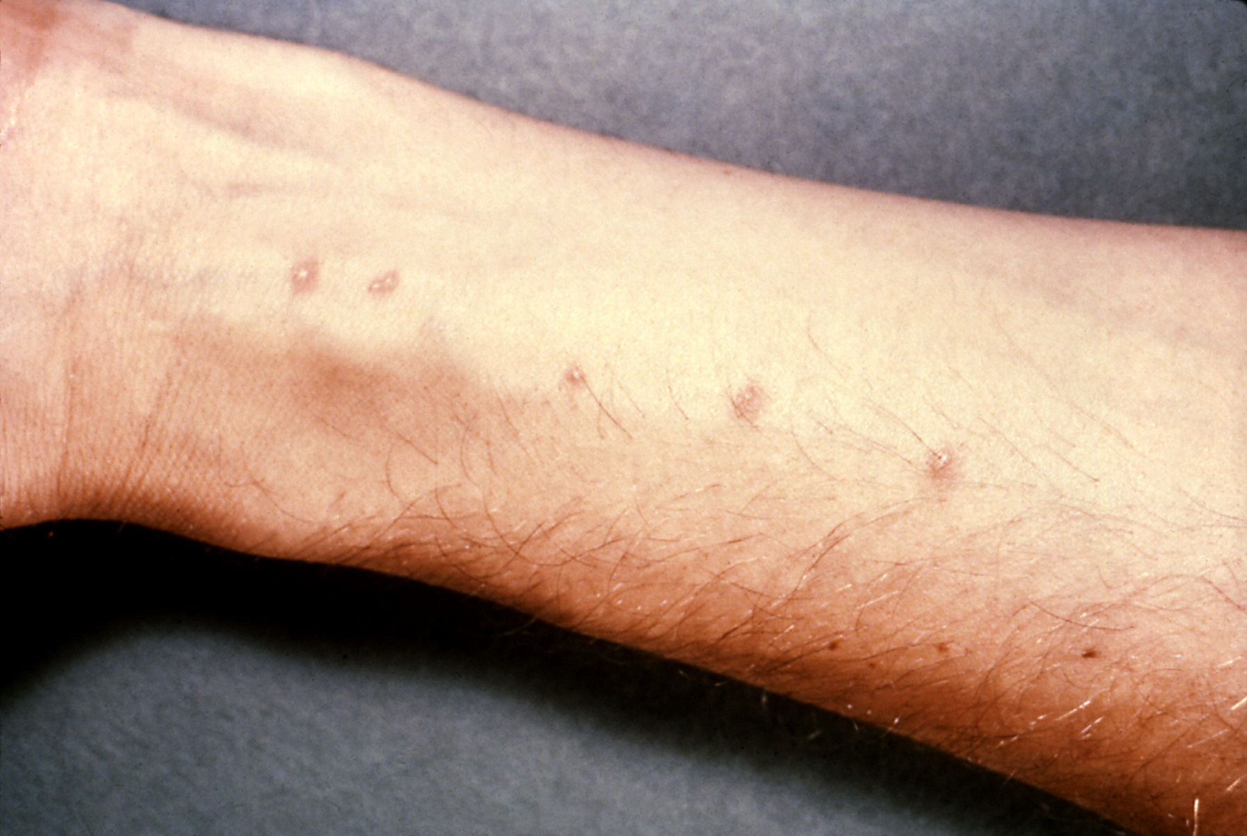
The first sign of infection may be 'swimmers itch'.  The MRCP exam may give a history of a patient who has been on holiday to Asia/Africa/South America and has been swimming in a river and is now itchy with a rash.  In part 2 there may also be a picture like the one below:


The rash tends of appear within hours of infection and disappear within a week.


The classical presentation of acute schistosomiasis itself occurs roughly 2 weeks after infection with:
  • fever
  • urticaria
  • diarrhoea
  • abdominal pain
  • cough
  • hepatosplenomegaly
This acute version of schistosomiasis is known as Katayama Fever


The treatment is praziquantel.


If schistosomiasis is not recognised and treated it can become a chronic granulomatous disease (granulomas form around the schistosomiasis eggs).  Which parts of the body are affected depends on the type.  For the purposes of MRCP the following associations are worth learning:
  • s.mansoni - liver disease, transverse myelitis
  • s.japonicum - liver disease
  • s.haematobium - urinary tract/bladder disease (haematuria)
    • --> increased risk of squamous cell carcinoma of bladder


The most practical way of diagnosing is by looking for eggs in stool/urine.


It goes without saying that any question with an eosinophillia in the blood results of someone who has been travelling should raise the possibility of this as a diagnosis.


Now on to the less exotic world of haematology and a recap of the clotting cascade!

MRCP revision battle 14.2: The clotting cascade

Before we start tackling clotting disorders a recap of the clotting cascade would be useful.


A schematic of the clotting cascade is:

Its really important to get this in your head as various questions are likely to be indirectly testing your knowledge of it.  I remember the intrinsic pathway into the common pathway as by a little jingle of     " 12,11,9 then 10 (pause) 2, 1 then back again " with the 'back again' bit referring to factor 13 getting in on the action right at the very end to stablise the clot and hence the numbers returning back to being higher.

Also note where VIIIa and Va are involved.


Now look at the same schematic again but with the addition of activated protein C and antithrombin:


So as well as learning the cascade itself make sure the following are etched in your brain:
  • activated protein C inhitbits VIIIa and Va
  • antithrombin inhibits Xa and IIa 
  • protein C is converted to activated protein C by protein S        and finally
  • factors II, VII, IX, X are vitamin-K dependent (method of remembering: '1972')


With all that groundwork covered, lets  move on to battle 14.3 and put a bit of it into practice.

MRCP revision battle 14.3: Factor V Leiden

A point mutation on the gene for clotting factor V results in a varient known as 'factor V Leiden'.
It is inherited in an autosomal dominant fashion.


The result of the mutation is that factor V is insensitive to protein C inactivation, resulting in hypercoagulability.


This leads to increased risk of PE/DVTs and in females increased miscarriage.


Merely being factor V leiden positive doesn't necessitate anticoagulation; warfarin etc would only be considered in response to events rather than prophylactically - indeed, up to 7% of all white europeans/north americans have factor V leiden, making it the commonest inherited thrombophilia.


So having looked at a cause of thrombophillia, lets switch to the opposite and consider low platelets...

MRCP revision battle 14.4: Thrombocytopenia

Thrombocytopenia refers to low numbers of platelets.
Generally the lower limit of normal for platelets is 150 000, with thrombocytopenia often defined as <50 000.


Low platelets increase the risk of bleeding/bruising, commonly manifesting as bleeding gums or nose bleeds.


Causes of thrombocytopenia can be broadly divided into causes that increase platelet destruction and causes that decrease production:
  1. increased platelet destruction (= DISC HH)
    • DIC
    • ITP
    • SLE
    • CLL
    • Heparin
    • Hypersplenism (portal hypertension, leukaemia)
  2. decreased platelet production (=CAB M)
    • Chemo/radiotherapy
    • aplastic anaemia
    • B12/folate deficiency
    • marrow infiltration
Isotretinoin and valproic acid have also been associated with thrombocytopenia.


Treatment is dependent on the cause/the symptoms.


Onwards to the last haematological battle of the day, ITP...

MRCP revision battle 14.5: Idiopathic Thrombocytopenia Purpura

ITP is a condition which results in low numbers of platelets.  Despite its title of 'idiopathic' it is often autoimmune mediated, with IgG autoantibodies against glycoprotein IIb/IIIa/Ib complex.


Acute ITP:
  • affects mainly children
  • boys and girls equally affected
  • often after infection/vaccination
  • tends to be self-limiting over 2 weeks

Chronic ITP:
  • females >males
  • relapsing-remitting


In ITP the bone marrow shows megakaryocytes


Treatment is only needed if the patient is symptomatic or the platelets are <20.
First line = oral prednisolone - 80% respond
If no response in 3 months --> splenectomy
Other options include IV IG or immunosupressive drugs.


One random MRCP type syndrome: Evan's syndrome = ITP +AIHA.


Nearly there, just a quick tricyclic overdose to go...

MRCP revision battle 14.6: TCA overdose

Lets end the day (and the working week) on an overdose.  I've always found it somewhat ironic (and at times in A&E irritating) that a medication which you give to depressed people to treat their depression is so incredibly dangerous in overdose...


TCA in overdose produce symptoms due to their:
  • anticholinergic effects (dry mouth, dilated pupils, tachycardia, constipation, urinary retention, raised intraocular pressure)
  • alpha blockade (hypotension)
  • sodium channel blockade (my personal favourite, cardiac arrhythmias)


On the ECG look out for:
  • long PR
  • wide QRS
  • long QT


In terms of managing these patients you need:
  • ECG and cardiac monitoring
  • consider charcoal if <2 hrs post ingestion
  • bloods for UEs and paracetamol and salicylate (always assume paracetamol taken until proven otherwise)
  • ABG
  • fluids if needed to support BP
  • if acidotic - bicarb
  • dialysis is NOT indicated
  • avoid class Ia/Ic/III antiarrythmics as they all prolong QT.  Also avoid flumazenil (also prolongs QT)
  • observe


Thats all for today folks, and I'm taking tomorrow off.  And possibly the day after too, depending on my mood.  But battles will definitely recommence on Monday.

Thursday, 16 September 2010

MRCP revision battle 13.1: Brugada Syndrome

I'm afraid I'm too tired to even attempt an insightful intro today so lets just go straight to listing the battles:

MRCP revision battle 13.1: Brugada Syndrome
MRCP revision battle 13.2: SVC obstruction
MRCP revision battle 13.3: Causes long PR
MRCP revision battle 13.4: Cauda equina syndrome
MRCP revision battle 13.5: Surviving Sepsis


MRCP revision battle 13.1: Brugada Syndrome


Brugada Syndrome is an autosomal dominant condition that causes abnormal sodium channel proteins, leading to a risk of VF/VT and sudden death.


It is commonest in Asians.

20-40% of cases are associated with a SCN5A mutation.


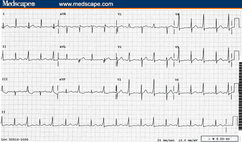
ECG shows partial/complete RBBB with ST elevation in V1 to V2.  The classical ECG is type 1 below, with the other 2 possible variations.

 Treatment is ICD.


Next up - SVC obstruction...

MRCP revision battle 13.2: SVC obstruction

SVC obstruction can be frightening for the doctor as well as the patient - the first lady I saw with it came in with a lovely slim face and as time went on it became more and more puffy and she became increasingly dyspnoeic.  Once seen it will never be forgotten; I can post this photo:
                                                                     Herbert L. Fred, MD and Hendrik A. van Dijk, Wiki Commons

which demonstrates the type of difference I saw in my patient (normal on right, SVC on left).  My patient however was female and smiled more!


Anyway, lets refocus on points to learn for the MRCP exam:


Features of SVC obstruction:
  • dyspnoea
  • orthopnoea
  • headache 
  • cyanosis
  • cough
  • swollen face/arm
  • engorged veins


There is a rather nice test called Pemberton's Test which involves asking the patient to lift their arms above their head for >1 min and watch for increasing plethora/cyanosis, raised JVP and listen for stridor (that sound every doctor wants to induce in their patient...)  If these things occur it is a positive Pembertons test and suggestive of SVC obstruction.


Causes of SVC obstruction include:
  • lung cancer
  • lymphoma
  • thymus malignancy
  • thrombus around central line


Treatment is surgical, with dexamethasone whilst awaiting this.


Lets return to the small-squared world of the ECG to consider causes of a long PR...

MRCP revision battle 13.3: Long PR interval

The PR interval represents the time taken for the electrical impulse to travel from the sino-atrial node and through the atrioventricular node to the ventricles.

Normal range is 0.12 to 0.2 milliseconds (= 3 to 5 small squares)

A long PR = first degree heart block.


Causes of a long PR ( can be remembered as DRIP SAL, easier to remember if you have a drippy friend called Sally):
  • Drugs (beta blockers, calcium channel blockers, digoxin)
  • Rheumatic fever
  • IHD (anterior MIs in particular can prolong the PR interval)
  • Potassium - either high or low levels
  • Sarcoidosis
  • Aortic root pathology
  • Lyme disease


So that was short and sweet.  Onto another brief battle that should be familiar, cauda equina syndrome...

MRCP revision battle 13.4: Cauda Equina Syndrome

Cauda equina syndrome refers to a set of features caused by compression of the cauda equina.

The cauda equina begins in most people at around L1/L2.


Features of cauda equina syndrome include:
  • low back pain
  • sciatica
  • urinary incontinence/retention
  • faecal incontinence/retention
  • variable motor/sensory loss
  • classically saddle/perianal paraesthesia


Causes of cauda equina are logical when you think about it, you just need to consider what could be there to press on it:
  • commonest = large central disc herniation at L4/L5 or L5/S1
  • tumours
  • trauma
  • epidural abscess (consider this one in MRCP questions that mention a 'post op patient')
  • post op haematoma (another reason to look out for that 'post-op patient')
  • IVC thrombus
  • ankylosing spondylitis
  • sarcoidosis

Treatment is urgent surgery.


So to the final (and juiciest) battle of the day, surviving sepsis...

MRCP revision battle 13.5: Surviving sepsis

There has been a concerted push in recent years to improve the prognosis of patients presenting with sepsis, led by the international 'Surviving Sepsis' campaign.  But what are the key points to pick up for MRCP?


Firstly, a few definitions.


SIRS = systemic inflammatory response = 2 or more of:
  • temperature >38 or <36
  • WCC >12 or <4
  • RR>20 or pCO2 <4.3
  • pulse >90 (note this is lower than you'd probably have expected)


Sepsis is then defined as SIRS plus infection.


Severe sepsis is then defined as sepsis with evidence of organ dysfunction (hypoxia, anuria, raised lactate... so many possible options for the evidenve)

And then septic shock is defined as severe sepsis plus hypotension.


Now we know what sepsis is, what are the key messages from the surviving sepsis campaign?

  1. resuscitate
  • if hypotensive or lactate >4 give fluids
  • aim 
    • CVP 8-12mmHg
    • MAP>65
    • urine output >0.5mls/kg/hr
    • central venous O2 >70% or mixed venous >65%
  • if fluid alone not achieving goals, consider:
    • packed RBC to haematocrit >30% or
    • dobutamine infusion
  1.  =
  2. give antibiotics
  3. keep MAP >65mmHg
    1. norepinephrine 
    2. dopamine
  4. consider hydrocortisone if fluids and vasopressors aren't working
  5. consider recombinant human activated protein c 
    • if apache >25 or multiple organ failure
  6. remember DVT prophylaxis
  7. consider peptic ulceration prophylaxis


As Bugs Bunny would say, thaatts all folks... unless you fancy participating in today's war to check your recall of yesterday's topics...

MRCP questions: War 12

As with previous 'wars' after 'battles' these are just a few quick questions to see if your brain cells have retained the information provided in battles 11.1 to 11.4.

Grab a piece of paper, jot down your answers then compare them to my answers here


Question 1:
Which atria are atrial myxomas found in 75% of the time?


Question 2:
What murmur is associated with aortic dissection?


Question 3:
What % of patients with aortic dissection will have a pulse deficit?


Question 4:
What is the commonest form of aortic dissection?


Question 5:
How are type A dissections managed?


Question 6:
What is the classic cell associated with CLL on blood film?


Question 7:
List 5 features that suggest a poorer prognosis in CLL


Question 8:
What condition does tartrate resistant acid phosphatase positive suggest?


Question 9:
What is Richters syndrome?


Question 10:
How often is the mediastinum wide on a CXR if the patient has aortic dissection?


The answers are here

MRCP revision battle 13


MRCP revision battle 13


Wednesday, 15 September 2010

MRCP revision battle 12.1: Atrial myxoma

Today's battles are a mixed bag; usually they are generated from past questions I've done while today's bunch include some motivated by gaps in clinical knowledge revealed during a recent take.  So the battles are:

MRCP revision battle 12.1: Atrial Myxoma
MRCP revision battle 12.2: CLL
MRCP revision battle 12.3: Hairy cell leukaemia
MRCP revision battle 12.4: Aortic dissection


MRCP revision battle 12.1: Atrial Myxoma

Atrial myxoma are usually benign tumours found in the atria.
75% are in the left atrium.

0.3% of the population are found to have an atrial myxoma at post mortem.
Females >males, 2:1


Atrial myxoma are usually sporadic but an autosomal dominant inheritance also exists.


They are usually benign but as they grow rapidly and embolise they require surgery.


Features of atrial myxoma include:
  • clubbing
  • weight loss
  • fever
  • AF
  • emboli
  • mid diastolic murmur - an 'atrial plop'


Investigations may show:
  • raised wcc
  • low platelets
  • raised ESR in 60%


After that brisk whizz through atrial myxoma, onward to CLL!

MRCP revision battle 12.2: CLL

I keep clerking patients who say they've got 'CLL, doctor' and I nod sagely and scribble it down, while desperately trying to recall any of my med school haematology lectures.  My brief summary for MRCP means that now hopefully my sage nod is slightly more appropriate!!


CLL (=chronic lymphocytci leukaemia) is a monoclonal proliferation of mature lymphocytes.

99% of cases of CLL involve B cells.

CLL is important clinically as it is the commonest cause of lymphocytosis in the elderly.
Males are more often affected than females, 2:1


Presentation:
  • no symptoms in 25%
  • recurrent infections (due to hypogammaglobulinaemia)
  • anaemia (warm AIHA in 10-15%)
  • decreased weight
  • sweats
  • anorexia


Signs includer:
  • enlarged rubbery, non-tender nodes
  • hepatomegaly
  • splenomegaly


Blood tests will show:
  • raised lymphocytes
  • low Hb, neutrophils and platelets
  • smear/smudge cells on blood film


Staging is done by the Rai classification, which can also be used to give the prognosis:
  • Stage 0 - lymphocytosis alone - mean survival >13 yrs
  • Stage 1 - lymphocytosis plus lymphadenopathy - mean survival 8 yrs
  • Stage 2 - lymphocytosis plus spleno/hepato megaly - mean survival 5 yrs
  • Stage 3 - lymphocytosis plus anaemia (defined as Hb <11) - mean survival 2 yrs
  • Stage 4 - lymphocytosis plus platelets <100 - mean survival 1 yr


Indicators of poorer prognosis include:
  • being male
  • age >70
  • lymphocytes >50
  • prolymphocytes >10%
  • doubling rate <12 months
  • raised LDH
  • CD 38 positive
  • ZAP 70 + (if ZAP 70 -ive, mean survival 25 years)


Management of CLL is:
  1. monitoring only if asymptomatic
  2. chlorambucil to decrease lymphocyte count, improve marrow function and decrease node size
  3. fludarabine (a chemo agent, purine analog that works by inhibiting DNA synthesis) if:
    • bulky disease
    • cytopenias due to marrow failure
    • short lymphocyte doubling time


CLL may transform to Richters syndrome.
Richters syndrome is the transformation of CLL to agressive B cell lymphoma.  LDH is raised.


Now onwards to a subtype of CLL, hairy-cell leukaemia.

MRCP revision battle 12.3: Hairy Cell Leukaemia

Hairy cell leukaemia is a subtype of CLL characterised by (suprise, suprise) cells that appear 'hairy' on microscopy.


Hairy cell leukaemia is 4 times more common in men than women.


Features of hairy cell leukaemia include:
  • pancytopenia
  • splenomegaly
  • skin vasculitis in 1/3 cases
  • 'dry tap' 


A key phrase to look out for in MRCP is 'tartrate resistant acid phosphatase positive'


Treatment of hairy cell leukaemia is chemotherapy.  Second line options include alpha interferon and rituximub.


Thats quite enough haematology for one day... lets move on for a 'rippingly' good time with aortic dissection...

MRCP revision battle 12.4: Aortic dissection

Aortic dissection is a scary condition with a mortality rate of 1% per hour.  But before we jump into it, lets begin with a quick recap of some basic anatomy.

The layers of the aorta are, from inside out:
  • tunica intima = epithelial cells
  • tunica media = smooth muscle and elastic fibres
  • tunica adventitia = connective tissue

Aortic dissection usually involves blood collecting in the media.


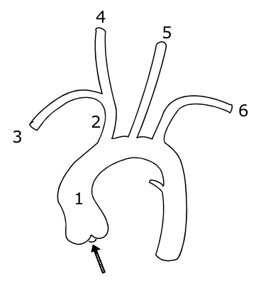
The aorta itself leaves the heart, goes up into an arch and then down again, giving off branches as it goes.  The picture below shows the vessels coming off the aorta:

1 indicates the ascending aorta, 2 the brachiocephalic trunk, 3 the right subclavian artery, 4 the right common carotid, 4 the right common carotid, 5 the left common carotid and 6 the left subclavian artery.

A knowledge of this anatomy aides understanding of the signs and symptoms of aortic dissection.
So what are the features of an aortic dissection?
  • sudden onset of 'tearing' or 'ripping' pain
  • if pain intrascapular, likely a descending dissection
  • MI
  • stroke
  • parathesia of arms
  • hemiparesis
  • many others, depending on the part of the aorta affected

Signs to look out for include:
  • aortic regurgitation - present in 1/3 ascending dissections
  • pulse deficit in an arm - present in 15%
  • BP difference in arms >20mmHg (but note 'normal' individuals may have a BP difference)


Aortic dissections may be classified using either the Stanford or DeBakey systems:
  • Stanford A = ascending dissection - accounts for 2/3 of dissections
    • this covers DeBakey I - ascending and descending aorta involved and
    • DeBakey II - ascending aorta only involved
  • Stanford B = descending aorta only dissected (distal to left subclavian artery) - accounts for 1/3 of dissections
    • = DeBakey III


 Management of aortic dissection:
  • if type A - IV labetalol and urgent surgery
  • if type B - IV labetalol only


Complications associated with type A dissections include:
  • aortic regurgitation
  • inferior MI
  • pericardial effusion
  • carotid dissection
  • loss of pulses


Investigations for dissection:
  • CXR 
    • shows widened mediastinum in 70% of cases
    •  Ring sign - calcification of aorta with wall displaced >5mm
  • CT
  • TOE

Risk factors for dissection include:
  • hypertension (80%)
  • Marfans
  • Noonans
  • Turners
  • trauma
  • pregnancy
  • coarctation
  • congenital bicuspid valve
  • giant cell artertitis
  • cocaine use

If you feel motivated to read more, I'd recommend this webpage: http://www.aorticdissection.com/Aortic%20Diagnosis.htm


A 'war' for yesterday's questions is here


MRCP questions: War 11

As with previous 'wars' after 'battles' these are just a few quick questions to see if your brain cells have retained the information provided in battles 10.1 to 10.6.

Grab a piece of paper, jot down your answers then compare them to my answers here

Question 1
List 4 factors that lead to a worse prognosis in GBS.


Question 2
What percentage of patients with GBS will make a complete or near complete recovery?


Question 3
What regular investigation do patients with GBS require?


Question 4
What is the classic triad of Miller-Fisher syndrome?


Question 5
What antibodies are usually associated with Miller-Fisher syndrome?


Question 6
What is syringomyelia?


Question 7
What are the cardinal symptoms of syrinomyelia?


Question 8
What is the inheritance of Kallmans syndrome?


Question 9
List 5 symptoms/signs of carcinoid syndrome


Question 10
What is the treatment of carcinoid syndrome?


answers here

Tuesday, 14 September 2010

MRCP revision battle 11.1: Guillain Barre Syndrome

When you're working 12 hr days (plus commute) fitting in revision can feel a bit like trying to squeeze that extra towel into the suitcase - you may manage it, but the suitcase is then stretched and prone to snap open at unfortunate times.  If you extend this metaphor to the suitcase being your sanity it explains why junior doctors can often become more emotionally incontinent/irrational as MRCP exams get nearer.  So whats the solution?  Well, in the suitcase scenario you'd add some of those rubber bands round to keep it closed.  In real life, your friends, family and managing to keep some form of hobby/interest going are your rubber bands.

Since I don't want to overpressure my 'rubber bands' I'm going to limit today's battles to

MRCP revision battle 11.1: Guillain Barre Syndrome
MRCP revision battle 11.2: Miller Fisher Syndrome
MRCP revision battle 11.3: Syringomyelia
MRCP revision battle 11.4: Kallmans
MRCP revision battle 11.5: Carcinoid Syndrome


MRCP revision battle 11.1: Guillain Barre Syndrome


Guillain Barre Syndrome is a rare, post-infective demyelinating polyneuropathy.


It tends to occur a few weeks after 'the flu' or a minor illness.  The most commonly associated pathogens are:
  • campylobacter jejuni
  • mycoplasma
  • CMV
  • EBV
  • HIV
  • zoster

Guillain Barre Syndrome is characterised by an ascending, symmetric muscle weakness.  Proximal muscles are more affected than distal ones.  Cranial nerves may also be involved, with CN VII being the most common.


There is usually a 'progressive' phrase for around 4 weeks, after which the patient starts to improve.


Features associated with GBS that appear in MRCP questions include:
  • papilloedema
  • urinary retention
  • arrhythmias

Diagnostically GBS is characterised by:
  • very high protein in CSF
  • decreased nerve velocity on EMG
  • areflexia

The prognosis for patients with GBS is worse if:
  • rapid onset of symptoms
  • older age
  • axomal neuropathoes
  • preceeded by campylobacter infection

Management is:
  • 4 hourly FVC, to check the patient's respiratory muscles aren't affected
  • ventilation if needed
  • plasma exchange
  • IV IG

Overall, 85% of patients make a complete/new complete recovery.
10% will relapse
Less than 5% will die.


Now to to meet a close relative of GBS, Miller Fisher Syndrome.

MRCP revision battle 11.2: Miller Fisher Syndrome

Miller Fisher Syndrome is essentially a rarer variant of Guillain Barre Syndrome.


It's classical triad is:
  • areflexia
  • ataxia
  • opthalmoplegia

Anti-ganglioside antibodies, especially to GQ1b, will be positive in 90% of cases.


On to syringomyelia...

MRCP revision battle 11.3: Syringomyelia and Syringobulbia

Syringomyelia and syringobulbia have always confused me, partly because for some reason I even have problems pronouncing their names!  However, in terms of MRCP there isn't too much you need to know...


Syringomyelia is a cyst/cavity forming in or close to the central canal of the spinal cord.


The cause may be anything that is hindering CSF circulation, but the classical cause is an Arnold Chiari malformation (=condition where the cerebellum herniates through the foramen magnum.)


The essential features are:
  • wasting and weakness of muscles of the hands (possibly 'claw hand')
  • loss of pain and temperature sensation in hands

Other possible signs/symptoms are varied and include Horner's syndrome, eye movement abnormalities and UMN signs in the legs.


Treatment is surgery.


If the cavity/cyst extends from the cord into the brain stem it is known as syringobulbia and causes bulbar palsy type symptoms, facial pain and dizziness.


After that neurology binge onto a slightly lighter topic, Kallmans...

MRCP revision battle 11.4: Kallmans

Kallmans syndrome is an x-linked recessive condition caused by failure of GnRH secreting neurones to migrate to the hypothalamus.


Its features are:
  • anosia
  • 'delayed puberty'
  • hypogonadism, crytoorchidism
  • may be tall or of normal height

Investigations: blood LH/FSH/testosterone and all either normal or low


The most important complication to remember for MRCP is the increased risk of osteoporosis

Next up: Carcinoid syndrome

MRCP revision battle 11.5: Carcinoid Syndrome

Carcinoid tumours are common - 1% of the population will have one at post mortem.

They primarily secrete serotonin (5HT) but may also produce histamine/adrenal hormones/insulin...


45% of carcinoid tumours are in the appendix and a further 30% in the ileum.
They can cause complications such as appendicitis, obstruction or intussception.


Having said carcinoid tumours are common, it's important to understand that carcinoid syndrome is rare - it only occurs when the liver is involved, as tumours in other places will have their 5HT metabolised rather than it have an effect.


Features of carcinoid syndrome are:
  • flushing
  • diarrhoea
  • bronchospasm
  • pellegra
  • hypotension

Diagnosis is by:
  1. urinary 5-HIAA
  2. plasma chromograffin - the new way which, depending on the leve,  also gives an indication of the mass of tumours

Treatment is:
  • surgery
  • ocreotide

Survival from diagnosis tends to be 5 to 8 years.
Patients with carcinoid heart disease have a worse prognosis.


It is also worth remembering that if these tumours are over-handed (eg during surgery) or out-grow their blood supply, a massive 'hormone hit' can be released in one go, causing a carcinoid crisis.


Features of a carcinoid crisis:
  • vasodilation
  • hypotension
  • tachycardia 
  • bronchoconstriction
  • hyperglycaemia 


So those are today's battles fought, and hopefully won.  Click here if you want to partake of the 'war' of questions to check your recall of yesterday's topics.

    MRCP questions: War 10

    As with previous 'wars' after 'battles' these are just a few quick questions to see if your brain cells have retained the information provided in battles 9.1 to 9.6.


    Grab a piece of paper, jot down your answers then compare them to my answers here

    Question 1:
    What are the diagnostic criteria for hereditary haemorrhagic telangiectasia?


    Question 2:
    List 3 possible causes of increased shortness of breath on sitting up compared to lying down.


    Question 3:
    Which form of MND affects only LMN?


    Question 4:
    Name the only drug treatment available for MND


    Question 5:
    List 5 causes of bulbar palsy


    Question 6:
    What condition is associated with a 'Donald Duck' voice?


    Question 7:
    Which genetic condition causes an increase in uric acid levels?


    Question 8:
    List 5 causes of low voltage ECG complex


    Question 9:
    What causes electrical alternans on ECG?


    Question 10:
    How is multifocal motor neuropathy treated?


    answers here

    Monday, 13 September 2010

    MRCP revision battle 10.1: Hereditary Haemorrhagic Telangiectasia

    Once again I can see a sunny day... through the window my desk sits in front of.  Although today I've found the sun a catalyst for working rather than a mocking presence as my lovely other half has promised me a picnic later, so the sooner I finish the sooner I get jam sandwiches!

    Today's topics are:

    MRCP revision battle 10.1: Hereditary haemorrhagic telangiectasia
    MRCP revision battle 10.2: Motor Neurone Disease
    MRCP revision battle 10.3: Bulbar Palsy
    MRCP revision battle 10.4: Pseudobulbar palsy
    MRCP revision battle 10.5: Multifocal Motor Neuropathy
    MRCP revision battle 10.6: Lesch-Nyhan Syndrome
    MRCP revision battle 10.7: Low voltage ECG complexes

    I promise that's less than it looks...


    MRCP revision battle 10.1: Hereditary haemorrhagic telangiectasia


    Hereditary haemorrhagic telangiectasia (HHT), also known as Osler-Weber-Rendu syndrome, is an autosomal dominant condition associated with abnormal blood vessel formation.


    Features of HHT include:
    • telangiectasia on skin
    • telangiectasia on mucosal membranes (leading to nose bleeds/GI bleeds)
    • arterio-venous malformations (AVMs)
      • commonest in lung
      • also in liver
      • brain
      • spine (rarest)

    The diagnostic criteria are:
    1. spontaneous and recurrent nosebleeds
    2. telangiectasia
    3. AVMs
    4. positive family history - a first degree relative
    The international consensus  states if 3 or 4 of the above features are present HHT is definite.  If 2 are present it is possible.  Less than that is unlikely.


    A rare symptom the MRCP exam may throw up is platypnoea, which is difficulty breathing when sitting up/standing which is relieved on lying down.  Platypnoea is therefore the direct opposite of orthopnoea!  To briefly diversify, apart from lung AVM two other possible causes of platynoea are left atrial tumour or left atrial thrombus.


    A rare sign associated with lung AVM is a 'humming sound' on auscultation over that area of the chest.


    Management of HHT is symptomatic.


    After that forray into the world of bleeding bits we're about to dive into the murky world of neurology with motor neurone disease...

    MRCP revision battle 10.2: Motor Neurone Disease

    Most people associated motor neurone disease (MND) with Stephen Hawking, that incredible physicist who has lived with MND for over 40 years.  However, in the same way he is not a stereotype for normal intelligence he is also not a stereotype for MND - most people with it die within 5 years.  More stereotypical for the course of the disease was Lou Gehrig, an American baseball player in the 1920s/1930s who was diagnosed with the disease and died within 3 years.  I mention him not just for random interest but also because in America MND is sometimes known as Lou Gehrig disease.  But what is MND?


    MND is a group of neurological disorders affecting neurones in motor cortex, CN nuclei and anterior horn cells.


    MND affects UMN and LMN but there is no sensory involvement.


    As a quick recap, UMN signs include:
    • weakness
    • spasticity
    • brisk reflexes
    • upgoing plantars

    LMN signs include:
    • weakness
    • wasting
    • fasciculation
    • depressed reflexes

    There are several patterns of MND, including:
    1. Amyotrophic lateral sclerosis
      • presents as UMN signs in legs and LMN signs in arms
      • may be familial - in which case often associated with Ch 21
      • accounts for 50% of cases of MND
      • has an 'intermediate' prognosis
    2. Bulbar palsy
      • accounts for around 25% of cases of MND
      • has the worst prognosis of the presentations
      • more details in the next battle
    3. Progressive muscular atrophy
      • only LMN affected
      • begins distally
      • has a (relatively) good prognosis
    4. Primary lateral sclerosis
      • affects UMN only
    10-35% of patients with MND will have fronto-temporal dementia.


    Diagnosis of MND is primarily clinical.
    EMG will show preserved motor conduction velocities
    CSF analysis may show raised protein.


    Treatment is with riluzole, which prolongs life by around 3 months and requires monitoring of LFTs.  There is no cure.

    As the first paragraph stated, the prognosis is very poor and most patients are dead within 5 years.


    So on to battle 10.3, to learn more about bulbar palsy.

    MRCP revision battle 10.3: Bulbar Palsy

    Bulbar palsy refers to palsy of the nerves coming from the 'bulbar' part of the brain, ie the pons, medulla and cerebellum.  These are VII, VIII, IX, X, XI and XII.


    Classically bulbar palsy presents as palsy of tongue, muscles of chewing and swallowing and facial muscles.


    Signs are LMN:
    • flaccid tongue
    • fasciculating tongue
    • absent or normal jaw jerk
    • loss of gag reflex
    • quiet/horse voice

    Causes of bulbar palsy include:
    • MND
    • syringobulbia (more about this condition tomorrow!)
    • GBS (also lined up for battle tomorrow)
    • polio
    • Lyme disease
    • brain-stem tumours

    Next up: battle 10.4, pseudobulbar palsy

    MRCP revision battle 10.4: Pseudobulbar palsy

    Pseudobulbar palsy is a UMN palsy of the muscles affecting eating, swallowing and talking above the midpons.


    Signs are:
    • spastic tongue
    • brisk jaw jerk
    • emotional incontinence
    • 'Donald Duck' voice.

    Causes include:
    • MS
    • MND
    • PD
    • stroke
    • high brain stem tumour


    Just one more skirmish with neurology to go today, in the form of multifocal motor neuropathy.

    MRCP revision battle 10.5: Multifocal motor neuropathy

    I must confess I'd never come accross multifocal motor neuropathy until I started doing MRCP questions, but I'm now rather glad that I have, as essentially it is a treatable differential diagnosis for some presentations that appear like MND (which, as we've just covered, is nasty and deadly)


    Multifocal motor neuropathy is an acquired immune-mediated demyelinating neuropathy.


    Features of multifocal motor neuropathy include:
    • slowly progressive, asymmetric distal muscle weakness
    • cramps and twitching
    • no sensory loss
    • no/minimal muscle atrophy
    • possibly positive anti GM1 antibodies (NB also + in GBS)
    • no UMN signs
    • normal or decrease tone
    • absent reflexes

    The joy of multifocal motor neuropathy is that it can be successfully treated with IV IG!


    Well thats quite enough neurology for today, lets now look briefly at Lesch-Nyhan Syndrome, as promised yesterday

    MRCP revision battle 10.6: Lesch-Nyhan Syndrome

    Lesch-Nyhan Syndrome is a rare inherited x-linked disorder caused by an abnormality of an enzyme involved in purine metabolism resulting in increased uric acid.  Only boys tend to be affected.


    Features of Lesch-Nyhan Syndrome include:
    • orange crystals in nappies
    • low IQ
    • self-mutilation
    • fits
    • recurrent gout (probably the most important relationship to remember in MRCP)
    • megaloblastic anaemia

    Patients usually die by the age of 25, often of renal failure.


    So to the final battle of the day, a nice gentle ending with a breeze through the causes of low-voltage complexes on ECG

    MRCP revision battle 10.7: Causes of low-voltage complexes on ECG

    'Low voltage complex' on ECG is defined as:
    • QRS amplitude <5mm in all limb leads and/or
    • QRS amplitude <10mm in all chest leads


    The causes of low voltage complexes can be split into 2 main groups: increased distance from leads to heart or infiltrative disease of heart.

    Using these divisions:
    1. increased distance from leads to heart:
      • emphysema/COPD
      • pericardial effusion
      • severe obesity
      • pleural effusion
    2. infiltrative disease of heart/problems with heart itself: 
      • amyloid
      • haemochromatosis
      • cardiomyopathies
      • global ischaemia

    Myxoedema can also cause low complex ECGs but my search for why this is (?caused by myxoedema or ?caused by effects of myxoedema) proved fruitless... please let me know if you have the answer!


    If you see an ECG with alternating normal complexes and low-voltage complexes, you are looking at electrical alternans which tends to be a sign of pericardial effusion/cardiac tamponade.  Click here to see an image of it.


    So today's battles are over.  If you want to undertake the war to see how much of yesterday's battles you recall, please click here 

    MRCP questions: War 9

    As with previous 'wars' after 'battles' these are just a few quick questions to see if your brain cells have retained the information provided in battles 9.1 to 9.5.


    Grab a piece of paper, jot down your answers then compare them to my answers here


    Question 1:
    Name the classical rash associated with Lyme disease


    Question 2:
    What is the treatment for uncomplicated Lyme disease?


    Question 3:
    A joint aspiration shows negatively birefrigent crystals.  What condition does this suggest?


    Question 4:
    What XR appearance may you see in gout?


    Question 5:
    Which joint is classically affected by pseudogout?


    Question 6:
    What is a craniopharyngioma?


    Question 7:
    What is a catamenial pneumothorax?

    .


    Question 8:
    What is Hammans sign?  What does it signify?


    Question 9:
    A 19 yr old boy presents with shortness of breath.  A CXR shows a 3cm rim of air around his left lung.  How would you manage him?


    Question 10:
    You have a patient with a painful joint and the XR shows chondocalcinosis.  What condition does this suggest?


    answers here

    Sunday, 12 September 2010

    MRCP revision battle 9.1: Lyme Disease

    Working and revising really does feel rather relentless.  I keep thinking of the pop song "and the beat goes on..." - somehow it seems appropriate for the continuous dedication work plus MRCP revision requires!  Anyway, what revision battles does today bring?

    MRCP revision battle 9.1: Lyme disease
    MRCP revision battle 9.2: Gout
    MRCP revision battle 9.3: Pseudogout
    MRCP revision battle 9.4: Craniopharyngioma
    MRCP revision battle9.5: Pneumothorax


    MRCP revision battle 9.1: Lyme disease

    Lyme disease, named after the place in America where the condition was first noted, is a condition caused by the spirochaete borrelia burdorferi.  


    It is spread by tic bites, with certain areas (such as northern America) being higher risk than, for example, Surrey.  However, it is found in the UK and it is important to note the majority of patients will not remember the bite itself.


    Signs, symptoms and complications associated with Lyme disease are multitude and vague, including:
    • malaise
    • fever
    • muscle pain
    • joint swelling
    • decreased cognition
    • encephalitis
    • meningitis
    • lymphadenopathy
    • cranial nerve palsies (MRCP likes bilateral facial nerve palsy for some reason)
    • neuropathy
    • skin conditions


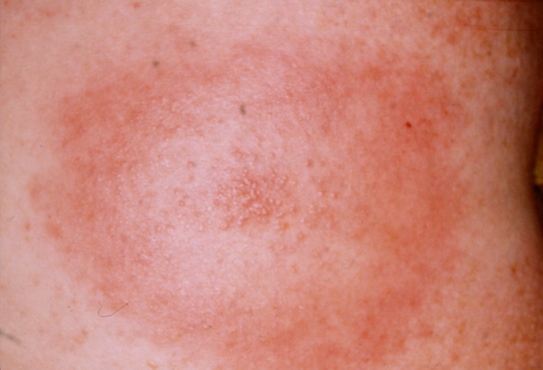
    The classical skin condition associated with Lyme disease is erythema chronicum migrans, which is a spreading erythematous rash which then clears centrally but leaves a spot in the middle (click here for a picture).


    A rarer skin condition is acrodermatitis chronica atrophicans, which occurs late in infection and eventually results in atrophic skin that is thin like cigarette paper.


    Another rarer skin manifestation is borrelial lymphocytoma, which is a blue-red discoloration of the ear lobe.


    Investigation is usually ELISA/PCR.
    If the question throws in a CSF sample look for slightly raised protein and lymphocytosis.


    Treatment of the rash only is doxycycline.  More serious disease may require IV ben pen/a 3rd generation cephalosporin. 


    There is of course far more to learn about Lyme disease should you be so inclined, but if you're content with the barer MRCP-bones of it lets move on to battle 9.2, gout.

    MRCP revision battle 9.2: Gout

    Ah Gout... that classical disease of red-faced old men...

    In terms of MRCP, you need to expand the above knowledge to include the following...


    Gout usually presents as a single, swollen, hot, painful joint.  If the joint affected is the big toe, it is called a podagra.


    Microscopy of joint fluid would reveal negatively birefrigent needle-shaped monosodium urate crystals.

    Uric acid may be >450micromols - but equally may be normal in an acute attack.

    CRP is usually raised, but if the question shows a raised WCC too start thinking about different diagnoses, for example septic arthritis.


    Gout may be either primary or secondary.

    Primary gout may be:
    • idiopathic - usually due to decreased excretion of uric acid 
    • associated with Lesch-Nyhan Syndrome (to be covered in a very brief battle tomorrow)


    Secondary gout can broadly be divided into things which cause increased production/intake of urate, and things which decrease its excretion.


    Causes of increased production/intake include:
    • myeloproliferative/lymphoproliferative disorders
    • psoriasis
    • cytotoxic drugs
    • food - beer, yeast, seafood, liver, kidney
    • exercise
    • fits
    • acidosis

    Causes of decreased excretion include:
    • renal failure
    • diuretics
    • low dose aspirin
    • alcohol
    • lead poisoning

    XR should show a normal joint space (may be decreased in late disease) and large punched-out erosions distant from the joint margins.


    Treatment is NSAIDs - if these are contraindicated, colchine can be used.


    Prophylaxis is with allopurinol (a xanthine oxidase inhibitor).  It should be started 2 to 3 weeks after an acute attack if:
    • >1 attack
    • tophi present
    • renal disease
    • urate stones
    • Lesch-Nyhan syndrome
    • cytotoxic drugs used (in which case it should be given before any episodes of gout)

    So after that quick whizz through gout, lets embrace is near-cousin pseudo-gout...

    MRCP revision battle 9.3: Pseudogout

    Pseudogout involves the deposition of positively birefrigent calcium pyrophosphate dihydrate crystals in joints.


    Pseudogout is associated with a variety of conditions, including:
    • hyperparathyroidism
    • hypothyroidism
    • haemochromatosis
    • Wilsons disease
    • acromegaly
    • old age
    • OA

    It tends to cause a monoarthritis of a larger joint, classically a knee.


    An XR may show chondrocalcinosis = soft tissue calcium deposition.


    Treatment is with NSAIDs.  If this is unsuccessful steroids or hydroxychloriquine can be considered.


    Now on to revision battle 9.4, a treat left over from yesterday, craniopharyngioma...

    MRCP revision battle 9.4: Craniopharyngioma

    Craniopharyngiomas are benign tumours that arise from the remnants of Rathke's pouch (= area between pituitary and 3rd ventricle floor).

    Technically it is not a pituitary tumour as it is derived from the embryonic remnants of the pituitary gland rather than the gland itself.   However, in terms of the mass effects/how it presents it is very similar.  Amenorrhoea and decreased libido are often sited as symptoms.


    Craniopharyngiomas frequently calcify (50%)


    Investigation of choice is MRI, treatment is surgery.


    After that brief squirmish its on to the last battle of the day, the pneumothorax

    MRCP revision battle 9.5: Pneumothorax

    A pneumothorax, as I'm sure you all know, is a collection of gas in the pleural cavity of the chest between the lung and chest wall.


    I'm not about to insult you by going through the basic signs/symptoms of pneumothorax, but I will just briefly mention a rare sign that occasionally pops up in MRCP exams: a clicking sound at the sternal edge synchronised with the heart beat.  This rare sign of a pneumothorax is most commonly assocaited with small left sided pneumothoraces.


    As a quick recap, its important to establish if the pneumothorax is primary (ie no underlying lung disease), secondary (=underlying lung disease) or traumatic (in which case as a medic you shouldn't be treating it!)


    BTS guidelines state that in primary pneumothorax, if the patient is not breathless and the rim of air is <2cm you do not need to necessarily do anything and may consider sending the patient home.  If the rim of air is >2cm or the patient is breathless you should aspirate; try x2, if still unsuccessful insert a drain.


    The guidelines for secondary pneumothorax are slightly more complex:
    • if >50yrs old AND breathless AND rim of air >2cm, insert a drain first line
    • if the above 3 criteria do not apply, try aspirating first
    • regardless of which category your patient falls into, patients with secondary pneumothorax should be admitted for 24 hours observation.


    A random type of pneumothorax to be aware of is catamenial pneumothorax - this is a pneumothorax associated with menstruation, which is due to endometriosis of the lung.  90% of these occur on the right side.


    As a complete aside, as well as recognising the 'click synchronised with heartbeat' as a possible pneumothorax it is worth learning that a 'crunching sound' in the chest can be due to a pneumomediastinum  and is called Hamman's sign.


    Well those are todays battles over, but for those with residual enthusiasm the beat goes on with a war to test your memory of yesterday's topics...

    MRCP questions: War 8

    As with previous 'wars' after 'battles' these are just a few quick questions to see if your brain cells have retained the information provided in battles 8.1 to 8.6.

    Grab a piece of paper, jot down your answers then compare them to my answers here


    Question 1:
    What is the treatment for cluster headache?


    Question 2:
    List 3 symptoms of cluster headache.

    Question 3:
    What odour is associated with acute arsenic poisoning?

    Question 4:
    List 3 causes of Mees lines

    Question 5:
    State the 4 H's and 4 T's which are known as the reversible causes of cardiac arrest.


    Question 6: 
    What kind of visual disturbance is classically associated with a pituitary tumour

    Question 7:
    What histological type of pituitary tumour is most likely to cause a pressure effect?

    Question 8:
    What is Nelson's syndrome?


    Question 9:
    What are the similarities and differences between Romano Ward and Jervell Lange Nielson syndromes?

    Question 10:
    What is the commonest hormone produced by a pituitary tumour?


    answers here