Sunday 12 September 2010

MRCP revision battle 9.1: Lyme Disease

Working and revising really does feel rather relentless.  I keep thinking of the pop song "and the beat goes on..." - somehow it seems appropriate for the continuous dedication work plus MRCP revision requires!  Anyway, what revision battles does today bring?

MRCP revision battle 9.1: Lyme disease
MRCP revision battle 9.2: Gout
MRCP revision battle 9.3: Pseudogout
MRCP revision battle 9.4: Craniopharyngioma
MRCP revision battle9.5: Pneumothorax


MRCP revision battle 9.1: Lyme disease

Lyme disease, named after the place in America where the condition was first noted, is a condition caused by the spirochaete borrelia burdorferi.  


It is spread by tic bites, with certain areas (such as northern America) being higher risk than, for example, Surrey.  However, it is found in the UK and it is important to note the majority of patients will not remember the bite itself.


Signs, symptoms and complications associated with Lyme disease are multitude and vague, including:
  • malaise
  • fever
  • muscle pain
  • joint swelling
  • decreased cognition
  • encephalitis
  • meningitis
  • lymphadenopathy
  • cranial nerve palsies (MRCP likes bilateral facial nerve palsy for some reason)
  • neuropathy
  • skin conditions


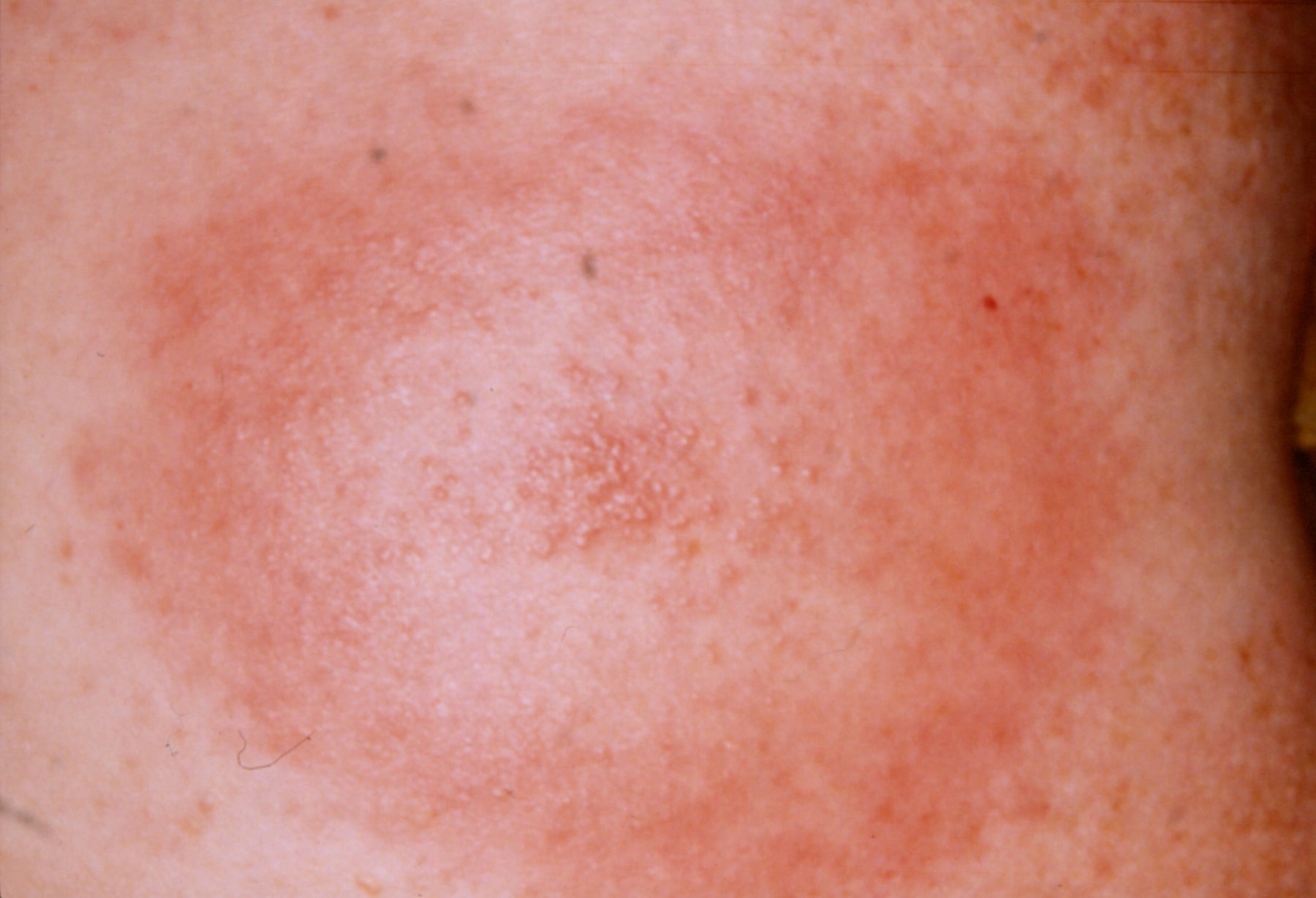
The classical skin condition associated with Lyme disease is erythema chronicum migrans, which is a spreading erythematous rash which then clears centrally but leaves a spot in the middle (click here for a picture).


A rarer skin condition is acrodermatitis chronica atrophicans, which occurs late in infection and eventually results in atrophic skin that is thin like cigarette paper.


Another rarer skin manifestation is borrelial lymphocytoma, which is a blue-red discoloration of the ear lobe.


Investigation is usually ELISA/PCR.
If the question throws in a CSF sample look for slightly raised protein and lymphocytosis.


Treatment of the rash only is doxycycline.  More serious disease may require IV ben pen/a 3rd generation cephalosporin. 


There is of course far more to learn about Lyme disease should you be so inclined, but if you're content with the barer MRCP-bones of it lets move on to battle 9.2, gout.

MRCP revision battle 9.2: Gout

Ah Gout... that classical disease of red-faced old men...

In terms of MRCP, you need to expand the above knowledge to include the following...


Gout usually presents as a single, swollen, hot, painful joint.  If the joint affected is the big toe, it is called a podagra.


Microscopy of joint fluid would reveal negatively birefrigent needle-shaped monosodium urate crystals.

Uric acid may be >450micromols - but equally may be normal in an acute attack.

CRP is usually raised, but if the question shows a raised WCC too start thinking about different diagnoses, for example septic arthritis.


Gout may be either primary or secondary.

Primary gout may be:
  • idiopathic - usually due to decreased excretion of uric acid 
  • associated with Lesch-Nyhan Syndrome (to be covered in a very brief battle tomorrow)


Secondary gout can broadly be divided into things which cause increased production/intake of urate, and things which decrease its excretion.


Causes of increased production/intake include:
  • myeloproliferative/lymphoproliferative disorders
  • psoriasis
  • cytotoxic drugs
  • food - beer, yeast, seafood, liver, kidney
  • exercise
  • fits
  • acidosis

Causes of decreased excretion include:
  • renal failure
  • diuretics
  • low dose aspirin
  • alcohol
  • lead poisoning

XR should show a normal joint space (may be decreased in late disease) and large punched-out erosions distant from the joint margins.


Treatment is NSAIDs - if these are contraindicated, colchine can be used.


Prophylaxis is with allopurinol (a xanthine oxidase inhibitor).  It should be started 2 to 3 weeks after an acute attack if:
  • >1 attack
  • tophi present
  • renal disease
  • urate stones
  • Lesch-Nyhan syndrome
  • cytotoxic drugs used (in which case it should be given before any episodes of gout)

So after that quick whizz through gout, lets embrace is near-cousin pseudo-gout...

MRCP revision battle 9.3: Pseudogout

Pseudogout involves the deposition of positively birefrigent calcium pyrophosphate dihydrate crystals in joints.


Pseudogout is associated with a variety of conditions, including:
  • hyperparathyroidism
  • hypothyroidism
  • haemochromatosis
  • Wilsons disease
  • acromegaly
  • old age
  • OA

It tends to cause a monoarthritis of a larger joint, classically a knee.


An XR may show chondrocalcinosis = soft tissue calcium deposition.


Treatment is with NSAIDs.  If this is unsuccessful steroids or hydroxychloriquine can be considered.


Now on to revision battle 9.4, a treat left over from yesterday, craniopharyngioma...

MRCP revision battle 9.4: Craniopharyngioma

Craniopharyngiomas are benign tumours that arise from the remnants of Rathke's pouch (= area between pituitary and 3rd ventricle floor).

Technically it is not a pituitary tumour as it is derived from the embryonic remnants of the pituitary gland rather than the gland itself.   However, in terms of the mass effects/how it presents it is very similar.  Amenorrhoea and decreased libido are often sited as symptoms.


Craniopharyngiomas frequently calcify (50%)


Investigation of choice is MRI, treatment is surgery.


After that brief squirmish its on to the last battle of the day, the pneumothorax

MRCP revision battle 9.5: Pneumothorax

A pneumothorax, as I'm sure you all know, is a collection of gas in the pleural cavity of the chest between the lung and chest wall.


I'm not about to insult you by going through the basic signs/symptoms of pneumothorax, but I will just briefly mention a rare sign that occasionally pops up in MRCP exams: a clicking sound at the sternal edge synchronised with the heart beat.  This rare sign of a pneumothorax is most commonly assocaited with small left sided pneumothoraces.


As a quick recap, its important to establish if the pneumothorax is primary (ie no underlying lung disease), secondary (=underlying lung disease) or traumatic (in which case as a medic you shouldn't be treating it!)


BTS guidelines state that in primary pneumothorax, if the patient is not breathless and the rim of air is <2cm you do not need to necessarily do anything and may consider sending the patient home.  If the rim of air is >2cm or the patient is breathless you should aspirate; try x2, if still unsuccessful insert a drain.


The guidelines for secondary pneumothorax are slightly more complex:
  • if >50yrs old AND breathless AND rim of air >2cm, insert a drain first line
  • if the above 3 criteria do not apply, try aspirating first
  • regardless of which category your patient falls into, patients with secondary pneumothorax should be admitted for 24 hours observation.


A random type of pneumothorax to be aware of is catamenial pneumothorax - this is a pneumothorax associated with menstruation, which is due to endometriosis of the lung.  90% of these occur on the right side.


As a complete aside, as well as recognising the 'click synchronised with heartbeat' as a possible pneumothorax it is worth learning that a 'crunching sound' in the chest can be due to a pneumomediastinum  and is called Hamman's sign.


Well those are todays battles over, but for those with residual enthusiasm the beat goes on with a war to test your memory of yesterday's topics...

MRCP questions: War 8

As with previous 'wars' after 'battles' these are just a few quick questions to see if your brain cells have retained the information provided in battles 8.1 to 8.6.

Grab a piece of paper, jot down your answers then compare them to my answers here


Question 1:
What is the treatment for cluster headache?


Question 2:
List 3 symptoms of cluster headache.

Question 3:
What odour is associated with acute arsenic poisoning?

Question 4:
List 3 causes of Mees lines

Question 5:
State the 4 H's and 4 T's which are known as the reversible causes of cardiac arrest.


Question 6: 
What kind of visual disturbance is classically associated with a pituitary tumour

Question 7:
What histological type of pituitary tumour is most likely to cause a pressure effect?

Question 8:
What is Nelson's syndrome?


Question 9:
What are the similarities and differences between Romano Ward and Jervell Lange Nielson syndromes?

Question 10:
What is the commonest hormone produced by a pituitary tumour?


answers here