Wednesday, 15 September 2010

MRCP revision battle 12.1: Atrial myxoma

Today's battles are a mixed bag; usually they are generated from past questions I've done while today's bunch include some motivated by gaps in clinical knowledge revealed during a recent take.  So the battles are:

MRCP revision battle 12.1: Atrial Myxoma
MRCP revision battle 12.2: CLL
MRCP revision battle 12.3: Hairy cell leukaemia
MRCP revision battle 12.4: Aortic dissection


MRCP revision battle 12.1: Atrial Myxoma

Atrial myxoma are usually benign tumours found in the atria.
75% are in the left atrium.

0.3% of the population are found to have an atrial myxoma at post mortem.
Females >males, 2:1


Atrial myxoma are usually sporadic but an autosomal dominant inheritance also exists.


They are usually benign but as they grow rapidly and embolise they require surgery.


Features of atrial myxoma include:
  • clubbing
  • weight loss
  • fever
  • AF
  • emboli
  • mid diastolic murmur - an 'atrial plop'


Investigations may show:
  • raised wcc
  • low platelets
  • raised ESR in 60%


After that brisk whizz through atrial myxoma, onward to CLL!

MRCP revision battle 12.2: CLL

I keep clerking patients who say they've got 'CLL, doctor' and I nod sagely and scribble it down, while desperately trying to recall any of my med school haematology lectures.  My brief summary for MRCP means that now hopefully my sage nod is slightly more appropriate!!


CLL (=chronic lymphocytci leukaemia) is a monoclonal proliferation of mature lymphocytes.

99% of cases of CLL involve B cells.

CLL is important clinically as it is the commonest cause of lymphocytosis in the elderly.
Males are more often affected than females, 2:1


Presentation:
  • no symptoms in 25%
  • recurrent infections (due to hypogammaglobulinaemia)
  • anaemia (warm AIHA in 10-15%)
  • decreased weight
  • sweats
  • anorexia


Signs includer:
  • enlarged rubbery, non-tender nodes
  • hepatomegaly
  • splenomegaly


Blood tests will show:
  • raised lymphocytes
  • low Hb, neutrophils and platelets
  • smear/smudge cells on blood film


Staging is done by the Rai classification, which can also be used to give the prognosis:
  • Stage 0 - lymphocytosis alone - mean survival >13 yrs
  • Stage 1 - lymphocytosis plus lymphadenopathy - mean survival 8 yrs
  • Stage 2 - lymphocytosis plus spleno/hepato megaly - mean survival 5 yrs
  • Stage 3 - lymphocytosis plus anaemia (defined as Hb <11) - mean survival 2 yrs
  • Stage 4 - lymphocytosis plus platelets <100 - mean survival 1 yr


Indicators of poorer prognosis include:
  • being male
  • age >70
  • lymphocytes >50
  • prolymphocytes >10%
  • doubling rate <12 months
  • raised LDH
  • CD 38 positive
  • ZAP 70 + (if ZAP 70 -ive, mean survival 25 years)


Management of CLL is:
  1. monitoring only if asymptomatic
  2. chlorambucil to decrease lymphocyte count, improve marrow function and decrease node size
  3. fludarabine (a chemo agent, purine analog that works by inhibiting DNA synthesis) if:
    • bulky disease
    • cytopenias due to marrow failure
    • short lymphocyte doubling time


CLL may transform to Richters syndrome.
Richters syndrome is the transformation of CLL to agressive B cell lymphoma.  LDH is raised.


Now onwards to a subtype of CLL, hairy-cell leukaemia.

MRCP revision battle 12.3: Hairy Cell Leukaemia

Hairy cell leukaemia is a subtype of CLL characterised by (suprise, suprise) cells that appear 'hairy' on microscopy.


Hairy cell leukaemia is 4 times more common in men than women.


Features of hairy cell leukaemia include:
  • pancytopenia
  • splenomegaly
  • skin vasculitis in 1/3 cases
  • 'dry tap' 


A key phrase to look out for in MRCP is 'tartrate resistant acid phosphatase positive'


Treatment of hairy cell leukaemia is chemotherapy.  Second line options include alpha interferon and rituximub.


Thats quite enough haematology for one day... lets move on for a 'rippingly' good time with aortic dissection...

MRCP revision battle 12.4: Aortic dissection

Aortic dissection is a scary condition with a mortality rate of 1% per hour.  But before we jump into it, lets begin with a quick recap of some basic anatomy.

The layers of the aorta are, from inside out:
  • tunica intima = epithelial cells
  • tunica media = smooth muscle and elastic fibres
  • tunica adventitia = connective tissue

Aortic dissection usually involves blood collecting in the media.


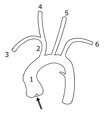
The aorta itself leaves the heart, goes up into an arch and then down again, giving off branches as it goes.  The picture below shows the vessels coming off the aorta:

1 indicates the ascending aorta, 2 the brachiocephalic trunk, 3 the right subclavian artery, 4 the right common carotid, 4 the right common carotid, 5 the left common carotid and 6 the left subclavian artery.

A knowledge of this anatomy aides understanding of the signs and symptoms of aortic dissection.
So what are the features of an aortic dissection?
  • sudden onset of 'tearing' or 'ripping' pain
  • if pain intrascapular, likely a descending dissection
  • MI
  • stroke
  • parathesia of arms
  • hemiparesis
  • many others, depending on the part of the aorta affected

Signs to look out for include:
  • aortic regurgitation - present in 1/3 ascending dissections
  • pulse deficit in an arm - present in 15%
  • BP difference in arms >20mmHg (but note 'normal' individuals may have a BP difference)


Aortic dissections may be classified using either the Stanford or DeBakey systems:
  • Stanford A = ascending dissection - accounts for 2/3 of dissections
    • this covers DeBakey I - ascending and descending aorta involved and
    • DeBakey II - ascending aorta only involved
  • Stanford B = descending aorta only dissected (distal to left subclavian artery) - accounts for 1/3 of dissections
    • = DeBakey III


 Management of aortic dissection:
  • if type A - IV labetalol and urgent surgery
  • if type B - IV labetalol only


Complications associated with type A dissections include:
  • aortic regurgitation
  • inferior MI
  • pericardial effusion
  • carotid dissection
  • loss of pulses


Investigations for dissection:
  • CXR 
    • shows widened mediastinum in 70% of cases
    •  Ring sign - calcification of aorta with wall displaced >5mm
  • CT
  • TOE

Risk factors for dissection include:
  • hypertension (80%)
  • Marfans
  • Noonans
  • Turners
  • trauma
  • pregnancy
  • coarctation
  • congenital bicuspid valve
  • giant cell artertitis
  • cocaine use

If you feel motivated to read more, I'd recommend this webpage: http://www.aorticdissection.com/Aortic%20Diagnosis.htm


A 'war' for yesterday's questions is here


MRCP questions: War 11

As with previous 'wars' after 'battles' these are just a few quick questions to see if your brain cells have retained the information provided in battles 10.1 to 10.6.

Grab a piece of paper, jot down your answers then compare them to my answers here

Question 1
List 4 factors that lead to a worse prognosis in GBS.


Question 2
What percentage of patients with GBS will make a complete or near complete recovery?


Question 3
What regular investigation do patients with GBS require?


Question 4
What is the classic triad of Miller-Fisher syndrome?


Question 5
What antibodies are usually associated with Miller-Fisher syndrome?


Question 6
What is syringomyelia?


Question 7
What are the cardinal symptoms of syrinomyelia?


Question 8
What is the inheritance of Kallmans syndrome?


Question 9
List 5 symptoms/signs of carcinoid syndrome


Question 10
What is the treatment of carcinoid syndrome?


answers here