Thursday 18 November 2010

MRCP revision battle 53.1: Genital ulcers

A day of randomness that starts with the delights of genital ulcers and ends in a cat's scratch...


MRCP revision battle 53.1: Genital ulcers
MRCP revision battle 53.2: Vertebral artery dissection
MRCP revision battle 53.3: Intracranial venous thrombosis
MRCP revision battle 53.4: Infective endocarditis
MRCP revision battle 53.5: Upper GI bleeds
MRCP revision battle 53.6: Meckel's diverticulum
MRCP revision battle 53.7: Cat scratch disease


MRCP revision battle 53.1: Genital ulcers


Genital ulcers.  Such a lovely topic.  If you really can't face it there is a summary table at the end.


1. Chancroid
  • This is a sexually transmitted infection, most prevalent in third world countries.
  • Chancroid begins as a small lump that then turns into an ulcer.  
  • It is painful
  • The ulcer bleeds easily when rubbed 
  • The ulcer has a greeney-yellow base
  • One third of affected people will develop inguinal lymph node involvement, with half of these developing abscesses after the lymph nodes become so big they break through the skin.
  • Chancroid is caused by the gram negative bacteria haemophillus ducreyi
  • Treatment options are:
    • 1g azithromycin orally or
    • IM ceftriaxone or
    • 7 days erythromycin
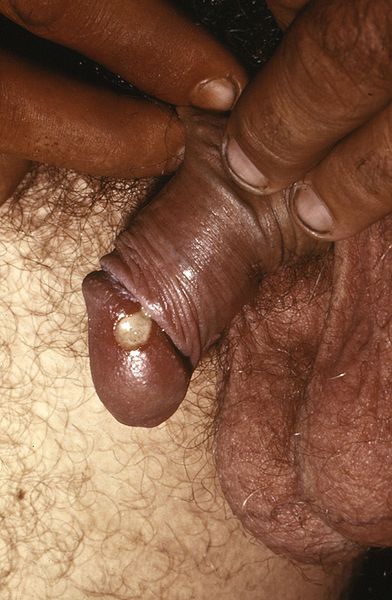
Chancroid lesion filled with pus prior to rupture.  From wiki commons, uploaded by Joe Miller


2.  Granuloma inguinale = Donovanosis
  • This is a sexually transmitted infection mainly found in 3rd world countries.
  • Granuloma inguinale begins as a small lump which then bursts into an ulcer/open lesion that continues to spread until treated
  • The ulcer is painless and has a 'beefy red' appearence
  • There is not usually inguinal lymphadenopathy
  • Granuloma inguinale is caused by klebsiella granulomatis
  • Donovan bodies are rod-shaped klebsiella granulomatis found in the cytoplasm of phagocytes in infected individuals.  They stain dark purple with Wright's stain.
  • Treatment is
    • 3 weeks erthyromycin or tetracyline


3. Lymphogranuloma venereum
  • This is a sexually transmitted infection
  • It is caused by chlamydia trachomatis (L type)
  • There are several stages of infection:
    • Primary:
      • painless pustule which bursts into a painless ulcer
      • often not noticed by women as may be internal
      • 10% of patients will have accompanying erythema nodosum
    • Secondary
      • tender inguinal lymphadenopathy
    • Tertiary
      • up to 20 yrs later - protocolitis, tenesmus
  • Treatment options:
    • doxycycline or
    • erythromycin

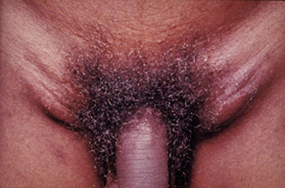
 Image from wiki commons, uploaded by Dr Fred


4. Genital herpes
  • This is a sexually transmitted infection
  • Up to 8 in 10 people who contract it have no symptoms
  • Those who have symptoms tend to develop groups of painful ulcers
  • Primary infection may last up to 3 weeks
  • Subsquent infections tend to be less severe
  • It is highly infective when ulcers are present
  • It is classically caused by HSV 2 but can be caused by HSV 1.
  • Oral aciclovir may be given within the first 5 days of symptoms starting as a 5 day course but there is no cure
  • Subsequent recurrences tend to be less severe.


5. Behcet's disease
  • This is NOT sexually transmitted
  • It is associated with oral ulcers and anterior uveitis
  • See battle 25.2 for more information


Summary of sexually transmitted causes of genital ulcers:


Now for something completely different...

MRCP revision battle 53.2: Vertebral artery dissection

Vertebral artery dissection is an important recognised cause of stroke in patients under 45 years of age.

The vertebral arteries themselves arise from the subclavian and join at the base of the medulla oblongata to form the basilar artery.


Presentation of vertebral artery dissection tends to be:
  • several occipital headache
  • cerebellar signs
  • brainstem signs
    •  CN IX, X, XI and XII:
      • dysarthria
      • dysphagia
    • hiccups
    • loss of sensation to ipilateral face
    • ipsilateral Horner's syndrome in 1/3 of patients
    • 'crossed signs':
      • ipisilateral cranial nerve palsy with contralateral hemiparesis or hemiplegia

Causes include:
  • trauma
  • stretching of neck
    • in MRCP questions look for trips to hairdresser, painting ceiling etc
  • connective tissue disorders

Investigation:
  • CT 
  • 4 vessel angiogram


Treatment:
  • ?neurosurgery
  • anticoagulate if no associated subarachnoid haemorrhage


Now for some intracranial thrombosis...

MRCP revision battle 53.3: Intracranial venous thrombosis

The presentation of intracranial venous thrombosis depends on which venous sinus is affected.

All locations of intracranial venous thrombosis can cause headache.


Isolated sagittal sinus thrombosis (=nearly half of intracranial venous thrombosis) or lateral sinus thrombosis present with:
  • headache
  • vomiting
  • seizures
  • papilloedema
  • potentially focal neurological signs


Cavernous sinus thrombosis can cause:
  • headache
  • oedematous eyelids
  • proptosis
  • painful eye movements/opthalmoplegia
Cavernous sinus thrombosis is often associated with infection spreading from the face or paranasal sinuses.
Remember: cavernous sinus contains CN III, IV, V1, V2 and VI, plus the internal carotid artery.


Sigmoid sinus thrombosis:
  • headache
  • cerebellar signs


Inferior petrosal sinus thrombosis:
  • 5th and 6th nerve palsies

Risk factors for developing intracranial venous thrombosis include:
  • pregnancy
  • head injury
  • recent LP
  • oral contraceptive pill

Investigation is:
  • MRI/CT
    • CT may show 'absent delta sign' which implies a filling defect and therefore a thrombosis
  • ?MRV

Management is by specialists, ?heparin


On to the more familiar topic of infective endocarditis...

MRCP revision battle 53.4: Infective endocarditis

Infective endocarditis is diagnosed by Dukes criteria, which requires:
  • 2 major criteria OR
  • 1 major and 2 minor criteria OR
  • all 5 minor criteria.
Major criteria are:
  • positive blood culture
    • typical organism in 2 separate cultures OR
    • persistently +blood cultures
  • endocardial involvement
    • positive echo
    • new valvular regurgitation
Minor criteria are:
  • fever >38C
  • vascular/immunological signs
  • predisposition (IVDU, valve replacemen)
  • positive blood culture that doesn't mean major criteria
  • positive echo that doesn't meet major criteria

Vascular/immunological signs include:
  • Janeway lesions
  • Oslers nodes
  • splinter haemorrhages
  • Roth spots


40% of cases of endocarditis occur in patients with no previous problems.
30% have had rheumatic heart disease.


Commonest causative organism is streptococcus viridans (alpha haemolytic)
Commonest organism in IVDUs is staphlycoccus aureus.
Commoenst organism within 6 weeks of valve surgery is staph epidermidis.


Usually bicuspid valve affected, except in IVDUs where the tricuspid valve is most commonly affected.


Mortality from strep in 5%, staph around 30%

Poor prognostic factors:
  • s. aureus
  • prosthetic valve
  • culture negative
  • low complement


Treatment:
  • initial blind therapy: fluclox and gent
  • prosthetic valve/penicillin allergy: vancomycin and rifampicin and gent
  • staph: fluclox/vanc and gen
  • strep: benzylpenicillin and gent


Surgery if:
  • abscess
  • recurrent emboli
  • severe valve incompetence
  • cardiac failure


Random MRCP facts:
  • prolongation of PR suggests aortic valve abscess
  • if causative organism is found to be strep bovis look for an associated bowel malignancy.


On to upper GI bleeds...

MRCP revision battle 53.5: Upper GI bleeds

Upper GI bleeds may present with:
  • haematemesis (=vomiting blood)
  • coffee ground vomiting or
  • malaena (=black, tar-like motions)

Commonest causes of upper GI bleeds are:
  • 35% duodenal ulcers
  • 20% gastric ulcers
  • 18% gastric erosins
  • 10% Mallory-Weiss tear
 

Rarer causes of GI bleeds include:
  • variceal haemorrhage
  • aorto-enteric fistula
  • Meckel's diverticulum
  • Peutz-Jeghers syndrome

Risk of rebleeding and mortality from upper GI bleeds is calculated using the Rockall score.
Pre-endoscopy Rockall score is calculated based on:
  • age
    • 0pt <60yrs
    • 1pt 60-79
    • 2pts >80
  • degree of shock
    • 0pt: BP >100 sys and HR <100
    • 1pt: BP >100 sys but HR >100
    • 2pts: BP <100 sys
  • co-morbidities
    • 0pt: none
    • 1pt: heart problems
    • 2pts: liver/renal failure
    • 3pts: mets

Mortality is roughly:
  • 1 in 20 with 2 pts
  • 1 in 10 with 3 points
  • 1 in 4 with 4 points
  • 1 in 2 with 7 points

Post endoscopy mortality is calculated based on the initial score, the diagnosis and the signs of haemorrhage seen.

Below is a summary of the Rockall score from the SIGN guidelines:


Management of upper GI bleeds is:
  • classic ABC resuscitation
  • if cause suspected to be variceal haemorrhage: IV terlipressin 2mg then 2mg/4hr
  • urgent endoscopy
  • surgery if endoscopy unsuccessful
  • if cause is ulcer: omeprazole after endoscopy
  • if cause is variceal haemorrhage: consider transjugular intrahepatic portosystemic shunt to prevent rebleeding (note: nearly 25% of people with a TIPS get hepatic encephalopathy)


Now to Meckel's diverticulum...

MRCP revision battle 53.6: Meckel's diverticulum

Meckel's diverticulum is the vestigial remnant of the vitellointestinal duct.


Its features are often recalled as a series of '2s':
  • found in 2% of the population
  • around 2 inches long
  • at 2 foot from the ileocaecal valve

It may present with:
  • painless rectal bleeding
  • GI obstruction
  • abdominal pain due to faeces trapped inside diverticulum

Investigation is with radionucleotide scan.

Treatment for complications is surgical.


Now to end on an unusual condition - cat scratch disease.

MRCP revision battle 53.7: Cat scratch disease

Cat scratch disease is  usually caused by bartonella henselae (a gram negative rod)

Features:
  • several erythematous, crusted lesions at site of scratch
  • regional lymphadenopathy
  • up to half of patients will feel systemically unwell

If confirmation of diagnosis is required an indirect fluorescent antibody test for bartonella is avaliable.


Treatment is generally supportive and resolution tends to occur within 2 months.

Prevention is keeping cats flea- free!