Monday 22 November 2010

MRCP revision battle 57.1: Stroke thrombolysis

Now we're at utterly last minute topics...

MRCP revision battle 57.1: Stroke thrombolysis
MRCP revision battle 57.2: Cardiac tamponade
MRCP revision battle 57.3: Systemic sclerosis
MRCP revision battle 57.4: Tapeworms
MRCP revision battle 57.5: Brown-Sequard Syndrome
MRCP revision battle 57.6: Trigeminal neuralgia


MRCP revision battle 57.1: Stroke thrombolysis

Contraindications to stroke thrombolysis:
  • evidence of intracranial haemorrhage on CT/MRI
  • minor neurological defect
  • symptoms rapidly improving
  • seizures at onset of stroke
  • any prior history of stroke and diabetes
  • stroke within 3 months
  • systolic BP >185 or diastolic BP >110
  • hyperglycaemia >25mmol
  • weight >15 stones
  • platelets <100

MRCP revision battle 57.2: Cardiac tamponade

Cardiac tamponade occurs when pericardial fluid collects to a degree that prevents the heart pumping.

On echo you see RA+/- RV collapse during diastle.


The classic triad is Becks:
  • falling BP
  • rising JVP
  • muffled heart sounds

Other possible signs include:
  • Kussmauls = rising JVP on inspiration
  • pulsus paradoxus
  • JVP - loss of y descent, x descent prominant


ECG may show electrical alternanas


Treatment is ugern pericardiocentesis

MRCP revision battle 57.3: Systemic sclerosis

Systemic sclerosis is a connective tissue disease characterised by thickening and fibrosis of skin (scleroderma) and involvement of internal organs.


Female: male 4:1

Commonest in 5th/6th decades


Limited systemic sclerosis = skin involvement is limited to face, hands and feet.
Associated with anti-centromere antibodies in 70-80%

Diffuse systemic sclerosis = more skin involvement
Anti Scl 70 in 40%


Features of sclerosis:
  • Raynaulds - 1st presentation in 70% of cases
  • arthralgia
  • renal failure
  • scleroderma
  • GI problems
  • classical skin presentation:
    • telangiectasia
    • peri-oral puckering
    • smooth shiny skin
    • livedo reticularis
  • pulmonary fibrosis/hypertension

ESR/CRP are raised


CREST is a type of limited systemic sclerosis:
- calcinosis, raynaulds, eosophageal dysmotility, sclerodalyl, telangectasia
- renal crsis is rare but pulmonary hypertension is common


Linear scleroderma = coup de sabre

Treatment:
  • supportive
  • d-penicillamine
  • steroids if lung disease
  • ACE-i to help kidneys

MRCP revision battle 57.4: Tapeworms

Cysticercosis:
  • taenia solum - found in pork
  • taenia saginata - found in beef
  • treatment:albendazole

Hydatid disease
:
  • caused by echinococcus
  • treatment: albendazole

MRCP revision battle 57.5: Brown-Sequard Syndrome

Brown-Sequard Syndrome is caused by a lesion in one half of the cord.

Commonest cause = MS
Other causes:
  • trauma
  • tumour
  • degenerative disease

Result is:
  • ipsilateral UMN weakness below
    • increased reflexes
    • spastic paralysis
  • ipsilateral dorsal column loss
    • loss of proprioception and vibration
  • contralateral spinothalmic loss
    • loss of pain and temperature sensation

MRCP revision battle 57.6: Trigeminal neuralgia

Trigeminal neuralgia is intense stabbing pain in the trigeminal root distribution.

It is unilateral and tends to affect the mandibular or maxillary divisions

Most common in females over 50.


Over 1 in 10 cases will be secondary (to aneuysm, tumour) so ?MRI


Treatment is carbamazepine

Sunday 21 November 2010

MRCP revision battle 56.1: Amyloidosis

7 non-waffling battles


MRCP revision battle 56.1: Amyloidosis
MRCP revision battle 56.2: Plasma exchange
MRCP revision battle 56.3: P450 inducers and inhibitors
MRCP revision battle 56.4: Therapeutic drug monitoring
MRCP revision battle 56.5: Vaccines
MRCP revision battle 56.6: Acute lymphoid leukaemia
MRCP revision battle 56.7: Nematodes = roundworms


MRCP revision battle 56.1: Amyloidosis

Amyloidosis is a pathological process characterised by extracellular accumulation of fibrils of insoluble protein.


Inherited forms of amyloidosis are rare but amyloid deposition is central to many diseases including Alzheimers and type 2 diabetes.


Types of amyloid:

1: AL = light chains
  • secreted by plasma B cells
  • occurs in 15% of myeloma patients
  • also occurs in:
    • Waldenstroms
    • MGUS
  • features:
    • cardiac deposition = 'sparkling' appearence on echo
    • GI 
    • macroglossia
    • peri orbital
  • median survival is 12 months
  • treatment is to treat the underlying condition, ?prednisolone

2: AA = serum amylase A
  • this is secondary amyloidosis, with the amyloid derived from serum amylase A which is an acute phase protein
  • seen in:
    • RA
    • TB
    • bronchiectasis
    • familial med fever
    • IBD
  • commonest organ involved = kidney  (congo red stain -->red-green birefrigence under polarised light)
  • median survival is 24 months

3:

MRCP revision battle 56.2: Plasma exchange

A summary of the indications for plasma exchange:
  • GBS
  • myathesnia gravis
  • Goodpastures
  • ANCA + vasculitis
  • TTP/HUS
  • cryoglobulinaemia
  • some hyperviscosity eg myeloma

MRCP revision battle 56.3: P450 inducers and inhibitors

P450 inducers include:
  • rifampicin
  • antiepileptics except sodium valproate
  • griseofulvin
  • smoking
  • st johns wort
  • barbituates
  • chronic alcohol

P450 inhibitors include:
  • anti-infection meds
    • ciprofloxacin
    • erythromycin
    • ketoconazole
    • isoniazid
  • Anti reflux
    • cimetidine
    • omeprazole
  • Pysch meds
    • SSRIs
    • antipyschotics
    • TCA
  •  sodium valproate
  • acute alcohol
  • class IV antiarrhymics
    • diltiazen
    • verapamil

MRCP revision battle 56.4: Therapeutic drug monitoring

When to take the sample, that is the question....

Lithium: 12 hrs post dose

Digoxin: at least 6 hours post dose

Ciclosporin and pheytoin: immediately post-dose

MRCP revision battle 56.5: Vaccines

Live attenuated vaccines:
  • BCG
  • MMR
  • polio
  • yellow fever
  • oral typhoid

Whole killed:
  • rabies
  • petussis
  • influenza

Fragment:
  • tetanus
  • diptheria
  • meningococcus
  • pneumococcus

MRCP revision battle 56.6: Acute lymphoid leukaemia

ALL is commonest in children.

There is >60% cure

Treatment is by non-myelosupressive chemo.

Prognosis is worse if:
  • <1yr or >10yrs
  • higher WCC
  • male
  • philidelphia chromosome +

Better if >47 chromosomes

MRCP revision battle 56.7: Nematodes = roundworms

2 main groups to consider for MRCP:


1. Strongyloides stercoralis
  • AKA threadworm
  • acquired percutaneously
  • causes migratory urticaria = larva currens
  • presents with itching
  • if it reaches lungs -->pneumonitis
  • treatment: mebendazole

2. Toxocara canis
  • caused by ingesting eggs from soil contaminated with dog poo
  • treatmet: mebendazole

Saturday 20 November 2010

MRCP revision battle 55.1: Trypanosomiasis

More randomness...


MRCP revision battle 55.1: Trypanosomiasis
MRCP revision battle 55.2: Hypokalaemia
MRCP revision battle 55.3: Cryoglobulins
MRCP revision battle 55.4: Cold agglutinins
MRCP revision battle 55.5: Lateral medullary syndrome
MRCP revision battle 55.6: Purtscher retinopathy
MRCP revision battle 55.7: Optic atrophy


MRCP revision battle 55.1: Trypanosomiasis

Trypanosomiasis = diseases causes by parasitic protozoa trypanosoma.


2 main divisions:


1. African = sleeping sickness
  • spread by tsetse fly
  •  caused by
    • t. rhodesiense - east africa - quicker
    • t.gambiense - west africa - slower
  • classical presentation 
    • fever, rigors, headache, hepatosplenomegaly, lymphadenopathy
    • sleepy during day, awake at night
    •  meningoencephalitis
  • look for chancre and posterior cervical nodes (=Winterbottom's sign)
  • diagnosis is by microscopy
  • treatment:
    • IV pentamide
    • IV melarsoprol
Below is an image of t.gambiense:


2. American - Chagas disease
  • carried by triatomine bugs
  • caused by t. cruzi
  • classical presentation
    • fever, rash, hepatosplenomegaly, lymphadenopathy 
    • myocarditis
    • meningoencephalitis
    • megaoesophagus/megacolon
  • look for:
    • chagoma (=indurated, erythamatous nodule at site of infection)
    • periorbital oedema (=Romana's sign)
  •  diagnosis is by blood culture
  • treatment: benznidazole


Now for some low potassium....

MRCP revision battle 55.2: Hypokalaemia

Hypokalaemia is defined as K <3.5mmol; if <2.5mmol it is severe.


Symptoms/signs include:
  • muscle weakness
  • hypotonia
  • cramps
  • tetany
  • worsening of dig toxicity
  • cardiac arrhythmias

ECG may show:
  • small/inverted T waves
  • U waves
  • long PR
  • depressed ST

Causes of hypokalaemia


Low K with hypertension
  • raised plasma renin
    • Cushings
    • renin-secreting tumour
  • low plasma renin

Low K without hypertension


On to cryoglobulins...

    MRCP revision battle 55.3: Cryoglobulins

    Cryoglobulins are immunoglobulins that undergo reversible precipitation at 4c and dissolve when warmed to 37c.

    Cryoglobulins may be IgG, IgM or IgA.

    One third of cases are idiopathic.


    Type 1 cryoglobulinaemia
    • monoclonal
    • high titres, usually >5mg/ml
    • associated with:
      • myeloma
      • Waldenstroms
    • features include vasculitis, ulceration and Raynaud's

    Type 2 cryoglobulinaemia
    • mixed mono and polyclonal
    • titres usually >1mg/ml
    • associated with:
      • RA
      • sjogrens
      • lympoma
      • CLL
      • hep C
      • HIV
    • features include vasculitis, mesangiocapillary GN


    Type 3 cryoglobulinaemia
    • mixed polyclonal
    • associated with:
      • RA
      • sjogrens
      • SLE
    • features include purpura, GN, arthritis


    Now on to a similar but distinct topic...

    MRCP revision battle 55.4: Cold agglutinins

    Cold agglutinins are molecules, usually IgM, which act against red cells agglutinising and haemolysing them at low temperatures.

    Symptoms of cold agglutinins include:
    • Raynauds
    • acrocyanosis
    • mild haemolytic anaemia

    Causes of cold agglutinins include:
    • idiopathic
    • CLL
    • lymphoma
    • mycoplasma
    • coxsachie
    • mononucleosis
    • HIV

    Management is to avoid cold weather.


    Now to diversify to neurology....

    MRCP revision battle 55.5: Lateral medullary syndrome

    Lateral medullary syndrome is a condition in which a patient has a set of symptoms as a result of insult to the medulla.

    Lateral medullary syndrome is usually due to PICA or vertebral artery occlusion.


    Features include:
    • cerebellar signs
    • contralateral pain and temperature sensation loss
    • ipsilateral Horners
    • ipsilateral loss of pain and temperature sensation on face
    • ipsilateral paralysis of palate, pharynx and cords


    On to some vision loss...

    MRCP revision battle 55.6: Purtscher retinopathy

    Purtscher's retinopathy is characterised by cotton wool spots on fundoscopy with sudden detioration in vision.


    It is classically associated with head trauma or chest trauma.

    Other associations include:
    • pancreatitis
    • fat embolism
    • amniotic fluid embolism
    • vascular diseases

    There is no treatment.


    Finally - optic atrophy...

    MRCP revision battle 55.7: Optic atrophy

    Optic atrophy is loss of fibres of the optic disc.  It is characterised by decreased vision (particulary loss of colour vision) and a pale disc on fundoscopy.


    Causes include:


    Congenital:

    Acquired:
    • MS
    • papilloedema
    • glaucoma
    • ischaemia
    • B6, B12, folate deficieny
    • lead poisoning
    • arsenic poisoning
    • methanol
    • ethambutol
    • isoniazid
    • chloramphenicol

    Friday 19 November 2010

    MRCP revision battle 54.1: Primary sclerosing cholangitis

    Today is in pairs: a pair of gastro battles, a pair of cardiology battles, a pair of haematology battles then a strange inherted disorder to finish on....


    MRCP revision battle 54.1: Primary sclerosing cholangitis
    MRCP revision battle 54.2: Jaundice
    MRCP revision battle 54.3: Patent ductus arteriosus
    MRCP revision battle 54.4: Cardiac axis
    MRCP revision battle 54.5: Haemolysis
    MRCP revision battle 54.6: Leukoerythroblastic change
    MRCP revision battle 54.7: Gaucher's disease


    MRCP revision battle 54.1: Primary sclerosing cholangitis

    Primary sclerosing cholangitis is a condition of unknown aetiology in which there is inflammation and fibrosis of bile ducts.


    Presentation:
    • patients may be asymptomatic with an incidental finding of raised ALP
    • may present as:
      • jaundice
      • pruritus
      • abdo pain
      • fatigue

    Diseases associated with primary sclerosing cholangitis include:
    • IBD (UC>crohns; 70% of patients with PSC have UC but only 5% of patients with UC have PSC)
    • HIV
    • HLA A1, B8, DR3

    Males are more frequently affected than females.


    Diagnosis is by:
    • ERCP - shows strictures of the biliary tree and a 'beaded' appearence
    • liver biopsy - fibrous, obliterative cholangitis
    • ANA, SMA and ANCA may be positive

    Treatment is:
    • colestyramine for pruritis
    • ursodeoxycholic acid to improve cholestatis
    • ?stenting of strictures 
    • ?liver transplant

    Complications include:
    • decreased absorption of fat-soluble vitamins A,D,E and K
      • clotting abnormalities and easy bruising
      • osteoporosis
    • cholangiocarcinoma (10-30%)
    • bacterial cholangitis
    • liver cirrhosis
      • portal hypertension

    Survival tends to be around 10 yrs


    As an aside, the other 2 conditions associated with cholangiocarcinoma are:
    • liver flukes
    • Caroli's disease = dilation of hepatic ducts


    Into the yellow...

    MRCP revision battle 54.2: Jaundice

    Jaundice is yellow pigmentation of the skin caused by raised bilirubin.

    It is generally clinically visible above 35micromol/l.


    The 'story' of bilirubin is illustrated in the diaphragm below:
    • phagocytes break haemoglobin down into unconjugated (=insoluble) bilirubin
    • unconjugated bilirubin is joined with glucoronic acid in the liver, making it conjugated (=soluble)
    • conjugatde bilirubin passes into the gallbladder and on to the small intestine, where it is converted into urobilinogen and excreted in urine by the kidneys or to stercobilinogen and excreted in faeces


    The types of jaundice are:

    1. Pre-hepatic = unconjugated
    • caused by:
      • haemolysis
      • lack of UDP: Gilberts, Crigler Najjar
    • urine/faeces colours normal

    2. Hepatic = both conjugated and unconjugated
    • caused by:
      • infection: HBV, HCV, EBV
      • Wilsons
      • Budd-Chiari
      • Dubin-Johnson/Rotor syndromes
      • cirrhosis
      • drugs:
        • anti-TB meds
        • statins
        • sodium valproate
        • MAOIs
        • halothane
        • paracetamol OD
    • urine dark, faeces normal

    3. Post-hepatic = conjugated = obstructive/cholestatic
    • gallstones
    • pancreatic cancer
    • cholangiocarcinoma
    • primary bilary sclerosis
    • sclerosing cholangitis
    • Mirrizi's syndrome = obstructive jaundice secondary to compression of the common hepatic duct by a gallstone impacted in the cystic duct
    • Drugs
      • antibiotics - co-amoxiclav, nitrofurantoin, flucloxacillin
      • OCP
      • chlorperazine
      • sulphonylureas
      • anabolic steroids
    • urine pale and faeces pale


    Now for something different... PDA...

    MRCP revision battle 54.3: Patent ductus arteriosus

    The ductus arteriosus is a connection between the pulmonary trunk and the descending aortic arch allowing blood flow in the fetus to avoid the fluid-filled lungs.

    It should close shortly after birth, being mostly closed within 48 hrs and fully closed within 3 weeks, leaving the ligamentum arteriosum in its place.


    Closure is due to 2 mechanisms:
    1. exposure of infants lungs to oxygen promotes bradykinin production which promotes PDA closure
    2. loss of maternal prostaglandins encourages closure

    Closure is more likely to fail (=patent ductus arteriosus) in:
    • premature infants
    • infants exposed to rubella in 1st trimester
    • infants at high altitude

    Features of patent ductus arteriosus include:
    • machinery murmur
    • wide, collapsing pusle
    • apical heave

    If it does not close the infant may fail to thrive or develop heart failure.
    Many cases however will be asymptomatic.
    5% of patients with PDA go to develop pulmonary hypertension and Eisenmengers.


    Treatment:
    • indomethacin (a prostaglandin antagonist) causes closure in 90%
    • surgical treatment is also an option

    In some cases (eg transposition of the great vessels) keeping the ductus arteriosus open may be beneficial to the infant, in which case IV prostaglandin E1 is given.


    On to the cardiac axis...

    MRCP revision battle 54.4: Cardiac axis

    The normal cardiac axis is -30 to +90 degrees.

    More negative = left axis deviation
    More positive = right axis deviation.


    Causes of left axis deviation:
    • LBBB
    • LVH
    • ASD
    • cardiomyopathies
    • aortic stenosis
    • can be normal in:
      • ascites
      • pregnancy
      • obesity

    Causes of right axis deviation:
    • RBBB
    • RVH --> lung disease
    • PE
    • can be normal in:
      • infancy
      • thin people


    Next: haemolysis

    MRCP revision battle 54.5: Haemolysis

    Haemolysis (=breakdown of red blood cells) may be intravascular or extravascular.
    It causes a microcytic anaemia and raised reticulocyte count.


    Intravascular haemolysis

    Intravascular haemolysis is characterised by:
    • low haptoglobin (as all used up trying to 'recycle' the broken down red cells)
    • raised free plasma haemoglobin
    • haemoglobuinuria: red-brown urine
    • haemosiderinuria: once haptoglobin binding capacity is surpassed free Hb is filtered by the kidneys and can be detected in the urine in sloughed tubular cells using Prussian blue staining

    Causes of intravascular haemolysis include:

    Extravascular haemolysis

    Extravascular haemolysis may cause splenomegaly.

    Causes of extravascular haemolysis include:
    • warm AIHA (remember battle 39.7?)
    • cold haemagglutination disease
    • spherocytosis
    • haemoglobinopathies
    • haemolytic disease of the newborn


    Now to the second haem battle of the day...

    MRCP revision battle 54.6: Leukoerythroblastic change

    Leukoerythroblastic change is the term used to describe nucleated red cells and primative white cells in the peripheral blood.


    Causes of leukoertythroblastic change can be split into 2 catagories:

    1. Invasion of bone marrow space
    • metastatic carcinoma
    • leukaemia
    • myeloma
    • lymphoma
    • myelofibrosis
    • Gaucher's disease (covered in next battle)
    2. Severe illness


    Leukoerythroblastic change may be indicated by:
    • high leucocyte alkaline phosphatase
    • Dohle bodies = blue leucocyte inclusions in peripheral cytoplasm of neutrophils
    • left shift of neutrophils = younger neutrophils than normal -->less nuclei


    As an aside, Dohle bodies are also seen in burns, infection and trauma.


    Now for the final short skirmish, Gaucher's disease...

    MRCP revision battle 54.7: Gaucher's disease

    Gaucher's disease is the most common of the lysosomal storage disorders.

    It is a recessive inherited disorder on chromosome 1 in which lipid accumulates in organs (spleen, liver, brain, kidneys, lungs) and bone marrow due to lack of an enzyme.

    Ashkenazi Jews are more affected.


    Possible features include:
    • splenomegaly
    • hepatomegaly
    • yellow-brown skin pigmentation
    • seizures
    • low IQ
    • osteoporosis

    Treatment can include splenectomy and replacement of the missing enzyme.

    Thursday 18 November 2010

    MRCP revision battle 53.1: Genital ulcers

    A day of randomness that starts with the delights of genital ulcers and ends in a cat's scratch...


    MRCP revision battle 53.1: Genital ulcers
    MRCP revision battle 53.2: Vertebral artery dissection
    MRCP revision battle 53.3: Intracranial venous thrombosis
    MRCP revision battle 53.4: Infective endocarditis
    MRCP revision battle 53.5: Upper GI bleeds
    MRCP revision battle 53.6: Meckel's diverticulum
    MRCP revision battle 53.7: Cat scratch disease


    MRCP revision battle 53.1: Genital ulcers


    Genital ulcers.  Such a lovely topic.  If you really can't face it there is a summary table at the end.


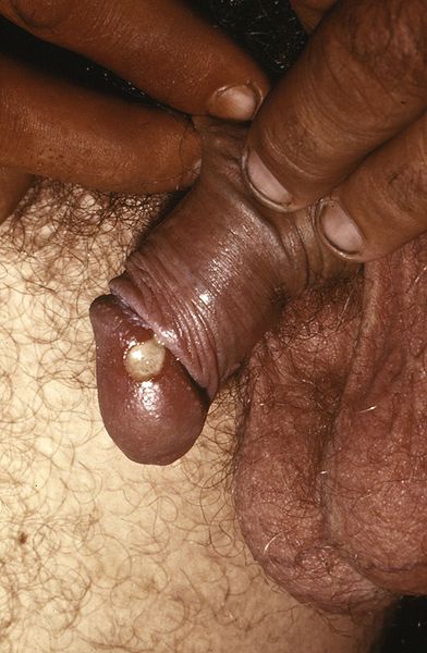
    1. Chancroid
    • This is a sexually transmitted infection, most prevalent in third world countries.
    • Chancroid begins as a small lump that then turns into an ulcer.  
    • It is painful
    • The ulcer bleeds easily when rubbed 
    • The ulcer has a greeney-yellow base
    • One third of affected people will develop inguinal lymph node involvement, with half of these developing abscesses after the lymph nodes become so big they break through the skin.
    • Chancroid is caused by the gram negative bacteria haemophillus ducreyi
    • Treatment options are:
      • 1g azithromycin orally or
      • IM ceftriaxone or
      • 7 days erythromycin
    Chancroid lesion filled with pus prior to rupture.  From wiki commons, uploaded by Joe Miller


    2.  Granuloma inguinale = Donovanosis
    • This is a sexually transmitted infection mainly found in 3rd world countries.
    • Granuloma inguinale begins as a small lump which then bursts into an ulcer/open lesion that continues to spread until treated
    • The ulcer is painless and has a 'beefy red' appearence
    • There is not usually inguinal lymphadenopathy
    • Granuloma inguinale is caused by klebsiella granulomatis
    • Donovan bodies are rod-shaped klebsiella granulomatis found in the cytoplasm of phagocytes in infected individuals.  They stain dark purple with Wright's stain.
    • Treatment is
      • 3 weeks erthyromycin or tetracyline


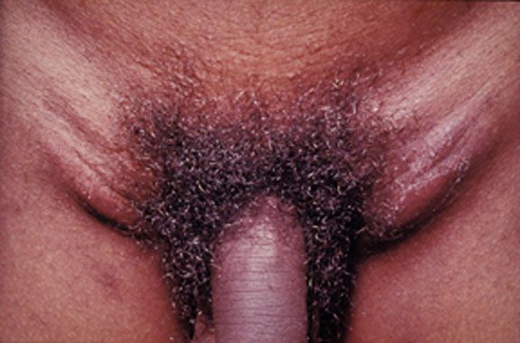
    3. Lymphogranuloma venereum
    • This is a sexually transmitted infection
    • It is caused by chlamydia trachomatis (L type)
    • There are several stages of infection:
      • Primary:
        • painless pustule which bursts into a painless ulcer
        • often not noticed by women as may be internal
        • 10% of patients will have accompanying erythema nodosum
      • Secondary
        • tender inguinal lymphadenopathy
      • Tertiary
        • up to 20 yrs later - protocolitis, tenesmus
    • Treatment options:
      • doxycycline or
      • erythromycin

     Image from wiki commons, uploaded by Dr Fred


    4. Genital herpes
    • This is a sexually transmitted infection
    • Up to 8 in 10 people who contract it have no symptoms
    • Those who have symptoms tend to develop groups of painful ulcers
    • Primary infection may last up to 3 weeks
    • Subsquent infections tend to be less severe
    • It is highly infective when ulcers are present
    • It is classically caused by HSV 2 but can be caused by HSV 1.
    • Oral aciclovir may be given within the first 5 days of symptoms starting as a 5 day course but there is no cure
    • Subsequent recurrences tend to be less severe.


    5. Behcet's disease
    • This is NOT sexually transmitted
    • It is associated with oral ulcers and anterior uveitis
    • See battle 25.2 for more information


    Summary of sexually transmitted causes of genital ulcers:


    Now for something completely different...

    MRCP revision battle 53.2: Vertebral artery dissection

    Vertebral artery dissection is an important recognised cause of stroke in patients under 45 years of age.

    The vertebral arteries themselves arise from the subclavian and join at the base of the medulla oblongata to form the basilar artery.


    Presentation of vertebral artery dissection tends to be:
    • several occipital headache
    • cerebellar signs
    • brainstem signs
      •  CN IX, X, XI and XII:
        • dysarthria
        • dysphagia
      • hiccups
      • loss of sensation to ipilateral face
      • ipsilateral Horner's syndrome in 1/3 of patients
      • 'crossed signs':
        • ipisilateral cranial nerve palsy with contralateral hemiparesis or hemiplegia

    Causes include:
    • trauma
    • stretching of neck
      • in MRCP questions look for trips to hairdresser, painting ceiling etc
    • connective tissue disorders

    Investigation:
    • CT 
    • 4 vessel angiogram


    Treatment:
    • ?neurosurgery
    • anticoagulate if no associated subarachnoid haemorrhage


    Now for some intracranial thrombosis...

    MRCP revision battle 53.3: Intracranial venous thrombosis

    The presentation of intracranial venous thrombosis depends on which venous sinus is affected.

    All locations of intracranial venous thrombosis can cause headache.


    Isolated sagittal sinus thrombosis (=nearly half of intracranial venous thrombosis) or lateral sinus thrombosis present with:
    • headache
    • vomiting
    • seizures
    • papilloedema
    • potentially focal neurological signs


    Cavernous sinus thrombosis can cause:
    • headache
    • oedematous eyelids
    • proptosis
    • painful eye movements/opthalmoplegia
    Cavernous sinus thrombosis is often associated with infection spreading from the face or paranasal sinuses.
    Remember: cavernous sinus contains CN III, IV, V1, V2 and VI, plus the internal carotid artery.


    Sigmoid sinus thrombosis:
    • headache
    • cerebellar signs


    Inferior petrosal sinus thrombosis:
    • 5th and 6th nerve palsies

    Risk factors for developing intracranial venous thrombosis include:
    • pregnancy
    • head injury
    • recent LP
    • oral contraceptive pill

    Investigation is:
    • MRI/CT
      • CT may show 'absent delta sign' which implies a filling defect and therefore a thrombosis
    • ?MRV

    Management is by specialists, ?heparin


    On to the more familiar topic of infective endocarditis...

    MRCP revision battle 53.4: Infective endocarditis

    Infective endocarditis is diagnosed by Dukes criteria, which requires:
    • 2 major criteria OR
    • 1 major and 2 minor criteria OR
    • all 5 minor criteria.
    Major criteria are:
    • positive blood culture
      • typical organism in 2 separate cultures OR
      • persistently +blood cultures
    • endocardial involvement
      • positive echo
      • new valvular regurgitation
    Minor criteria are:
    • fever >38C
    • vascular/immunological signs
    • predisposition (IVDU, valve replacemen)
    • positive blood culture that doesn't mean major criteria
    • positive echo that doesn't meet major criteria

    Vascular/immunological signs include:
    • Janeway lesions
    • Oslers nodes
    • splinter haemorrhages
    • Roth spots


    40% of cases of endocarditis occur in patients with no previous problems.
    30% have had rheumatic heart disease.


    Commonest causative organism is streptococcus viridans (alpha haemolytic)
    Commonest organism in IVDUs is staphlycoccus aureus.
    Commoenst organism within 6 weeks of valve surgery is staph epidermidis.


    Usually bicuspid valve affected, except in IVDUs where the tricuspid valve is most commonly affected.


    Mortality from strep in 5%, staph around 30%

    Poor prognostic factors:
    • s. aureus
    • prosthetic valve
    • culture negative
    • low complement


    Treatment:
    • initial blind therapy: fluclox and gent
    • prosthetic valve/penicillin allergy: vancomycin and rifampicin and gent
    • staph: fluclox/vanc and gen
    • strep: benzylpenicillin and gent


    Surgery if:
    • abscess
    • recurrent emboli
    • severe valve incompetence
    • cardiac failure


    Random MRCP facts:
    • prolongation of PR suggests aortic valve abscess
    • if causative organism is found to be strep bovis look for an associated bowel malignancy.


    On to upper GI bleeds...

    MRCP revision battle 53.5: Upper GI bleeds

    Upper GI bleeds may present with:
    • haematemesis (=vomiting blood)
    • coffee ground vomiting or
    • malaena (=black, tar-like motions)

    Commonest causes of upper GI bleeds are:
    • 35% duodenal ulcers
    • 20% gastric ulcers
    • 18% gastric erosins
    • 10% Mallory-Weiss tear
     

    Rarer causes of GI bleeds include:
    • variceal haemorrhage
    • aorto-enteric fistula
    • Meckel's diverticulum
    • Peutz-Jeghers syndrome

    Risk of rebleeding and mortality from upper GI bleeds is calculated using the Rockall score.
    Pre-endoscopy Rockall score is calculated based on:
    • age
      • 0pt <60yrs
      • 1pt 60-79
      • 2pts >80
    • degree of shock
      • 0pt: BP >100 sys and HR <100
      • 1pt: BP >100 sys but HR >100
      • 2pts: BP <100 sys
    • co-morbidities
      • 0pt: none
      • 1pt: heart problems
      • 2pts: liver/renal failure
      • 3pts: mets

    Mortality is roughly:
    • 1 in 20 with 2 pts
    • 1 in 10 with 3 points
    • 1 in 4 with 4 points
    • 1 in 2 with 7 points

    Post endoscopy mortality is calculated based on the initial score, the diagnosis and the signs of haemorrhage seen.

    Below is a summary of the Rockall score from the SIGN guidelines:


    Management of upper GI bleeds is:
    • classic ABC resuscitation
    • if cause suspected to be variceal haemorrhage: IV terlipressin 2mg then 2mg/4hr
    • urgent endoscopy
    • surgery if endoscopy unsuccessful
    • if cause is ulcer: omeprazole after endoscopy
    • if cause is variceal haemorrhage: consider transjugular intrahepatic portosystemic shunt to prevent rebleeding (note: nearly 25% of people with a TIPS get hepatic encephalopathy)


    Now to Meckel's diverticulum...

    MRCP revision battle 53.6: Meckel's diverticulum

    Meckel's diverticulum is the vestigial remnant of the vitellointestinal duct.


    Its features are often recalled as a series of '2s':
    • found in 2% of the population
    • around 2 inches long
    • at 2 foot from the ileocaecal valve

    It may present with:
    • painless rectal bleeding
    • GI obstruction
    • abdominal pain due to faeces trapped inside diverticulum

    Investigation is with radionucleotide scan.

    Treatment for complications is surgical.


    Now to end on an unusual condition - cat scratch disease.

    MRCP revision battle 53.7: Cat scratch disease

    Cat scratch disease is  usually caused by bartonella henselae (a gram negative rod)

    Features:
    • several erythematous, crusted lesions at site of scratch
    • regional lymphadenopathy
    • up to half of patients will feel systemically unwell

    If confirmation of diagnosis is required an indirect fluorescent antibody test for bartonella is avaliable.


    Treatment is generally supportive and resolution tends to occur within 2 months.

    Prevention is keeping cats flea- free!

    Wednesday 17 November 2010

    MRCP revision battle 52.1: Zollinger-Ellison Syndrome

    Finally I have a topic to put under the 'z' catagory in my A to Z index!

    Today we have:

    MRCP revision battle 52.1: Zollinger-Ellison Syndrome
    MRCP revision battle 52.2: Congential Hyperbilinrubinaemia
    MRCP revision battle 52.3: Metformin
    MRCP revision battle 52.4: Flecainide
    MRCP revision battle 52.5: Arrhythmogenic right ventricular cardiomyopathy
    MRCP revision battle 52.6: Pulmonary hypertension and cor pulmonale
    MRCP revision battle 52.7: Scromboid poisoning


    MRCP revision battle 52.1: Zollinger-Ellison Syndrome


    Zollinger-Ellison syndrome is a rare condition in which there are multiple gastric and duodenal ulcers in association with gastrin-secreting adenoma. 

    The adenoma is usually pancreatic in origin but may be found in the stomach or duodenum.


    50-60% of the adenomas in Zollinger-Ellison syndrome are malignant.
    10-30% are associated with MEN-1.
    (quick recap: MEN 1 = pancreatic tumours, parathyroid tumours and pituitary tumours)


    Presentation:
    • epigastric pain from the ulcers
    • diarrhoea
    • malabsorption/steatorrhoea from inactivation of pancreatic enzymes

    Diagnosis is by a raised fasting gastrin level.
    Note gastrin levels will also be raised in achlorhydria - this differential can be eliminated by doing a secretin stimulation test which will cause a raised gastrin level in Zollinger-Ellison syndrome but no rise with achlorhydria.


    Treatment is with high dose PPI (eg 60mg/day).
    Ocreotide (somatostatin analogue) may help with symptom relief.


    On to consider some congenital causes of jaundice...

    MRCP revision battle 52.2: Congential Hyperbilinrubinaemia

    This battle will briefly run through 4 conditions associated with congenital hyperbilinrubinaemia....


    1. Gilberts


    This is the absolute classic.  Gilberts is inherted in an autosomal recessive fashion and affects 1-2% of the population.


    Gilberts is due to low levels of UDP glucuronosyltransferase.
    It results in a rise in unconjugated bilirubin.

    Gilberts is entirely benign and many sufferers only take on a yellow tinge when they have a concurrent illness.


    2. Crigler Najjar

    There are 2 types of Crigler Najjar - type 1, which is autosomal recessive, and type 2, which is autosomal dominant.


    It is due to there being no UDP glucuronosyltransferase
    This results in a catastophic rise in unconjugated bilirubin.


    Unless the sufferer has a liver transplant they are likely to die as a baby.


    3. Dubin Johnson

    Dubin Johnson is an autosomal recessive condition in which there is a mutation in the cMOAT transport protein resulting in a defect of hepatic excretion of bilirubin and a rise in conjugated bilirubin.


    This manifests as intermittent jaundice with RUQ pain.


    Tests to confirm Dubin Johnson include:
    • coproporphyrin I levels being 3-4x higher than coproporphyrin III- in normal subjects this is reversed
    • normal levels of urine coproporphyrin  but 80% being the I isomer, when normally this would be 25%
    • at postmortem: liver has black pigmentation

    Happily Dubin-Johnson is a benign condition


    4. Rotor syndrome

    Rotor syndrome is an autosomal recessive disorder which is similar to Dubin-Johnson and is also due to a defective mechanism of excretion of conjugated bilirubin.

    It is also benign.

    Rotor syndrome can be differentiated from Dubin-Johnson as:
    • Dubin-Johnson has normal levels of urinary coproporphyrin while Rotor syndrome has high levels
    • liver in Dubin-Johnson has black pigmentation whereas in Rotor syndrome it is normal


    Now on to metformin...

    MRCP revision battle 52.3: Metformin

    Metformin's mechanism of action is:
    • increasing gluconeogenesis
    • increasing insulin sensitivity and therefore increasing peripheral utilisation of glucose
    • possibly by decreasing GI absorption of carbohydrates

    Metformin works only in the presence of endogenous insulin and is therefore only effective if there is residual functioning pancreatic islet cells.


    Adverse effects of metformin include:
    • GI upset - to an intolerable degree in up to 20% of subjects
    • decreased B12 absorption - rarely clinically relevant but popular in MRCP questions
    • rarely it may provoke lactic acidosis - usually only if there is also renal impairment.

    Contra-indications to metformin include:
    • eGFR <30 
    • IV contrast
    • stop of morning of general anaesthesia
    • pregnancy
    • breast feeding

    Now on to a cardiac drug as we enter the cardiology section of today's battles...

    MRCP revision battle 52.4: Flecainide

    Flecainide is a class Ic antiarrhythmic which works by blocking the sodium channels.

    Flecainide causes a wider QRS and longer PR.


    Indications for flecainide are:
    • AF
    • SVT with accessory pathway

    Its half life is 16 hrs.


    Flecainide is contraindicated post MI.
    It should be used with caution in those with pacemakers as it can raise the potential needed for stimulation.


    Adverse effects of flecainide include:
    • negatively inotropic
    • bradycardic
    • proarrhythmic
    • oral parasthesia
    • visual disturbances
    • rarely: pneumonitis

    Next - Arrhythmogenic right ventricular cardiomyopathy

    MRCP revision battle 52.5: Arrhythmogenic right ventricular cardiomyopathy

    Arrhythmogenic right ventricular cardiomyopathy is the second most common cause of sudden cardiac death in young people after HCM.

    It is characterised by fatty and fibrofatty tissue infiltrating predominantly the right ventricle.

    It may be inherited in an autosomal dominant fashion with variable penetrance.


    Presentation may be:
    • palpitations
    • syncope
    • sudden death

    ECG may be normal, or:
    • T wave inversion V1-V3
    • epsilon wave = terminal notch in QRS

    The investigation of choice is MRI.


    Treatment includes:
    • sotolol
    • ablation
    • ICD


    An MRCP-gem to remember is Naxos disease, which is the association of arrhythmogenic right ventricular cardiomyopathy with palmoplantar keratosis and wooley hair.


    Now for some pulmonary hypertension...

    MRCP revision battle 52.6: Pulmonary hypertension and cor pulmonale

    Definition of pulmonary hypertension

    Normal pulmonary artery pressure is 12 to 16 mmHg.

    Pulmonary hypertension is defined as pulmonary artery pressure:
    • >25mmHg at rest OR
    • >30mmHg on exercising

    Note that technically a Swan-Ganz catheter/cardiac catherisation is needed to diagnose pulmonary hypertension; however, cardiac echo is often used.  Echo reports tend to quote systolic pulmonary artery pressure rather than mean - to convert to mean multiply by 0.61 and add 2.


    Cor pulmonale is right heart failure caused by pulmonary hypertension.


    Features of pulmonary hypertension


    Tends to present with progressive shortness of breath.


    On examination look for signs of right heart failure:
    • right ventricular heave
    • raised JVP, a waves
    • loud P2
    • pansystolic murmur - tricuspid regurgitation


    Types of pulmonary hypertension


    Primary - accounts for <1% pulmonary hypertension
    10% of cases are familial and inherited in an autosomal dominant fashion


    Secondary  - commonest cause is COPD
    Other secondary causes of pulmonary hypertension include:
    • IHD
    • MV disease
    • left to right shunts
    • chronic hypoxia


    Treatment of pulmonary hypertension
    • If secondary, treat cause
    • Give diuretics
    • anticoagulate
    • vasodilators:
      • calcium channel blockers
      • IV prostaglandins
      • bosentan = endothelial antagonist
      • sildenafil = PDE-5 inhibitor
    • transplant

    5 yr survival is less than 50%


    On to a fishy battle...

    MRCP revision battle 52.7: Scromboid poisoning

    Scromboid poisoning is caused by the ingestion of amines, mainly histamines, which are produced by bacterial decarboxylation of histadine in fish meat (mainly tuna, mackeral, sardines, anchoives, marlin)

    The commonest cause of scromboid poisoning is ingestion of spoiled fish following improper refridgeration.  Cooking well will not inactivate the hisatmines that have been produced.

    Degree of symptoms correlates to amount of fish consumed.

    Symptoms include:
    • nausea
    • abdo pain
    • diarrhoea
    • flushing
    • rash
    • headache 
    • palpitations
    • hypo or hypertension
    Onset of symptoms tends to be within 10 to 30 mins but may take up to 2 hours
    Symptoms tend to settle within 36 hours

    Treatment is with antihistamine; corticosteroids are not indicated.


    People with asthma or on isoniazid may be more severely affected.

    Tuesday 16 November 2010

    MRCP revision battle 51.1: Rheumatic fever

    Today includes a triple helping of cardiology with some random topics mixed in...


    MRCP revision battle 51.1: Rheumatic fever
    MRCP revision battle 51.2: Hypothermia
    MRCP revision battle 51.3: Dilated cardiomyopathy
    MRCP revision battle 51.4: Coeliac's Disease
    MRCP revision battle 51.5: Botulism
    MRCP revision battle 51.6: Hypertrophic cardiomyopathy
    MRCP revision battle 51.7: Bacterial vaginosis


    MRCP revision battle 51.1: Rheumatic fever


    Rheumatic fever is a systemic infection caused by group A beta haemolytic strep.


    Diagnosis is by the revised Jones criteria which stipulates:
    • evidence of preceeding streptococcl infection AND
    • 2 major criteria OR 1 major and 2 minor criteria

    Major criteria are:
    • carditis
    • polyarthritis
    • chorea
    • erythema marginatum
    • subcutaneous nodules

    Minor criteria are:
    • fever
    • arthralgia
    • raised ESR/CRP
    • long PR
    • previous rheumatic fever

    The cardiac histological marker is the aschoff nodule


    Treatment is:
    • bed rest until CRP normal for 2 weeks
    • aspirin
    • penicillin

    60% of patients with carditis develop chronic rheumatic heart disease, with the mitral valve most commonly affected.


    Rheumatic fever may recur so prophylaxic penicillin should be given until the age of 30 and when dental procedures are being carried out after that.


    Now for a cold topic...

    MRCP revision battle 51.2: Hypothermia

    Mild hypothermia is defined as a core body temperature less than 35C.
    Severe hypothermia is core body temperature less than 28C


    Signs of hypothermia include:
    • bradycardia
    • hypoventilation
    • hypotension
    • muscle stiffness
    • fixed and dilated pupils

    Metabolic acidosis is common and may predispose to pancreatitis.


    ECG changes in hypothermia include:
    • J waves
    • long PR, QT and QRS
    • under 28C increasing risk of VF


    Primary hypothermia is hypothermia due to exposure.
    Secondary hypothermia is hypothermia due to a medical illness, for example hypothyroidism or hypoglycaemia


    Treatment is passive rewarming for mild to moderate hypothermia and core rewarming (eg peritoneal lavage) for severe hypothermia.


    Next - dilated cardiomyopathy

    MRCP revision battle 51.3: Dilated cardiomyopathy

    Dilated cardiomyopathy is exactly what it says it is: a dilated heart.


    It is associated with:
    • alcohol excess
    • hypertension
    • coxsackie virus
    • HIV
    • doxorubicin
    • haemochromatosis
    • sarcoidosis

    Possible presentations include fatigue, dyspnoea or AF.


    Clinically there may be a displaced apex, S3 gallop, TR or MR


    The ECG may show poor R wave progression


    Treatment is as per heart failure.  ?cardiac transplant


    Now on to coeliac disease...

    MRCP revision battle 51.4: Coeliac's Disease

    Coeliac disease is a T cell mediated autoimmune disease of the small bowel.

    There is intolerance to prolamins (proteins found in wheat, barley, rye) which results in villous atrophy and malabsorption.

    Gliadin (found in gluten) is a form of prolamin.


    Presentation may be with:
    • abdominal pain
    • weight loss
    • nausea and vomiting
    • steatorrhoea
    • bloating

    Patients must eat gluten for 6 weeks prior to testing.

    NICE recommends diagnosis is by tissue transglutaminase (TTG) antibodies (IgA)
    Endomysial antibodies and anti gliadin antibodies may also be found but are not recommended by NICE.


    Jejenal biopsy may show:
    • villous atrophy
    • crypt hypoplasia
    • raised intraepithelial lymphocytes
    • lamina propria infiltrates with lymphocytes

    Associations with coelic disease include:
    • dermatitis herpetiformis
    • type 1 diabetes
    • autoimmune hepatitis


    Management is by careful diet.


    Now on to some botulism...

      MRCP revision battle 51.5: Botulism

      Botulism is caused by clostridium botulinum (gram positive rod)

      The toxin causes a descending flaccid paralysis by binding irreversibly to the presynaptic membranes of the neuromuscular junction, blocking acetylcholine release.


      Signs include:
      • flaccid paralysis
      • dysarthria
      • ptosis
      • fixed/dilated pupils
      • dry mouth
      • respiratory arrest

      Botulism may be caused by food or by wound infection.  Heroin users are at high risk.
      All commericial canned food has to undergo a 'botulum cook' at 121C.
      Honey can contain botulum and as a result it is not recommended to give honey to infants under 1 yr of age.


      Botulism may cause a false positive tensilon result.


      Treatment is with an antitoxin and ITU support.


      Next up: hypertrophic cardiomyopathy

      MRCP revision battle 51.6: Hypertrophic cardiomyopathy

      Hypertrophic obstructive cardiomyopathy is a condition in which there is left ventricular outflow tract obstruction due to asymmetric septal hypertrophy.

      It is inherited in an autosomal dominant fashion but 50% of cases are sporadic.
       
      Presentation may be with:
      • angina
      • syncope
      • shortness of breath
      • sudden death
      • palpitations


      Signs include:
      • jerky pulse
      • ESM radiating to axilla
        • increases with valsalva, decreases with squatting
      • large a waves
      • double apical beat
      • ?MR
      • ?split S2

      On ECG look for:
      • LVH
      • TWI
      • Q waves
      • possibly AF

      Associations include:
      • WPW
      • phaechromocytoma
      • Friedreichs ataxia

      Poorer prognosis if:
      • younger
      • family history of sudden death
      • syncope
      No correlation between degree of LVOT obstruction and prognosis.


      On angio look for a difference between the LV and aortic systolic pressures - in a normal patient they should be the same, in HCOM aortic pressure will be lower than LV pressure.


      Management is:
      • avoid 
        • nitrates
        • ACE-i
        • inotropes
        • atropin
      • give beta blockers
      • ?amiodarone
      • ?ICD
      • ?surgery

      Annual mortality is 2.5% in adults, 6% in children.


      Lets finish on the smelly topic of bacterial vaginosis...

      MRCP revision battle 51.7: Bacterial vaginosis

      Bacterial vaginosis is caused by an overgrowth of bacteria.  Lactobacillus tend to predominate.


      It produces a thin white-grey discharge with a fishy odour.
      It is not normally itchy or sore.


      Diagnosis is made by Amsel's criteria, which requires 3 of the following 4:
      • thin grey-white discharge
      • fishy odour on adding an alkali
      • pH >4.5 (normal pH 3.8-4.2)
      • clue cells on microscopy

      Treatment is given to symptomatic ladies.
      Options include metronidazole and clindamycin.

      Monday 15 November 2010

      MRCP revision battle 50.1: Gangrene and Necrotising Fasciitis

      A day dedicated to dermatology...


      MRCP revision battle 50.1: Gangrene and necrotising fasciitis
      MRCP revision battle 50.2: Acne Rosacea
      MRCP revision battle 50.3: Seborrhoeic dermatitis
      MRCP revision battle 50.4: Alopecia
      MRCP revision battle 50.5: Discoid lupus erythematosus
      MRCP revision battle 50.6: Bullous Pemphigoid and Pemphigus Vulgaris
      MRCP revision battle 50.7: Erythrasma


      MRCP revision battle 50.1: Gangrene and necrotising fasciitis


      A couple of grim topics: gangrene and necrotising fasciitis.


      Gangrene

      Gangrene is death of tissue due to ischaemia.  It can be subdivided into:
      • dry gangrene = ischaemia only
      • wet gangrene = ischaemia plus infection

      Management is surgical with debridement and IV antibiotics


      Necrotising fasciitis 


      Necrotising fasciitis is a rapidly spreading infection of the deep fascia.


      Type 1 is caused by mixed anaerobes and aerobes, classically occuring in diabetes and post surgery
      Type 2 is usually caused by streptococcus pyrogenes, which is a group A beta haemolytic strep.


      Fournier's gangrene is a subtype of necrotising fasciitis which affects male genitalia/the perineal area.
      Meleney's gangrene is a subtype of necrotising fasciitis which occurs after an operation


      Treatment of necrotising fasciitis is with debridement.


      Mortality is around 70%.


      Onwards to acne rosacea...

      MRCP revision battle 50.2: Acne Rosacea

      Acne rosacea is a skin condition that affects the face.

      The first sign is often facial flushing.


      Other features of acne rosacea include:
      • erythema of face
      • papules/pustules
      • telangiectasia
      • eye involvement - eg dry eyes
      • rarely rhinophyma 

      The cause is not known although  demodex follicularum is hypothesised to be involved as it occurs in larger numbers on suffers of acne rosacea than the general population.  Overuse of topical steroids can cause a condition like acne rosacea.


      If the condition is mild topical metronidazole is used.
      In severer cases oral oxytetracycline is given.


      On to some flakey skin....

      MRCP revision battle 50.3: Seborrhoeic dermatitis

      Seborrhoeic dermatitis, AKA seborrhoeic eczema, is a skin condition characterised by dandruff and sometimes dry, erythematous flakey skin.


      It is associated with a fungus called Malassezia furfur (previously known as Pityrosporum ovale).  However, it is not contagious.


      Treatment is with ketoconazole 2% shampoo and anti-fungal cream.


      Severe forms of seborrhoeic dermatitis are associated with Parkinson's disease and HIV.

      Complications include otitis externa and blepharitis.


      Now for some hair loss...

      MRCP revision battle 50.4: Alopecia

      This is a topic close to many men's hearts - alopecia (=hair loss).

      We will consider alopecia in 3 sections:
      1. autoimmune
      2. scarring
      3. non-scarring


      Alopecia areata
      • Alopecia areata is thought to be an autoimmune condition in which there are 'patches' of hair loss.
      • hair regrows in 50% of sufferers within a year
      • hair regrows in 80-90% of sufferers eventually
      • it is associated with:
        • autoimmune thyroid disease
        • pernicious anaemia
        • addisons
        • vitiligo
        • nail dystrophy
        • cataracts
      • alopecia totalis refers to complete loss of all hair on scalp
      • alopecia universalis refers to loss of all body hair


      Scarring alopecia

      Causes of scarring alopecia include:
      • infections: TB, syphyllis
      • radiotherapy
      • sarcoidosis
      • lichen planus

      Non-scarring alopecia

      Causes of non-scarring alopecia include:
      • areata
      • hypopituitarism
      • hypo/hyper thyroidism
      • pregnancy
      • OCP
      • carbimazole
      • thiouracil
      • lithium
      • iron deficiency
      • chronic illness

       Next up... some discoid lupus...

      MRCP revision battle 50.5: Discoid lupus erythematosus

      Discoid lupus erythematosus is a skin condition characterised by:
      • erythematous plaques with 
      • slight scaling with the 
      • inside hypopigmented compared to the edge.  As they age they develop
      • keratin plugs in the centre.

      Young females tend to be most commonly affected.

      The lesions are photosensitive and tend to occur on the face, neck and scalp.

      It is associated with SLE but less than 5% of cases progress to SLE.


      First line treatment is topical steroids.
      Second line treatment is hydroxychloroquine.


      Now to a pair of blistering diseases that have always confused me...

      MRCP revision battle 50.6: Bullous Pemphigoid and Pemphigus Vulgaris

      Personally I have always mixed up bullous pemphigoid and pemphigus vulgaris so I'm hoping by tackling them in the same battle the differences will become apparent...


      Note that pemphigus vulgaris can be drug-induced, with culprits including penicillamine, captopril and cephalosporin.

      Pemphigus vulgaris is more common in Ashkenazi Jews.


      On to something that glows coral red under woods lamp...

      MRCP revision battle 50.7: Erythrasma

      Erythrasma is a skin condition characterised by asymptomatic initially pink patches that become brown as the skin sheds.


      Erythrasma is caused by corynebacterium, a gram positive bacteria.


      If a woods lamp is held near a patch of erythrasma the erythrasma glows coral red due to porphyrins released by the bacteria.


      It is commonest amongst diabetics and the obese.


      Treatment is with topical fusidic acid/clindamycin or oral erythromycin if extensive.

      Sunday 14 November 2010

      MRCP revision battle 49.1: Homocystinuria

      Today is back to being a mixed set of battles with some familiar topics and some utterly unusual ones...


      MRCP revision battle 49.1: Homocystinuria
      MRCP revision battle 49.2: Nail-patella syndrome
      MRCP revision battle 49.3: Golfers and Tennis Elbow
      MRCP revision battle 49.4: Whipples disease
      MRCP revision battle 49.5: Hypermagnesaemia
      MRCP revision battle 49.6: Osteoporosis
      MRCP revision battle 49.7: Osteogenesis Imperfecta


      MRCP revision battle 49.1: Homocystinuria


      Homocystinuria is an autosomally recessive inherited condition in which there is decreased activity of cystathionine beta synthase resulting in accumulation of homocysteine and methionine which interferes with collagen cross-linkage.


      Features include:
      • Marfanoid body habitus
      • spontaneous retinal detachment
      • downwards lens dislocation
      • osteoporosis
      • venous and arterial thrombosis
      • low IQ

      Diagnosis is with the cyanide nitroprusside test which detects elevated urinary homocysteine.


      Treatment is:
      • methionine restriction
      • cysteine supplements
      • pyridoxine supplements

      Now to nail-patella syndrome...

      MRCP revision battle 49.2: Nail-patella syndrome

      Nail-patella syndrome occurs as a possible answer in a fair few MRCP questions so an insight into it is helpful...


      Nail-patella syndrome affects 1 in 50,000

      It is characterised by:
      • nail abnormalities, classically loss of the ulnar half of the thumb nail
      • small patella
      • boney spines over posterior iliac crests (70%)
      • renal abnormalities (30-50%)
      • over-extension of joints

      On to some elbows...

      MRCP revision battle 49.3: Golfers and Tennis Elbow

      Golfer's Elbow
      • medial epicondylitis
      • pain worse on pronating forearm/ wrist flexion
      • may have an assocaited ulnar neuropathy

      Tennis Elbow
      • lateral epicondylitis
      • pain worse on elbow/wrist extension
      • usually lasts 6 to 12 weeks
      • commoner than Golfer's elbow


      Both are treated with:
      • ice
      • NSAIDs
      • ? corticosteroid injection
      • ?physio

      Back to the world of esoteric medicine...

      MRCP revision battle 49.4: Whipples disease

      Whipple's disease is a rare cause of GI malabsorption.  It is caused by a gram positive bacterium Tropheryma whippelei.


      Presentation is:
      • weight loss
      • arthralgia
      • diarrhoea
      • malaise
      • fever

      Other symptoms may occur in relation to the malabsorption, for example signs of hypocalcaemia or vitamin C deficiency.


      Possible clinical signs include:
      • lymphadenopathy
      • perioral hyperpigmentation


      Cardiac involvement can result in an endocarditis, and CNS involvement can cause a reversible dementia.


      It is commonest in males (9:1) and is associated with HLA B27.


      Treatment is 2 weeks IV ceftriaxone then oral co-trimoxazole for one year.


      Now for some excess magnesium...

      MRCP revision battle 49.5: Hypermagnesaemia

      Hypermagnesaemia generally becomes clinically relevant above 4mmol


      Symptoms and signs include:
      • lethargy
      • nausea
      • areflexia
      • hypotension
      • heart block
      • bradycardia
      • double vision

      Treatment is with IV calcium.


      Next up: osteoporosis

      MRCP revision battle 49.6: Osteoporosis

      Osteoporosis literally means 'porous bone'.


      It is diagnosed by DEXA scan:
      • T to -1 SD = normal
      • T -1 to -2.5 SD = osteopenia
      • T below -2.5  SD = osteoporosis


      Fracture risk is increased by 2x with each standard deviation of the T score.

      Common fracture sites include:
      • vertebrae and neck of femur = trabecular bone
      • distal radius and humerus = cortical bone


      Factors increasing the risk of OP include:
      • being slim
      • smoker
      • alcoholic
      • inactive
      • old
      • steroid use
      • early menopause
      • thyrotoxicosis
      • primary biliary sclerosis
      • cushings

      Primary prevention of OP is with alendronate.


      Now to the final battle of the day, osteogenesis imperfecta

      MRCP revision battle 49.7: Osteogenesis Imperfecta

      Osteogenesis Imperfecta is an autosomal dominant inherited condition in which there is abnormal and or insufficient type 1 collagen synthesis.


      This results in:
      • bone fractures
      • blue sclerea
      • deafness secondary to otosclerosis
      • muscle weakness


      The photo below demonstrates the classical blue sclera  (from wiki commons, posted by Dr Fred)


      There is no cure, so treatment is trying to prevent bone fractures.

      Saturday 13 November 2010

      MRCP revision battle 48.1: Genital warts

      Today's battles have a distinctly dermatological flavour to them...

      MRCP revision battle 48.1: Genital warts
      MRCP revision battle 48.2: Livedo reticularis and erythema ab igne
      MRCP revision battle 48.3: Antiphospholipid syndrome
      MRCP revision battle 48.4: Skin conditions associated with malignancy
      MRCP revision battle 48.5: Sturge-Weber Syndrome
      MRCP revision battle 48.6: Erythema multiforme
      MRCP revision battle 48.7:  Toxic Epidermal Necrolysis and Steven-Johnson Syndrome


      MRCP revision battle 48.1: Genital warts


      Genital warts are not a topic I want to dwell on so lets make this very concise.


      Posh medical name for genital warts = condylomata accuminata

      Cause:
      HPV, most commonly strains HPV 6 and HPV 11.
      Less than 1% of people who become infected with HPV develop warts.


      Prevention: gardasil vaccine protects against HPV 6 and 11 (associated with warts) and 16 and 18 (associated with cervical cancer)


      Treatment:
      • 1st line: topical podophyllin/cryotherapy
      • 2nd line: imiquimod


      Lets move rapidly onwards...

      MRCP revision battle 48.2: livedo reticularis and erythema ab igne

      livedo reticularis is a mottled, reticulated vascular pattern that appears like 'purple lace' on the skin.

      Click here for an image
      Click here for another


      Causes of livedo reticularis include:
      • idiopathic - commonly occurs in young females
      • secondary
        • rheumatological: RA/SLE/PAN/dermatomyositis/antiphospholipid syndrome
        • pancreatitis
        • infections: TB/lyme disease/syphillis
        • lymphoma


      Note how similar it looks to erythema ab igne (AKA livedo recticularis e calore) which is a rash caused by prolonged heat exposure, for example by sitting by a radiator or holding a hot water bottle.  (photo form wiki commons, taken by Dr Heilman)


      Now lets move on to look at one of the causes of livedo reticularis, antiphospholipid syndrome

      MRCP revision battle 48.3: Antiphospholipid syndrome

      Antiphospholipid syndrome is characterised by:
      • recurrent venous/arterial thromboses
      • fetal loss
      • thrombocytopenia

      Livedo reticularis may also be a feature.


      Antiphospholipid syndrome is associated with:
      • lupus anticoagulant - an inhibitor of the coagulation pathway in vitro; pro-coagulant in vivo
      • anti cardiolipin antibodies - IgG and IgM

      Management is with aspirin post thrombus, or if recurrent thromboses despite aspirin warfarin.

      In pregnancy expert advice is needed; in general aspirin is taken once pregnant and LMWH started once the fetal heart is seen.  This is continued until 34 weeks.   This regime results in 70% success compared with 10% with nothing.


      Now for some pictures...

      MRCP revision battle 48.4: Skin conditions associated with malignancy

      A brief run-through of a few skin conditions that can be associated with malignancy.

      1.  Acanthosis nigricans

      Acanthosis nigricans is a brown, velvety pigmentation of the skin, as illustrated below.


      Acanthosis nigricans is associated with:
      • insulin resistance 
        • diabetes
        • acromegaly
        • cushings
        • obesity
        • hypothyroidism
      • PCOS
      • adenocarcinoma - particularly of GI tract/stomach


      2. Acanthosis palmaris

      Acanthosis palmaris, also known as 'tripe palms' is associated with lung or GI cancer.


      3. Erythema gyratum repens

      Erthema gyratum repens can be just the most beautiful swirly patterned erythema - click here to see a picture.  Unfortunately it is associated with lung cancer.


      4. Necrolytic migratory erythema

      From a beautiful rash to a truelly ugly one - necrolytic migratory erythema is associated with glucagonomas.  Click here for more information and images.


      5. Sweets syndrome

      These are tender purple-red plaques associated with haematological malignancy and IBD.  The patient is likely to have a fever.  The picture below is from wiki commons, posted by Cohen:


      On to a very brief battle, Sturge-Weber syndrome...

      MRCP revision battle 48.5: Sturge-Weber Syndrome

      Sturge-Weber syndrome is the association of a port wine stain on the face with ipsilateral vascular  malformations and epilepsy.


      Glaucoma and learning difficulties may also be features.


      Now for some target lesions...

      MRCP revision battle 48.6: Erythema multiforme

      Erythema multiforme is characterised by target lesions.

      Two pictures of erythema multuforme are below; the second is by Dr Heilmann.


      50% of cases of erythema multiforme are idiopathic.

      Secondary causes include:
      • drugs
        • penicillin
        • sulphonamides
        • phenytoin
        • carbamazepine
        • ACE-i
        • barbituates
        • NSAIDs
        • thiazides
      • infections
        • mycoplasma
        • herpes
        • orf

      Management is to treat the underlying cause and supportive treatment.


      For the final battle of the day on to what used to be considered as a severe form of erythema multiforme....

      MRCP revision battle 48.7: Toxic Epidermal Necrolysis and Steven-Johnson Syndrome

      Steven-Johnson Syndrome and Toxic Epidermal Necrolysis (TEN) are now consider to be varients of the same entity, with TEN being more severe.


      They are characterised by irregular purpuric macules with occasional blistering and most patients also have extensive mucosal involvement. 

      Although the appearence may initially seem similar to erythema multiforme (ableit more severe) histologically they are different:
      • erythema multiforme - cell-rich infiltrate, lots of T lymphocytes
      • SJS/TEN - cell-poor infiltrate, macrophages and dendrocytes

      The difference between SJS and TEN is arbitrarily defined as SJS involves <10% body surface while TEN involves >30%.


      The commonest cause of SJS/TEN is medication, with sulphonamides being the most commonly implicated drug.  

      Other drug precipitants include:
      • phenytoin
      • carbamazepine
      • barbituates
      • allopurinol
      • penicillin
      • NSAIDs

      The commonest time for SJS/TEN to develop is 9 to 14 days after starting a new drug.

      Infections can also cause SJS/TEN.


      In addition to the rash patients are systemically unwell and have a + Nikolsky's sign.


      Management is:
      • stop precipitating drug
      • often ITU
      • IV IG
      • ?immunosupression ?plasmophoresis

      Friday 12 November 2010

      MRCP revision battle 47.1: Membranous glomerulonephritis

      A final assault on renal medicine!

      MRCP revision battle 47.1: Membranous glomerulonephritis
      MRCP revision battle 47.2: Diffuse proliferative glomerulonephritis
      MRCP revision battle 47.3: Acute interstitial nephritis
      MRCP revision battle 47.4: Chronic interstitial nephritis
      MRCP revision battle 47.5: Alport's syndrome
      MRCP revision battle 47.6: Renal papillary necrosis
      MRCP revision battle 47.7: Mesangiocapillary glomerulonephritis


      MRCP revision battle 47.1: Membranous glomerulonephritis


      Membranous glomerulonephritis is the commonest cause of nephrotic syndrome in adults, accounting for around 30% of cases.

      Peaks in incidence of membranous glomerulonephritis occur in the 2nd and 6th decade of life.


      The key feature is IgG and C3 on the basemement membrane

      The complication to be aware of is renal vein thrombosis which occurs in 5% of cases.


      Secondary causes of membranous glomerulonephritis include:
      • malignancy
      • connective tissue disease: RA/SLE/sjogrens
      • infection: HBV/HCV/malaria/syphilis
      • drugs: NSAIDs/gold/penicillamine/captopril
      • other: GBS/sarcoid


      In terms of prognosis 1/3 recover spontaneously, 1/3 recover after treatment with immunosupression and 1/3 develop renal failure.


      Now on to another glomerulonephritis...

      MRCP revision battle 47.2: Diffuse proliferative glomerulonephritis


      Diffuse proliferative glomerulonephritis classically presents after a streptococcal infection or impetigo.  It tends to affect children and young adults.


      Diffuse proliferative glomerulonephritis tends to cause nephritic syndrome or acute renal failure.


      Immunofluorescence shows C3 and IgG.
      Serology shows decreased C3 and raised ASOT


      Treatment is supportive and >95% recover.


      Now lets consider nephritis...

      MRCP revision battle 47.3: Acute Interstitial Nephritis

      Interstitial nephritis is inflammation of the renal interstitium.


      Acute interstitial nephritis

      This is an immune reaction to drugs or infection.
      It accounts for 2% of all acute renal failure but 25% of drug-induced renal failure.


      Features include:
      • acute renal failure
      • hypertension
      • systemic symptoms
        • rash
        • fever
        • eosinophilia, raised IgE

      >1% urinary eosinophils suggests diagnosis


      Renal biopsy would show infiltration of the renal interstitium with:
      • T lymphocytes
      • macrophages
      • plasma cells

      Causes include:
      • infections: hanta virus, leptospirosis, mycobacterium, staph, strep
      • drugs: methicillin, NSAIDs, rifampacin, allopurinol, penicillin, cephalosporins, furosemide, thiazides, amphotericin, aspirin
      • sarcoid

      Lets move on to consider a hat-trick of causes of chronic interstitial nephritis