Monday 13 September 2010

MRCP revision battle 10.1: Hereditary Haemorrhagic Telangiectasia

Once again I can see a sunny day... through the window my desk sits in front of.  Although today I've found the sun a catalyst for working rather than a mocking presence as my lovely other half has promised me a picnic later, so the sooner I finish the sooner I get jam sandwiches!

Today's topics are:

MRCP revision battle 10.1: Hereditary haemorrhagic telangiectasia
MRCP revision battle 10.2: Motor Neurone Disease
MRCP revision battle 10.3: Bulbar Palsy
MRCP revision battle 10.4: Pseudobulbar palsy
MRCP revision battle 10.5: Multifocal Motor Neuropathy
MRCP revision battle 10.6: Lesch-Nyhan Syndrome
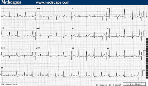
MRCP revision battle 10.7: Low voltage ECG complexes

I promise that's less than it looks...


MRCP revision battle 10.1: Hereditary haemorrhagic telangiectasia


Hereditary haemorrhagic telangiectasia (HHT), also known as Osler-Weber-Rendu syndrome, is an autosomal dominant condition associated with abnormal blood vessel formation.


Features of HHT include:
  • telangiectasia on skin
  • telangiectasia on mucosal membranes (leading to nose bleeds/GI bleeds)
  • arterio-venous malformations (AVMs)
    • commonest in lung
    • also in liver
    • brain
    • spine (rarest)

The diagnostic criteria are:
  1. spontaneous and recurrent nosebleeds
  2. telangiectasia
  3. AVMs
  4. positive family history - a first degree relative
The international consensus  states if 3 or 4 of the above features are present HHT is definite.  If 2 are present it is possible.  Less than that is unlikely.


A rare symptom the MRCP exam may throw up is platypnoea, which is difficulty breathing when sitting up/standing which is relieved on lying down.  Platypnoea is therefore the direct opposite of orthopnoea!  To briefly diversify, apart from lung AVM two other possible causes of platynoea are left atrial tumour or left atrial thrombus.


A rare sign associated with lung AVM is a 'humming sound' on auscultation over that area of the chest.


Management of HHT is symptomatic.


After that forray into the world of bleeding bits we're about to dive into the murky world of neurology with motor neurone disease...

MRCP revision battle 10.2: Motor Neurone Disease

Most people associated motor neurone disease (MND) with Stephen Hawking, that incredible physicist who has lived with MND for over 40 years.  However, in the same way he is not a stereotype for normal intelligence he is also not a stereotype for MND - most people with it die within 5 years.  More stereotypical for the course of the disease was Lou Gehrig, an American baseball player in the 1920s/1930s who was diagnosed with the disease and died within 3 years.  I mention him not just for random interest but also because in America MND is sometimes known as Lou Gehrig disease.  But what is MND?


MND is a group of neurological disorders affecting neurones in motor cortex, CN nuclei and anterior horn cells.


MND affects UMN and LMN but there is no sensory involvement.


As a quick recap, UMN signs include:
  • weakness
  • spasticity
  • brisk reflexes
  • upgoing plantars

LMN signs include:
  • weakness
  • wasting
  • fasciculation
  • depressed reflexes

There are several patterns of MND, including:
  1. Amyotrophic lateral sclerosis
    • presents as UMN signs in legs and LMN signs in arms
    • may be familial - in which case often associated with Ch 21
    • accounts for 50% of cases of MND
    • has an 'intermediate' prognosis
  2. Bulbar palsy
    • accounts for around 25% of cases of MND
    • has the worst prognosis of the presentations
    • more details in the next battle
  3. Progressive muscular atrophy
    • only LMN affected
    • begins distally
    • has a (relatively) good prognosis
  4. Primary lateral sclerosis
    • affects UMN only
10-35% of patients with MND will have fronto-temporal dementia.


Diagnosis of MND is primarily clinical.
EMG will show preserved motor conduction velocities
CSF analysis may show raised protein.


Treatment is with riluzole, which prolongs life by around 3 months and requires monitoring of LFTs.  There is no cure.

As the first paragraph stated, the prognosis is very poor and most patients are dead within 5 years.


So on to battle 10.3, to learn more about bulbar palsy.

MRCP revision battle 10.3: Bulbar Palsy

Bulbar palsy refers to palsy of the nerves coming from the 'bulbar' part of the brain, ie the pons, medulla and cerebellum.  These are VII, VIII, IX, X, XI and XII.


Classically bulbar palsy presents as palsy of tongue, muscles of chewing and swallowing and facial muscles.


Signs are LMN:
  • flaccid tongue
  • fasciculating tongue
  • absent or normal jaw jerk
  • loss of gag reflex
  • quiet/horse voice

Causes of bulbar palsy include:
  • MND
  • syringobulbia (more about this condition tomorrow!)
  • GBS (also lined up for battle tomorrow)
  • polio
  • Lyme disease
  • brain-stem tumours

Next up: battle 10.4, pseudobulbar palsy

MRCP revision battle 10.4: Pseudobulbar palsy

Pseudobulbar palsy is a UMN palsy of the muscles affecting eating, swallowing and talking above the midpons.


Signs are:
  • spastic tongue
  • brisk jaw jerk
  • emotional incontinence
  • 'Donald Duck' voice.

Causes include:
  • MS
  • MND
  • PD
  • stroke
  • high brain stem tumour


Just one more skirmish with neurology to go today, in the form of multifocal motor neuropathy.

MRCP revision battle 10.5: Multifocal motor neuropathy

I must confess I'd never come accross multifocal motor neuropathy until I started doing MRCP questions, but I'm now rather glad that I have, as essentially it is a treatable differential diagnosis for some presentations that appear like MND (which, as we've just covered, is nasty and deadly)


Multifocal motor neuropathy is an acquired immune-mediated demyelinating neuropathy.


Features of multifocal motor neuropathy include:
  • slowly progressive, asymmetric distal muscle weakness
  • cramps and twitching
  • no sensory loss
  • no/minimal muscle atrophy
  • possibly positive anti GM1 antibodies (NB also + in GBS)
  • no UMN signs
  • normal or decrease tone
  • absent reflexes

The joy of multifocal motor neuropathy is that it can be successfully treated with IV IG!


Well thats quite enough neurology for today, lets now look briefly at Lesch-Nyhan Syndrome, as promised yesterday

MRCP revision battle 10.6: Lesch-Nyhan Syndrome

Lesch-Nyhan Syndrome is a rare inherited x-linked disorder caused by an abnormality of an enzyme involved in purine metabolism resulting in increased uric acid.  Only boys tend to be affected.


Features of Lesch-Nyhan Syndrome include:
  • orange crystals in nappies
  • low IQ
  • self-mutilation
  • fits
  • recurrent gout (probably the most important relationship to remember in MRCP)
  • megaloblastic anaemia

Patients usually die by the age of 25, often of renal failure.


So to the final battle of the day, a nice gentle ending with a breeze through the causes of low-voltage complexes on ECG

MRCP revision battle 10.7: Causes of low-voltage complexes on ECG

'Low voltage complex' on ECG is defined as:
  • QRS amplitude <5mm in all limb leads and/or
  • QRS amplitude <10mm in all chest leads


The causes of low voltage complexes can be split into 2 main groups: increased distance from leads to heart or infiltrative disease of heart.

Using these divisions:
  1. increased distance from leads to heart:
    • emphysema/COPD
    • pericardial effusion
    • severe obesity
    • pleural effusion
  2. infiltrative disease of heart/problems with heart itself: 
    • amyloid
    • haemochromatosis
    • cardiomyopathies
    • global ischaemia

Myxoedema can also cause low complex ECGs but my search for why this is (?caused by myxoedema or ?caused by effects of myxoedema) proved fruitless... please let me know if you have the answer!


If you see an ECG with alternating normal complexes and low-voltage complexes, you are looking at electrical alternans which tends to be a sign of pericardial effusion/cardiac tamponade.  Click here to see an image of it.


So today's battles are over.  If you want to undertake the war to see how much of yesterday's battles you recall, please click here 

MRCP questions: War 9

As with previous 'wars' after 'battles' these are just a few quick questions to see if your brain cells have retained the information provided in battles 9.1 to 9.5.


Grab a piece of paper, jot down your answers then compare them to my answers here


Question 1:
Name the classical rash associated with Lyme disease


Question 2:
What is the treatment for uncomplicated Lyme disease?


Question 3:
A joint aspiration shows negatively birefrigent crystals.  What condition does this suggest?


Question 4:
What XR appearance may you see in gout?


Question 5:
Which joint is classically affected by pseudogout?


Question 6:
What is a craniopharyngioma?


Question 7:
What is a catamenial pneumothorax?

.


Question 8:
What is Hammans sign?  What does it signify?


Question 9:
A 19 yr old boy presents with shortness of breath.  A CXR shows a 3cm rim of air around his left lung.  How would you manage him?


Question 10:
You have a patient with a painful joint and the XR shows chondocalcinosis.  What condition does this suggest?


answers here