Monday, 13 September 2010

MRCP revision battle 10.2: Motor Neurone Disease

Most people associated motor neurone disease (MND) with Stephen Hawking, that incredible physicist who has lived with MND for over 40 years.  However, in the same way he is not a stereotype for normal intelligence he is also not a stereotype for MND - most people with it die within 5 years.  More stereotypical for the course of the disease was Lou Gehrig, an American baseball player in the 1920s/1930s who was diagnosed with the disease and died within 3 years.  I mention him not just for random interest but also because in America MND is sometimes known as Lou Gehrig disease.  But what is MND?


MND is a group of neurological disorders affecting neurones in motor cortex, CN nuclei and anterior horn cells.


MND affects UMN and LMN but there is no sensory involvement.


As a quick recap, UMN signs include:
  • weakness
  • spasticity
  • brisk reflexes
  • upgoing plantars

LMN signs include:
  • weakness
  • wasting
  • fasciculation
  • depressed reflexes

There are several patterns of MND, including:
  1. Amyotrophic lateral sclerosis
    • presents as UMN signs in legs and LMN signs in arms
    • may be familial - in which case often associated with Ch 21
    • accounts for 50% of cases of MND
    • has an 'intermediate' prognosis
  2. Bulbar palsy
    • accounts for around 25% of cases of MND
    • has the worst prognosis of the presentations
    • more details in the next battle
  3. Progressive muscular atrophy
    • only LMN affected
    • begins distally
    • has a (relatively) good prognosis
  4. Primary lateral sclerosis
    • affects UMN only
10-35% of patients with MND will have fronto-temporal dementia.


Diagnosis of MND is primarily clinical.
EMG will show preserved motor conduction velocities
CSF analysis may show raised protein.


Treatment is with riluzole, which prolongs life by around 3 months and requires monitoring of LFTs.  There is no cure.

As the first paragraph stated, the prognosis is very poor and most patients are dead within 5 years.


So on to battle 10.3, to learn more about bulbar palsy.

MRCP revision battle 10.3: Bulbar Palsy

Bulbar palsy refers to palsy of the nerves coming from the 'bulbar' part of the brain, ie the pons, medulla and cerebellum.  These are VII, VIII, IX, X, XI and XII.


Classically bulbar palsy presents as palsy of tongue, muscles of chewing and swallowing and facial muscles.


Signs are LMN:
  • flaccid tongue
  • fasciculating tongue
  • absent or normal jaw jerk
  • loss of gag reflex
  • quiet/horse voice

Causes of bulbar palsy include:
  • MND
  • syringobulbia (more about this condition tomorrow!)
  • GBS (also lined up for battle tomorrow)
  • polio
  • Lyme disease
  • brain-stem tumours

Next up: battle 10.4, pseudobulbar palsy

MRCP revision battle 10.4: Pseudobulbar palsy

Pseudobulbar palsy is a UMN palsy of the muscles affecting eating, swallowing and talking above the midpons.


Signs are:
  • spastic tongue
  • brisk jaw jerk
  • emotional incontinence
  • 'Donald Duck' voice.

Causes include:
  • MS
  • MND
  • PD
  • stroke
  • high brain stem tumour


Just one more skirmish with neurology to go today, in the form of multifocal motor neuropathy.

MRCP revision battle 10.5: Multifocal motor neuropathy

I must confess I'd never come accross multifocal motor neuropathy until I started doing MRCP questions, but I'm now rather glad that I have, as essentially it is a treatable differential diagnosis for some presentations that appear like MND (which, as we've just covered, is nasty and deadly)


Multifocal motor neuropathy is an acquired immune-mediated demyelinating neuropathy.


Features of multifocal motor neuropathy include:
  • slowly progressive, asymmetric distal muscle weakness
  • cramps and twitching
  • no sensory loss
  • no/minimal muscle atrophy
  • possibly positive anti GM1 antibodies (NB also + in GBS)
  • no UMN signs
  • normal or decrease tone
  • absent reflexes

The joy of multifocal motor neuropathy is that it can be successfully treated with IV IG!


Well thats quite enough neurology for today, lets now look briefly at Lesch-Nyhan Syndrome, as promised yesterday

MRCP revision battle 10.6: Lesch-Nyhan Syndrome

Lesch-Nyhan Syndrome is a rare inherited x-linked disorder caused by an abnormality of an enzyme involved in purine metabolism resulting in increased uric acid.  Only boys tend to be affected.


Features of Lesch-Nyhan Syndrome include:
  • orange crystals in nappies
  • low IQ
  • self-mutilation
  • fits
  • recurrent gout (probably the most important relationship to remember in MRCP)
  • megaloblastic anaemia

Patients usually die by the age of 25, often of renal failure.


So to the final battle of the day, a nice gentle ending with a breeze through the causes of low-voltage complexes on ECG

MRCP revision battle 10.7: Causes of low-voltage complexes on ECG

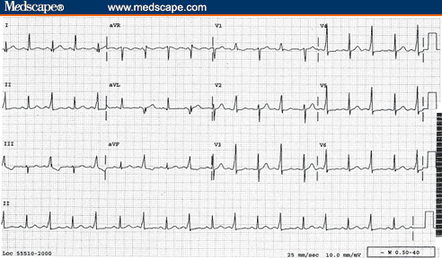
'Low voltage complex' on ECG is defined as:
  • QRS amplitude <5mm in all limb leads and/or
  • QRS amplitude <10mm in all chest leads


The causes of low voltage complexes can be split into 2 main groups: increased distance from leads to heart or infiltrative disease of heart.

Using these divisions:
  1. increased distance from leads to heart:
    • emphysema/COPD
    • pericardial effusion
    • severe obesity
    • pleural effusion
  2. infiltrative disease of heart/problems with heart itself: 
    • amyloid
    • haemochromatosis
    • cardiomyopathies
    • global ischaemia

Myxoedema can also cause low complex ECGs but my search for why this is (?caused by myxoedema or ?caused by effects of myxoedema) proved fruitless... please let me know if you have the answer!


If you see an ECG with alternating normal complexes and low-voltage complexes, you are looking at electrical alternans which tends to be a sign of pericardial effusion/cardiac tamponade.  Click here to see an image of it.


So today's battles are over.  If you want to undertake the war to see how much of yesterday's battles you recall, please click here 

MRCP questions: War 9

As with previous 'wars' after 'battles' these are just a few quick questions to see if your brain cells have retained the information provided in battles 9.1 to 9.5.


Grab a piece of paper, jot down your answers then compare them to my answers here


Question 1:
Name the classical rash associated with Lyme disease


Question 2:
What is the treatment for uncomplicated Lyme disease?


Question 3:
A joint aspiration shows negatively birefrigent crystals.  What condition does this suggest?


Question 4:
What XR appearance may you see in gout?


Question 5:
Which joint is classically affected by pseudogout?


Question 6:
What is a craniopharyngioma?


Question 7:
What is a catamenial pneumothorax?

.


Question 8:
What is Hammans sign?  What does it signify?


Question 9:
A 19 yr old boy presents with shortness of breath.  A CXR shows a 3cm rim of air around his left lung.  How would you manage him?


Question 10:
You have a patient with a painful joint and the XR shows chondocalcinosis.  What condition does this suggest?


answers here

Sunday, 12 September 2010

MRCP revision battle 9.1: Lyme Disease

Working and revising really does feel rather relentless.  I keep thinking of the pop song "and the beat goes on..." - somehow it seems appropriate for the continuous dedication work plus MRCP revision requires!  Anyway, what revision battles does today bring?

MRCP revision battle 9.1: Lyme disease
MRCP revision battle 9.2: Gout
MRCP revision battle 9.3: Pseudogout
MRCP revision battle 9.4: Craniopharyngioma
MRCP revision battle9.5: Pneumothorax


MRCP revision battle 9.1: Lyme disease

Lyme disease, named after the place in America where the condition was first noted, is a condition caused by the spirochaete borrelia burdorferi.  


It is spread by tic bites, with certain areas (such as northern America) being higher risk than, for example, Surrey.  However, it is found in the UK and it is important to note the majority of patients will not remember the bite itself.


Signs, symptoms and complications associated with Lyme disease are multitude and vague, including:
  • malaise
  • fever
  • muscle pain
  • joint swelling
  • decreased cognition
  • encephalitis
  • meningitis
  • lymphadenopathy
  • cranial nerve palsies (MRCP likes bilateral facial nerve palsy for some reason)
  • neuropathy
  • skin conditions


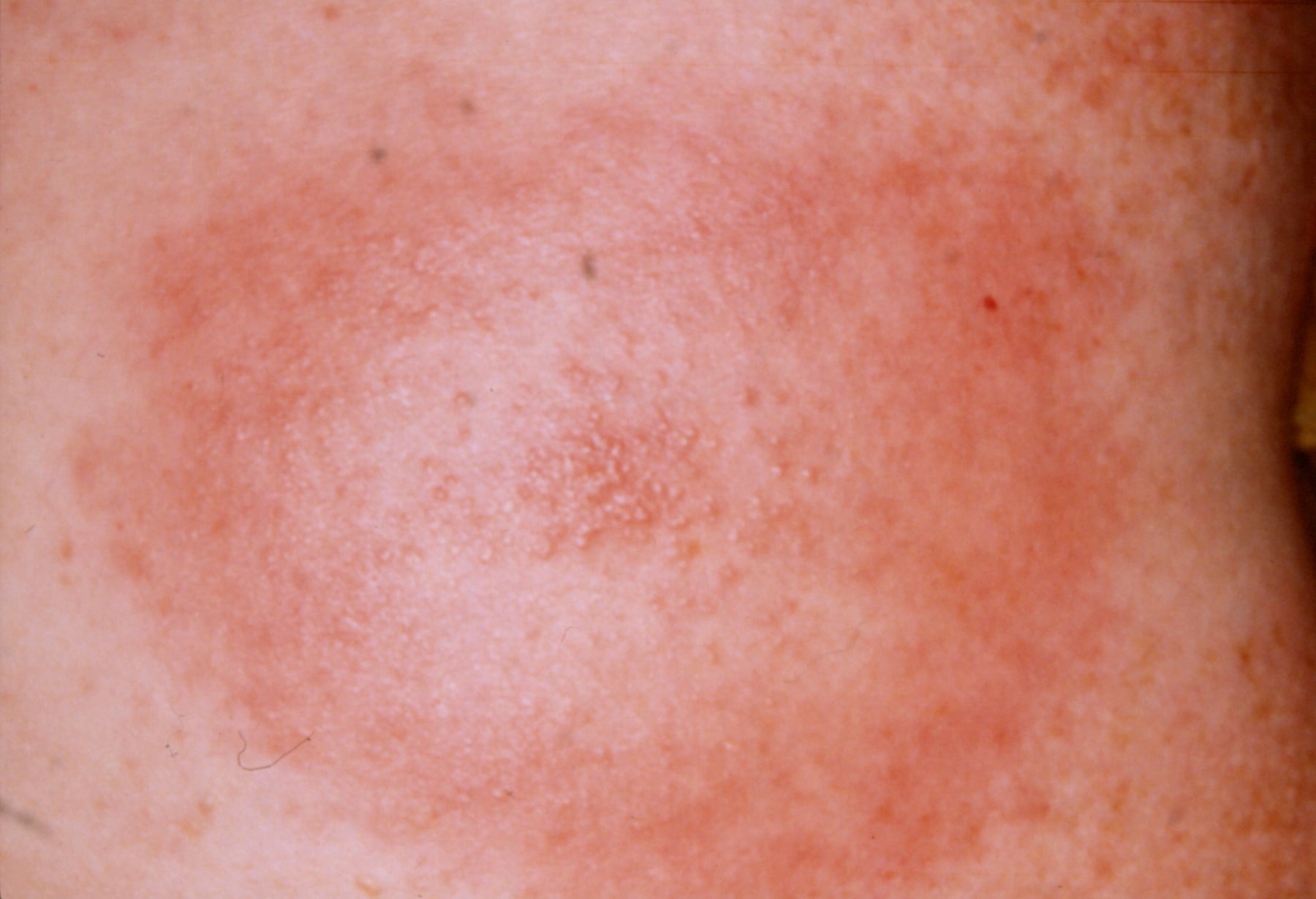
The classical skin condition associated with Lyme disease is erythema chronicum migrans, which is a spreading erythematous rash which then clears centrally but leaves a spot in the middle (click here for a picture).


A rarer skin condition is acrodermatitis chronica atrophicans, which occurs late in infection and eventually results in atrophic skin that is thin like cigarette paper.


Another rarer skin manifestation is borrelial lymphocytoma, which is a blue-red discoloration of the ear lobe.


Investigation is usually ELISA/PCR.
If the question throws in a CSF sample look for slightly raised protein and lymphocytosis.


Treatment of the rash only is doxycycline.  More serious disease may require IV ben pen/a 3rd generation cephalosporin. 


There is of course far more to learn about Lyme disease should you be so inclined, but if you're content with the barer MRCP-bones of it lets move on to battle 9.2, gout.

MRCP revision battle 9.2: Gout

Ah Gout... that classical disease of red-faced old men...

In terms of MRCP, you need to expand the above knowledge to include the following...


Gout usually presents as a single, swollen, hot, painful joint.  If the joint affected is the big toe, it is called a podagra.


Microscopy of joint fluid would reveal negatively birefrigent needle-shaped monosodium urate crystals.

Uric acid may be >450micromols - but equally may be normal in an acute attack.

CRP is usually raised, but if the question shows a raised WCC too start thinking about different diagnoses, for example septic arthritis.


Gout may be either primary or secondary.

Primary gout may be:
  • idiopathic - usually due to decreased excretion of uric acid 
  • associated with Lesch-Nyhan Syndrome (to be covered in a very brief battle tomorrow)


Secondary gout can broadly be divided into things which cause increased production/intake of urate, and things which decrease its excretion.


Causes of increased production/intake include:
  • myeloproliferative/lymphoproliferative disorders
  • psoriasis
  • cytotoxic drugs
  • food - beer, yeast, seafood, liver, kidney
  • exercise
  • fits
  • acidosis

Causes of decreased excretion include:
  • renal failure
  • diuretics
  • low dose aspirin
  • alcohol
  • lead poisoning

XR should show a normal joint space (may be decreased in late disease) and large punched-out erosions distant from the joint margins.


Treatment is NSAIDs - if these are contraindicated, colchine can be used.


Prophylaxis is with allopurinol (a xanthine oxidase inhibitor).  It should be started 2 to 3 weeks after an acute attack if:
  • >1 attack
  • tophi present
  • renal disease
  • urate stones
  • Lesch-Nyhan syndrome
  • cytotoxic drugs used (in which case it should be given before any episodes of gout)

So after that quick whizz through gout, lets embrace is near-cousin pseudo-gout...

MRCP revision battle 9.3: Pseudogout

Pseudogout involves the deposition of positively birefrigent calcium pyrophosphate dihydrate crystals in joints.


Pseudogout is associated with a variety of conditions, including:
  • hyperparathyroidism
  • hypothyroidism
  • haemochromatosis
  • Wilsons disease
  • acromegaly
  • old age
  • OA

It tends to cause a monoarthritis of a larger joint, classically a knee.


An XR may show chondrocalcinosis = soft tissue calcium deposition.


Treatment is with NSAIDs.  If this is unsuccessful steroids or hydroxychloriquine can be considered.


Now on to revision battle 9.4, a treat left over from yesterday, craniopharyngioma...

MRCP revision battle 9.4: Craniopharyngioma

Craniopharyngiomas are benign tumours that arise from the remnants of Rathke's pouch (= area between pituitary and 3rd ventricle floor).

Technically it is not a pituitary tumour as it is derived from the embryonic remnants of the pituitary gland rather than the gland itself.   However, in terms of the mass effects/how it presents it is very similar.  Amenorrhoea and decreased libido are often sited as symptoms.


Craniopharyngiomas frequently calcify (50%)


Investigation of choice is MRI, treatment is surgery.


After that brief squirmish its on to the last battle of the day, the pneumothorax

MRCP revision battle 9.5: Pneumothorax

A pneumothorax, as I'm sure you all know, is a collection of gas in the pleural cavity of the chest between the lung and chest wall.


I'm not about to insult you by going through the basic signs/symptoms of pneumothorax, but I will just briefly mention a rare sign that occasionally pops up in MRCP exams: a clicking sound at the sternal edge synchronised with the heart beat.  This rare sign of a pneumothorax is most commonly assocaited with small left sided pneumothoraces.


As a quick recap, its important to establish if the pneumothorax is primary (ie no underlying lung disease), secondary (=underlying lung disease) or traumatic (in which case as a medic you shouldn't be treating it!)


BTS guidelines state that in primary pneumothorax, if the patient is not breathless and the rim of air is <2cm you do not need to necessarily do anything and may consider sending the patient home.  If the rim of air is >2cm or the patient is breathless you should aspirate; try x2, if still unsuccessful insert a drain.


The guidelines for secondary pneumothorax are slightly more complex:
  • if >50yrs old AND breathless AND rim of air >2cm, insert a drain first line
  • if the above 3 criteria do not apply, try aspirating first
  • regardless of which category your patient falls into, patients with secondary pneumothorax should be admitted for 24 hours observation.


A random type of pneumothorax to be aware of is catamenial pneumothorax - this is a pneumothorax associated with menstruation, which is due to endometriosis of the lung.  90% of these occur on the right side.


As a complete aside, as well as recognising the 'click synchronised with heartbeat' as a possible pneumothorax it is worth learning that a 'crunching sound' in the chest can be due to a pneumomediastinum  and is called Hamman's sign.


Well those are todays battles over, but for those with residual enthusiasm the beat goes on with a war to test your memory of yesterday's topics...

MRCP questions: War 8

As with previous 'wars' after 'battles' these are just a few quick questions to see if your brain cells have retained the information provided in battles 8.1 to 8.6.

Grab a piece of paper, jot down your answers then compare them to my answers here


Question 1:
What is the treatment for cluster headache?


Question 2:
List 3 symptoms of cluster headache.

Question 3:
What odour is associated with acute arsenic poisoning?

Question 4:
List 3 causes of Mees lines

Question 5:
State the 4 H's and 4 T's which are known as the reversible causes of cardiac arrest.


Question 6: 
What kind of visual disturbance is classically associated with a pituitary tumour

Question 7:
What histological type of pituitary tumour is most likely to cause a pressure effect?

Question 8:
What is Nelson's syndrome?


Question 9:
What are the similarities and differences between Romano Ward and Jervell Lange Nielson syndromes?

Question 10:
What is the commonest hormone produced by a pituitary tumour?


answers here

Saturday, 11 September 2010

MRCP revision battle 8.1: Cluster headache

Its Saturday and sunny so I'm not particularly feeling the MRCP revision love.  But the battle must continue, and today's topics are:

Revision battle 8.1: Cluster headache
Revision battle 8.2: Arsenic poisoning
Revision battle 8.3: Mee's lines
Revision battle 8.4: Reversible causes of cardiac arrest
Revision battle 8.5 Pituitary tumours
Revision battle 8.6: Romano-Ward and Jervell-Lange-Nielson


Revision battle 8.1: Cluster headache


Cluster headaches appear in the MRCP exam (and occasionally real life) as:
  • severe unilateral pain focused around one eye with
  • redness of the eye and possibly
  • lid swelling with
  • lacrimation and
  • nasal congestion
It is worth noting that up to 20% will also get ptosis, which in a minority of cases can be permanent.


Cluster headaches are more common in males (5:1).
Risk of suffering from cluster headache is increased if you are a smoker.


Classically patients will suffer from severe 15 minute to 2 hour bouts of pain once or twice a day over a period of 4 to 12 weeks.


Treatment acutely is 100% oxygen.  S/C sumitriptan and nasal lidocaine are also proposed as treatments.

Prophylactic options include verapamil, prednisolone or lithium.


So onwards to a slightly curveball topic - arsenic poisoning...

MRCP revision battle 8.2: Arsenic poisoning

Arsenic poisoning used to be both a favourite method of murder (allegedly involved in the demise of Napoleon) and a substance commonly found in paint and water pipes.  It is now thankfully carefully controlled so it tends to be found mainly in MRCP exam questions...


Arsenic is still used in the glass, smelter and microelectronic industries, and is found in wood preservatives.


Acute poisoning tends to cause features in 30 mins to 2 hours.

Features of acute arsenic poisoning include:
  • severe gastroenteritis
  • garlic ordour
  • hypersalivation
Treatment for acute poisoning is DMPS.


Symptoms of chronic arsenic poisoning present at different times post-exposure; for example, skin symptoms tend to show clinically around 10 years afterwards.  There are multiple, multiple possible complications of chronic arsenic poisoning but a few which may pop up are:
  • keratoses
  • Mees lines (if you don't know what these are you'll find out in the next battle!)
  • hyperpigmentation
  • neuropathy
  • muscle fasciculation and wasting

So on to explore Mees lines...

MRCP revision battle 8.3: Mees lines

Mees lines are white transverse lines that go all the way across the nail.

An image of Mees lines can be found here


Mees lines are found in:
  • arsenic poisoning
  • thalium poisoning
  • heavy metal poisoning
  • renal failure

 Next up: battle 8.4!

MRCP revision battle 8.4: Reversible causes of cardiac arrest

A little bit of a random one to throw into MRCP revision but a quick recap of the reversible causes of cardiac arrest, famously recalled as the 4 Hs and 4 Ts, can never go amiss...


4 H's:
  • hypoxia
  • hypovolaemia
  • hypothermia
  • hypoglycaemia/hyper/hypokalaemia/ other electrolyte imbalance

4 T's:
  • Tension pneumothorax
  • cardiac Tamponade
  • Thombus
  • Toxins


I remember the 4 H's in the format of a little picture of a man standing on top of a snow-covered mountain.  He is struggling to breath as there's not much oxygen up there (hypoxia) and  is shivering as its so cold (hypothermia).  He is looking sad and in one hand is an empty water bottle (hypovolaemia) and in the other an empty lunchbox (hypoglycaemia... then remember the hypo/hyper kalaemia from that).

For the 4 T's I think of the less happy phrase of 'Thomas has Ten Toxic Tampons' (= Thombus, tension pneumothorax, toxins, tamponade)


So after a couple of nice straightforward battles let face a juicier one: pituitary tumours...

MRCP revision battle 8.5: Pituitary tumours

Most pituitary tumours are benign adenomas.


Symptoms from pituitary tumours are either due to:
  • local pressure
  • hormone secretion
  • hypopituitism

Local pressure symptoms include:
  • bitemporal hemianopia (pressure on CN III)
  • palsies of CN III/IV/VI (remember the cavernous sinus anatomy at the end of battle 1.1?)

The commonest hormone secreted by pituitary tumours is prolactin (35%) followed by GH (20%).

30% of pituitary tumours secrete no obvious hormone


Potentially a pituitary tumour can disturb hypothalamic control of temperature, sleep or appetite.

If the tumour erodes the floor of the sella the patient may get CSF rhinorrhoea


Histologically, pituitary tumours fall into the following catagories:
  • 70% - chromophobes
    • 30% cause a pressure effect
    • can produce prolactin, ACTH, GH
  • 15% - acidophils
    • 10% cause a pressure effect
    • can produce GH, prolactin
  • 15%  - basophils
    • rarely cause a pressure effect
    • can produce ACTH

The investigation of choice for suspected pituitary tumours  is MRI.


Finally, in this section we need to cover Nelsons Syndrome.  Nelsons syndrome is a condition that occurs after adrenalectomy for pituitary-dependent cushings.  The adrenalectomy removes the negative feedback on the pituitary which causes ACTH levels to rise massively, leading to hyperpigmentation +/- an invasive pituitary tumour.


Tomorrow we will look at craniopharyngioma, but to finish today on a lighter note onwards to Romano Ward and Jervell-Lange-Nielson Syndromes.

MRCP questions: War 7

As with previous 'wars' after 'battles' these are just a few quick questions to see if your brain cells have retained the information provided in battles 7.1 to 7.5.


Grab a piece of paper, jot down your answers then compare them to my answers here


Question 1:
An efflux of which ion terminates the cardiac action potential?

Question 2:
What is the mechanism of action of class III antiarrhythmics?


Question 3: 
Name 2 class IV antiarrhythmic drugs

Question 4:
What is the relationship between heart rate and QT?

Question 5:
How do you calculate QT using the Bazzett formula?

Question 6:
State the upper limit of a normal QT

Question 7: 
Name 5 drugs that prolong QT

Question 8:
State 5 metabolic conditions that can prolong QT

Question 9:
Name 2 drugs that increase the effect of adenosine and one that decreases it.

Question 10:
Does positive concordance in the chest leads make a broad complex tachycardia more or less likely to be VT?


answers here


MRCP revision battle 8.6: Romano Ward and Jervell-Lange-Nielson Syndomes

Both Romano-Ward and Jervell-Lange-Nielson Syndrome are congenitial causes of a long QT.

Both are believed to be caused by mutations in the potassium subunit.

The differences are:
  1. Romano-Ward is autosomal dominant while Jervell-Lange-Nielson is autosomal recessive
  2. Jervell-Lange-Nielson is associated with bilateral deafness; Romano Ward is not.

Thats all for today; if you want to test your recall of topics covered yesterday click here