Friday, 1 October 2010

MRCP revision battle 24.3: Acute Promyelocytic Leukaemia

Acute Promyelocytic Leukaemia is the M3 subtype of AML.

It is associated with the t(15;17) translocation and the RARA gene.


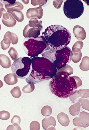
On microscopy you look for faggots (=collections of auer rods):

The cells are strongly sudan black/peroxidase positive.

Acute promyelocytic leukaemia tends to present in young adults (average approx 25 yrs) and the presentation is often DIC.


Despite this sounding bad, acute promyelocytic leukaemia actually has a good prognosis.


Remission can be induce with all-trans retinoic acid.


Thats enough heavy-going haematology for one day... lets move on to angioid retinal streaks...

MRCP revision battle 24.4: Angioid retinal streaks

Angioid retinal streaks are caused by breaks in Bruch's membrane


They look like dark red lines under the retina - almost like blood vessels.  I'd recommend using google to search for a selection of images to look at.


They are associated with SLAPPERS:
  • Sickle cell disease
  • Lead poisoning
  • Acromegaly
  • Pagets disease
  • Pseudoxanthoma elasticum
  • Ehlers-Danlos
  • Raised calicum/phosphate
  • Short people


 Onwards to SIADH...


MRCP revision battle 24.5: SIADH

SIADH = syndrome of inappropriate antidiuretic hormone secretion is a condition whose name really says it all: it is ADH being produced and released when it shouldn't be.


Quick bit of physiological background:
  • ADH is made in the magnocellular neurones in the supraoptic and paraventricular nuclei of the hypothalmus
  • ADH is stored in the posterior pituitary
  • it is released in response to raised plasma osmolality (osmoreceptors in hypothalamus) or decreased volume (baroreceptors in carotids, atria)
  • its effect is to increase the insertion of aquaporin 2 channels in the collecting ducts of the kidney, hence increasing water reabsorption.
  • it also increases peripheral vascular resistance


SIADH is diagnosed in the presence of:
  • concentrated urine ( sodium >20mmol/l and osmolality >500mosmol/kg) AND
  • hyponaturaemia (<125) or low plasma osmolality (<260mosmol/kg) AND
  • the absence of hypovolaemia, oedema or diuretics.


Causes of SIADH include:
  • malignancy: small cell lung cancer, pancreas, prostate, lymphoma
  • lung pathology: TB, pneumonia, aspergillosis
  • CNS pathology: injury, GBS, fits, subarachnoid/subdural haemorrhage, abscess, stroke
  • metabolic: porphyria
  • drugs: opiates, carbamazepine, SSRIs, TCA


So lets now finish todays battles by widening this topic out to hyponatraemia...

MRCP revision battle 24.6: Hyponatraemia

Hyponatraemia is low blood sodium.  The effect of this will depend on how low and for how long.

Possible signs and symptoms include:
  • confusion
  • seizures
  • hypertension
  • cardiac failrue
  • anorexia
  • nausea
  • muscle weakness
  • oedema
(=CASH MON)


My personal way of dealing with hyponatraemia is to begin by asking 2 cynical questions:
  1. is it actually just dilutional? - ?hyperglycaemia 
  2. is it actually just pseudo? - ?high protein ?high triglycerides


Once I've got negatives to my cynicism, I move on to ask:
  1. are they dehydrated or not?


The divisions from this question onwards are summarised in the diagram below:


If the low sodium is chronic, you treat by:
  • treating cause
  • giving cautious N.saline if dehydrated
  • fluid restricting if not dehydrated.


Occasionally demeclocycline might be needed; this is an antibiotic which has the side effect of inducing nephrogenic diabetes inspidus and hence helps treat SIADH.


Now on to neutrophils...


MRCP revision battle 24.7: Neutropenia and neutrophilia

Your humble neutrophil makes up 40-75% of your white blood cells.
It lives for a mere 5 days.
It migrates towards the 'bad guys' by following the signals of substances including IL-8.
It valiantly phagocytoses bacteria.

MRCP examiners enjoy asking questions about low and high levels of neutrophils...


Neutropenia = low neutrophil count

Causes:
  • viral infection
  • severe sepsis
  • bone marrow failure
  • neutrophil antibodies (eg SLE, haemolytic anaemia)
  • drugs - chemotherapy, carbimazole


Neutrophilia = raised neutrophil count.

Causes:
  • bacterial infection
  • MI
  • steroids
  • pregnancy
  • uraemia
  • acidosis
  • gout
  • myeloproliferative disorders


That's all the hard work done for today; for some light relief click this link

    Tuesday, 28 September 2010

    MRCP revision battle 23.1: Henoch Schonlein purpura

    No energy for an interesting introduction today... lets dive straight into the battles...


    MRCP revision battle 23.1: Henoch Schonlein purpura
    MRCP revision battle 23.2: Parinaud's syndrome
    MRCP revision battle 23.3: Polymyositis and Dermatomyositis
    MRCP revision battle 23.4: Trichomonas vaginalis
    MRCP revision battle 23.5: Atrial septal defects
    MRCP revision battle 23.6: Pyoderma gangrenosum


    MRCP revision battle 23.1: Henoch Schonlein purpura


    Henoch Schonlein Purpura (HSP) is a an IgA mediated small vessel vasculitis.

    Classically it affects mainly children post infection.


    Features include:
    • symmetrical macular rash over buttocks and extensor surfaces of legs
    • abdominal pain +/- blood diarrhoea
    • arthralgia

    Complications of HSP include:
    • renal failure (look for microhaematuria)
    • intussusception

    Management is supportive.


    Now for a very quick battle, Parinaud's Syndrome...

    MRCP revision battle 23.2: Parinaud's syndrome

    Parinaud's syndrome refers to the combination of:
    • upward gaze palsy
    • pseudo Argyll-Robertson pupils


    Causes include:
    • hydrocephalus
    • pineal tumours
    • stroke
    • multiple sclerosis


    Onwards to the longer 'buy-one-get-one-free' battle of dermatomyositis and polymyositis...

    MRCP revision battle 23.3: Polymyositis and Dermatomyositis

    Polymyositis is an idiopathic inflammatory disorder of skeletal muscle.  When it is associated with cutaneous lesions it is dermatomyositis.


    Features:
    • progressive proximal muscle weakness
    • dysphagia
    • interstitial lung disease
    • oesophageal dysfunction
    • weight loss 
    • fever
    • myocarditis
    • arthralgia

    To try and remember this list I think of the condition starting in the proximal muscles of the arm then creeping to the joint (causing arthralgia) and onwards to the oesophagus, causing dysphagia and oesphageal dysfunction.  I then imagine it seeping into the lungs (interstitial lung disease) and then onwards into the heart (myocarditis).


    Skin signs include:
    • heliotrope (liliac-purple) rash around eyelids/cheeks
    • macular rash over back and shoulders (=shawl sign)
    • Gottrons papules = scaly plaques on MCP/PIP joints
    • Gottrons sign = erythema over knees/elbows
    • periungal telangectasia
    • nail fold infarcts
    • photosensitivity


    There is a higher prevalence of malignancy with dermatomyositis.


    Investigations show:
    • raised CK/AST/LDH
    • abnormal EMG - shows fibrillation potentials
    • anti-Jo antibodies associated with a systemic form of disease


    Treatment is:
    • prednisolone
    • methotrexate
    • screen for maligancy.


    NB: juvenile dermatomyositis is different - it is more aggressive and includes a vasculitis and ectopic calcification.


    Now onwards for our first expedition into the world of sexually transmitted diseases...

    MRCP revision battle 23.4: Trichomonas vaginalis

    Trichomonas vaginalis is an anaerobic protozoan that is transmitted sexually.

    It needs an alkaline pH to thrive.


    It can cause:
    • thin, grey, fishy discharge
    • itchiness
    • dysparenia
    • dysuria


    As it inflames the endothelium it increases an individual's susceptibility to other sexually transmitted infections.


    Treatment is with metronidazole.


    Now on to atrial septal defects...

    MRCP revision battle 23.5: Atrial septal defects

    Atrial septal defects may present as:
    • dyspnoea on exertion
    • finding after AF diagnosed
    • finding after CVA
    • finding after heart failure


    Clinical signs include:
    • fixed splitting S2
    • ejection systolic murmur
    • left parasternal heave


    A cardiac echo may show paradoxical ventricular septal motion due to right sided overload.


    There are 3 main atrial septal defects to be aware of:


    1: Ostium secundum defect
    • Ostium secundum defects account for 70% of atrial septal defects
    • caused by an enlarged foramen ovale
    • ECG shows RBBB with right axis deviation and a long PR
    • 10-20% of cases will be associated with mitral valve prolapse
    • it tends to present in adulthood


    2: Ostium primum defect
    • ostium primum defects account for 15% of atrial septal defects
    • ECG shows RBBB with left axis deviation and a long PR
    • it tends to present earlier, in childhood
    • it is associated with Downs, Klinefelters and Noonans
    • some feel it should be classified as a atrioventricular septal defect as it is so low down in the atria, on the endocardial cushions
    • often associated with TR/MR.


    3: Sinus venous atrial septal defect
    • up to 15% of cases


    Treatment is closure.


    A very small atrial septal defect is patent foramen ovale.  This is different from the others as as it is so small it does not allow equilisation of atrial pressures.  It occurs in 25% of the population and can allow paradoxical emboli (see below) and is associated with migraine.


    Complications of ASDs include:
    • Eisenmengers Syndrome
      • left-to-right shunt causes increased blood flow through pulmonary vasculature and so pulmonary hypertension, which increases the pressure in the right side of the heart leading to reversal of the shun to right-to-left = eisenmnegers syndrome.
    • paradoxical emboli 
      • venous emboli ending up in arterial rather than venous system
      • --> strokes, intestinal infarcts, splenic infarcts 
    • ? migraines - controversial area of research - google if you are interested.


    Finally for ASDs a brief eponymous syndrome that occasionally appears in MRCP answer options: Holt-Oram.  Holt Oram is an inherited condition characterised by abnormalities of the heart and upper limb.  It is inherited in an autosomal dominant manner with incomplete penetrance.

    Now skip straight to last battle of the day

    MRCP revision battle 23.6: Pyoderma gangrenosum

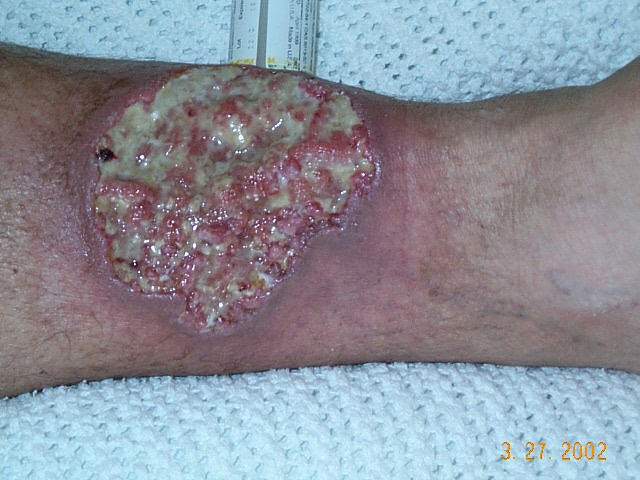
    Pyoderma gangrenosum is a nodulo-pustular ulcer, with a purulent surface and a tender bluish overhanging edge.

    Below is a picture from Wiki Commons showing pyoderma gangrenosum:


    In 50% of cases it is idiopathic.


    Associations with pyoderma gangrenosum include:
    • inflammatory bowel disease
    • RA/SLE/ank spond
    • lymphoma/leukaemia/myeloproliferative disorders
    • primary biliary sclerosis/sclerosing chloangitis
    • sarcoid
    • thyroid problems
    • diabetes
    • Wegeners granulomatosis


    Treatment includes high dose oral steroids.


    Another day's battling complete.  Congratulations!

    Monday, 27 September 2010

    MRCP revision battle 22.1: Goodpasture's Syndrome

    Today's battles cover a lot of the random conditions that frequently appear as differentials in MRCP questions but rarely feature in your day-to-day life.  As well as being random, or possibly as a consequence of them being random, their exact causes are often unknown, their treatments are debated and they are just downright complex.  Can you tell from this opening gambit how much fun I've had preparing today's battles?!  Hopefully I've whittled them down to digestible forms and have tried to give links if I've whittled away too much.  I've also thrown in aortic regurgitation for some light relief... when aortic regurgitation is light relief you know its going to be a bad day....

    So here goes, and good luck!!


    MRCP revision battle 22.1: Goodpasture's Syndrome
    MRCP revision battle 22.2: Neurofibromatosis
    MRCP revision battle 22.3: Tuberous sclerosis
    MRCP revision battle 22.4: Fanconi anaemia
    MRCP revision battle 22.5: Fanconi syndrome
    MRCP revision battle 22.6: Aortic regurgitation
    MRCP revision battle 22.7: Argyll Robertson Pupil


    MRCP revision battle 22.1: Goodpasture's Syndrome 


    Goodpasture's syndrome is a rare condition caused by anti GBM antibodies and is characterised by pulmonary haemorrhage and renal failure.


    There is less than 1 case per million people per year.
    Cases may be triggered by inhaled hydrocarbons, paraquat or viral infections.


    Pulmonary haemorrhage is treated by plasma exchange
    Renal involvement is treated by steroids, cyclophosphamide and plasma exchange.


    Renal biopsy would show:
    • IgG on basement membrane
    • crescent formation


    For the really keen:


    1 random battle down, several more to go...

    MRCP revision battle 22.2: Neurofibromatosis

    Neurofibromatosis is an autosomal dominant condition which causes lesions in the skin, nervous system and skeleton.

    It is quite complex so below are just a few key points to learn for MRCP:


    There are 2 types:


    Neurofibromatosis 1 = von Recklinghausen's disease
    • chromosome 17, 1:2500
    • features include:
      • cafe au lait spots
      • axillary/inguinal freckling
      • lisch nodules in iris (seen in >90%)
      • perpheral neurofibromas
      • optic gliomas (2%)
      • osseous lesions
    • may have low IQ or renal artery stenosis

    Neurofibromatosis 2
    • chromosome 22, rarer than NF 1
    • Features:
      • bilateral acoustic neuromas - sensorineural hearing loss is first sign
      • cafe au lait spots
      • juvenile posterior subcapsular lenticular opacity = a form of cataract


    For the really keen:


    So having got one complex, rare and unsatisfying battle out of the way lets jump straight into another...

    MRCP revision battle 22.3: Tuberous sclerosis

    This is going to be a very unsatisfying battle because tuberous sclerosis is rare and complex and essentially to grasp it properly goes far beyond what is required for MRCP, so please excuse my poor attempt at summarising some of the details which may be MRCP-relevant, particularly the skin manifestations...


    Tuberous sclerosis is a rare, complex autosomal dominant condition which causes multiple benign tumours.


    Features include:

    1. cutaneous features
      • ash leaf spots (appear under UV light)
      • shagreen patches
      • adenoma sebaceum
      • subungal fibromata
      • cafe au lait spots
    2. neurological features
      • epiplesy
      • low IQ
    3. other features
      • retinal haematoma
      • gliomas
      • renal cysts

    For the really keen:


    Now lets go on to the thankfully less complex topic of Fanconi anaemia...

    MRCP revision battle 22.4: Fanconi anaemia

    Fanconi anaemia is an autosomal recessive disorder which is characterised by:
    • pancytopenia secondary to bone marrow failure
    • dysmorphic features
      • short stature
      • microcephaly
      • absent radii
    • cafe au lait spots
    • susceptibility to cancers
      • AML
      • squamous cell cancers of head and neck
      • gynaecological  cancers

    Most patients die by 30.


    For the really keen:
    emedicine article on Faconi anaemia 


    For the rest of us lets move on to look at the similarly named, but completely different, Fanconi Syndrome

    MRCP revision battle 22.5: Fanconi Syndrome

    Fanconi syndrome is disturbance of proximal renal tubule function, resulting in defective reabsorption of:
    • amino acids
    • phosphate
    • potassium
    • glucose
    • bicarbonate

    It can cause vitamin D resistant rickets.

    Causes of Fanconi syndrome include:
    • idiopathic
    • inherited
      • cystinosis
      • Wilson's disease
    • acquired
      • heavy metal poisoning
      • light chains
        • amyloid
        • myloma

    Treatment is:
    • treat cause
    • replace losses (potassium, bicarb, phosphate, vit D supplements)


    Lets now move back mainstream with a revision battle with aortic regurgitation...

    MRCP revision battle 22.6: Aortic regurgitation

    Aortic regurgitation produces a fiendishly difficult to hear early diastolic murmur; I've heard it once and embarked on a most unprofessional victory dance once out of sight of the patient (even more embarrassingly it wasn't even me picking up the murmur, I had been sent to listen to the patient knowing they had the murmur...)


    Possibly because its so hard to hear there are a whole troop of eponymous signs that can suggest aortic regurgitation, and MRCP exams love throwing them into questions to excite you:
    • Corrigan's sign: visible carotid pulsation 
    • de Mussets sign: the head bobs with each pulse
    • Duroziez's sign: femoral artery is compressed and auscultated proximally and a diastolic murmur is heard as blood flows backwards during diastole
    • Millers sign: pulsation of uvula
    • Quincke's sign: capillary pulsations in nail be
    • Traube's sign: pistol shot sound over femoral arteries

    The less exciting but probably more important points to remember are:
    • the pulse is collapsing (=waterhammer)
    • wide pulse pressure
    • the apex is thrusting

    As well as the classic early diastolic murmur best heard down the left sternal edge which is loudest when the patient is leaning forward and in expiration, you need to be aware of the Austin Flint murmur.


    Austin Flint murmur only occurs in severe aortic incompetence.  It is a mid diastolic murmur and is probably due to the regurgitant jet interfering with the opening of the mitral valve.


    Symptoms of AR are:
    • dyspnoea
    • palpitations
    • heart failure


    Causes of AR include:
    • valve inflammation
      • rheumatic fever
      • IE
      • RA
      • SLE
      • appetite supressants
    • aortic root disease
      • hypertension
      • syphillis
      • aortic dissection
      • ankylosing spondylitis
      • psoriasis
    • collagen diseases
      • hurlers syndrome
      • marfans
      • psuedoxanthoma elasticum
    One way to remember these causes is SIR AA SHARP  plus collagen.


    Management: the aim is to replace the valve before significant LV dysfunction.  Indications for surgery are increasing symptoms, enlarging heart (CXR/echo), worsening ECG (TWI laterally)


    Onwards to the brief final battle - the Argyll Robertson Pupil

    MRCP revision battle 22.7: Argyll Robertson Pupil

    I love the Argyll Robertson pupil, and have done ever since a very serious elderly consultant intoned to us that "the Argyll Robertson pupil is like a prostitute... it will accommodate but will not react."


    Essentially the Argyll Roberston pupil is a pupil that:
    • will not react to light
    • will accommodate

    It is a sign of neurosyphillis, but a similar phenomenon occurs in diabetes.
    They are also seen in Parinaud's syndrome, which will be covered tomorrow.


    And on that teaser, adieu!

    Sunday, 26 September 2010

    MRCP revision battle 21.1: Erythema nodosum

    I'm feeling a rather keen bean today so I've decided to slightly modify the way I serve your daily MRCP battles to you.  From now on, as well as battles and wars, I'll try to add 'further reading' links to some of the battles as a form of desert wine for the really keen/anally retentive. 

    On that motivated note, today's battles will be:


    MRCP revision battle 21.1: Erythema nodosum
    MRCP revision battle 21.2: Glucagon and glucagonoma
    MRCP revision battle 21.3: Wegener's Granulomatosis
    MRCP revision battle 21.4: Rapidly progressive glomerulonephritis
    MRCP revision battle 21.5: Achalasia
    MRCP revision battle 21.6: Scabies
    MRCP revision battle 21.7: Pre eclampsia and HELLP syndrome
    MRCP revision battle 21.8: Cafe au lait spots


    MRCP revision battle 21.1: Erythema nodosum


    Erythema nodosum is inflammation of subcutaneous fat resulting in tender erythematous nodules, typically on the shins.

    It usually resolves without scars in around 6 weeks.


    It is associated with multiple conditions; below is a list of just a few that you really should remember:

    • infection
      • TB
      • streptococcus
      • viral infections
    • systemic
      • sarcoid
      • IBD
      • Behcets
    • malignancy
    • drugs
      • OCP
      • penicillin
      • sulphonamides
      • dapsone
      • tetracycline
    • pregnancy


    Treatment is to treat the underlying condition and give NSAIDs for the pain of the nodules.

    For the really keen:

    For everyone else, lets go to to battle 21.2!

    MRCP revision battle 21.2: Glucagon and glucagonoma

    Lets quickly recap glucagon from our med school days before we move on to the more exotic beast of glucagonoma...


    Glucagon is a peptide hormone secreaed from the alpha cells of the islets of langerhans.


    It results in raised blood glucose by:
    • increasing gluconeogenesis in the liver
    • increasing lipolysis

    Glucagon is secreted in response to low blood glucose, raised catecholamines and raised plasma amino acids.
    Glucagon secretion is inhibited by insulin, ketones in the blood, somatostatin and free fatty acids in the blood


    A glucagonoma is a very rare tumour of the alpha cells of the islets of langerhans which secretes glucagon.


    It results in:
    • massively raised levels of glucagon
    • diabetes mellitus
    • hypoaminoacidaemia (as GNG is using up protein)
    • anaemia
    • necrolytic migratory erythema


    Necrolytic migratory erythema is a red, blistering rash which is the presenting feature of a glucagonoma in 70% of cases.


    Glucagonomas can be associated with MEN 1.


    Treatment of a glucagonoma is ocreotide and surgery; prognosis is poor


    For the really keen:


    For the rest of us, its onwards to battle 21.3!

    MRCP revision battle 21.3: Wegener's Granulomatosis

    Wegener's Granulomatosis is a small/medium vessel necrotising granulomatous vasculitis which tends to affect the upper respiratory tract, lungs and kidneys.


    90% of cases present with respiratory symptoms.


    Associated features to be aware of include:
    • blood nasal discharge
    • haemoptysis
    • saddle-shaped nose

    75% get crescentic glomerulonephritis.


    cANCA is positive in 90%, pANCA is positive in 25%


    CXR may show large shadows.  Pulmonary haemorrhage is also possible.


    Treatment is with steroids, cyclophosphamide and possibly co-trimaoxazole to try and reduce risk of PCP.


    I guess this is a good opportunity to reconfront a personal demon in the form of a renal battle...

    MRCP revision battle 21.4: Rapidly progressive glomerulonephritis

    Rapidly progressive glomerulonephritis, AKA crescentic glomerulonephritis (don't you just love the way that in renal medicine everything has multiple names just to make it even more complicated than it already is?!) is, as the name suggests, a particularly aggressive form of glomerulonephritis.  It often presents with acute renal failure, and can progress to end stage renal failure in a matter of days.


    It causes crescent-shaped scarring of the glomeruli, hence its name.


    Causes of rapidly progressive glomerulonephritis include:
    • Wegeners 
    • microscopic polyangiitis
    • Goodpastures (a topic to be tackled tomorrow)
    • transformation from other glomerulonephritis'

    Treatment is with high dose steroids.


    The prognosis is poor, especially if the initial creatinine is higher than 600.


    Since I find anything renal hard to swallow, achalasia seemed an appropriate next battle...

    MRCP revision battle 21.5: Achalasia

    Achalasia is abnormal peristalsis and failure of relaxation of the lower oesophageal sphincter.


    It causes dysphagia, regurgitation, chest pain and weight loss.


    Females aged 30-50 are most commonly affected.


    Investigation is with oesophageal manometry and barium swallow.
    The barium swallow should show a characteristic 'birds beak' appearance (image below is from wiki commons):


    Treatment is with balloon dilation or hellers cardiomyotomy.    Nifedipine may be temporarily helpful.


    A rare and late complication of achalsia can be squamous carcinoma.


    Now on to battle 21.6, which is short but sure to make you itch...

    MRCP revision battle 21.6: Scabies

    Scabies is an intensely itchy condition caused by sarcoptes scabei, an arachnid which burrows into skin.


    In terms of MRCP the follow facts need to be committed to memory:
    • first line treatment is 5% permethrin which must be applied over whole body, face and scalp and washed off after 8 to 12 hrs and repeated after 7 days
    • second line treatment is 0.5% malatrion
    • pruritus persists for up to 6 weeks post erradication.
    • look out for pictures of lines (burrows) between web spaces of hands


    Now you're itching nicely, lets attack the penultimate battle of the day, pre-eclampsia and HELLP syndrome.

    MRCP revision battle 21.7: Pre eclampsia and HELLP syndrome

    Pre-eclampsia is defined as:
    • gestational hypertension
    • proteinuria
    • occurring after 20 weeks gestation
    Oedema is often a feature.
    Possible symptoms include blurred vision, headache and abdominal pain.


    Treatment is to lower blood pressure but ultimately the only way to treat pre-eclampsia which is advancing towards eclampsia (=fit) is to deliver the baby.


    HELLP syndrome is a severe form of pre-eclampsia characterised by:
    • haemolysis
    • elevated liver enzymes
    • low platelets

    It complicates 10-15% of cases of pre-eclapsia.
    Mortality is 20-25%


    The treatment for eclampsia is IV magnesium sulphate


    Now to the final battle of the day

    MRCP revision battle 21.8: Cafe au lait spots

    Cafe au lait spots are light brown patches on the skin (see picture below from wiki commons):


    Cafe au lait spots are associated with several conditions. 
    The top 4 associations I remember for MRCP are:
    • neurofibromatosis types I and II
    • tuberous sclerosis
    • Fanconi anaemia
    • Mc Cure-Albright syndrome

    Which serves as a teaser for conditions I will be covering tomorrow!  See you then.

    Saturday, 25 September 2010

    MRCP revision battle 20.1: MEN

    Yet another sunny day to try and tempt me outside (who am I kidding, it worked, I've been outside had some sunshine, beer and am now back inside trying to settle)

    Todays battles are going to be:

    MRCP revision battle 20.1: MEN
    MRCP revision battle 20.2: Folate
    MRCP revision battle 20.3: Subacute combined degeneration of the spinal cord
    MRCP revision battle 20.4: Vitamin B12
    MRCP revision battle 20.5: Pernicious anaemia
    MRCP revision battle 20.6: Churg Strauss
    MRCP revision battle 20.7: ANCA


    MRCP revision battle 20.1: MEN

    As any of my friends will tell you MEN have frequently been a problem in my life and the condition MEN (=multiple endocrine neoplasia) has been no less troublesome.  Yes, I get that they are genetic syndromes in which there are functioning hormone-producing tumours in multiple organs.  My problem lies in managing to associate the different patterns to the different classes of MEN, which MRCP seems to require you to do rather a lot.


    Firstly, 2 key facts to grasp:
    1. all MEN can be inherited or sporadic; if inherited they are autosomal dominant
    2. all are associated with hypercalcaemia, especially MEN 1


    So, to try and learn the subtypes... I've settled on learning "Para pits against the pan men, for which Phaeo gives a medal to the para"  (= parathyroid, pituitary, pancreas (men gene), phaechromocytoma, medullary thyroid, parathyroid)


    Which, in more conventional terms:

    MEN1
    • parathyroid (95%), pituitary (70%) and pancreas (50%)
    • caused by mutation of Menin gene (a tumour supressor) on chromosome 10

    MEN 2a
    • phaechromocytoma (95%), medullary thyroid cancer (70%) and parathyroid (60%)
    • ret gene on chromosome 11

    MEN 2b
    • MEN 2a but without the parathyroid tumours and with a Marfarnoid appearence and mucosal neuromas
    • also caused by the ret gene on chromosome 11


    Note that the medullary thyroid cancer in MEN is in general less aggressive than the sporadic forms but prophylactic thyroidectomy should still be considered.


    As a random aside it might be worth learning that in MEN the mutation in the ret gene is activating, whereas in Hirschsprung's disease, which is also caused by a ret gene mutation, the mutation is inactivating.


    So, have you got Para pits against the pan men, for which phaeo gives a medul to the para... not ideal but its the best I've come up with....


    Lets move on for a brief encounter with folate....

    MRCP revision battle 20.2: Folate

    Have you ever been confused by the seemingly random use of either 'folate' or 'folic acid' and wondered what the difference is?  Well, just in case you have, here is the answer: folate is naturally occurring vitamin B9, while folic acid is the artificial form of vitamin B9.   With that cleared up, lets briefly look at folate.


    Folate is important in DNA synthesis.  It is also vital in the remethylation of homocysteine, which is a current area of research I'll touch on at the end.


    Low folate levels result in a macrocytic anaemia.  In pregnancy they also predispose to neural tube defects.


    Good sources of folate include liver, green vegetables and nuts.


    Drugs that decrease absorption of folate include phenytoin
    Drugs that decrease its metabolism to its active form include trimethoprim, methotrexate and pyrimethamine


    If a patient has low folate you should never give folic acid without B12 as doing so may precipitate, or worsen, subacute combined degeneration of the spinal cord (wait for the next battle...)


    So just to round up by speaking about folate and homocysteine.  Raised homocysteine levels are associated with increased risk of cardiovascular events, cerebrovascular events and fractures.  Folate lowers homocysteine levels.  Unfortunately, despite this seemingly simple way to decrease risk trials so far have not shown lowering levels to decrease risk.


    On that wet blanket of an observation lets progress to subacute combined degeneration of the spinal cord...

    MRCP revision battle 20.3: Subacute combined degeneration of the spinal cord

    Subacute combined degeneration of the spinal cord is one of those conditions which does exactly what it says on the tin:
    • subacute - it's onset is insidious
    • combined degeneration - both dorsal and lateral columns affected
    • of the spinal cord

    The loss of dorsal columns causes sensory and LMN signs, while the lateral (corticospinal) column loss cause motor and UMN signs.


    Clinically the classical triad is:
    • extensor plantars (UMN)
    • absent knee jerks (LMN)
    • absent ankle jerks (LMN)

    Pain and temperature sensation are preserved as the spinothalamic tracts are preserved.


    Subacute combined degeneration of the spinal cord is caused by B12 deficiency.   Treatment is with B12, with varying levels of success.


    Now it seems only sensible to have a quick recap of B12....

    MRCP revision battle 20.4: Vitamin B12

    Vitamin B12 is essential in DNA synthesis.  The body has approximately 4 yrs worth of B12 stored, 'just in case.'

    B12 is found in meat and dairy, so other than vegans most people should get enough.


    Absorption of B12 is specific; it requires intrinsic factor (which is released from parietal cells in the stomach) to bind to it and it is then absorbed as a complex uniquely in the terminal ileum.


    Deficiency of B12 causes a variety of symptoms and signs:
    • anaemia
    • glossitis
    • dementia/depression
    • peripheral neuropathy
    • subacute combined degeneration of the spinal cord

    Investigations will show a macrocytic anaemia, and in severe cases WCC and platelets may also be decreased.


    Causes of B12 deficiency can be split into:
    • insufficient intake
      • vegans
      • alcoholics
      • anorexics
    • lack of absorption
      • lack of intrinsic factor
        • pernicious anaemia (see next battle)
        • gastrectomy
      • lack of terminal ileum/absorption space
        • crohns
        • resection
        • giardiasis
        • fish tapeworm, Diphyllobothrium 
        • drugs interfering with absorption, eg metformin


    Treatment is to treat the cause/give B12.


    Lets have a quick look at pernicious anaemia...

    MRCP revision battle 20.5: Pernicious anaemia

    Pernicious anaemia is an autoimmune atrophic gastritis which leads to achlorhydria and lack of intrinsic factor.


    It affects 1:1000 and is commonest in blood group A


    Diagnosis may be by:
    • parietal cell antibodies - present in 90% of patients with PA - but also 3-10% of people without
    • intrinsic factor antibodies - less common but more specific
    • the schilling test - click here if it wasn't drummed into you ad nauseum at med school!

    Treatment of pernicious anaemia is by B12 injections.


    Note gastric cancer is 3x more common in people with PA.


    Now for some diversification into the exciting area of small to medium vessel vasculitis...

    MRCP revision battle 20.6: Churg Strauss

    Churg Strauss is a rare vasculitic disease of unknown aetiology.


    The classic triad associated with it is:
    • asthma
    • eosinophilia
    • vasculitis of small and medium vessels
    The vasculitis usually affects the lungs, peripheral nerves and skin.


    Glomerulonephritis may occur with Churg Strauss but renal failure is rare.


    It is pANCA positive.


    Treatment is with high-dose steroids.


    Now to the final battle of the day, a bit about ANCA...

    MRCP revision battle 20.7: ANCA

    I had never heard of ANCA until I started my MRCP revision then it seemed to pop up everywhere!


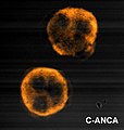
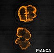
    ANCA stands for anti-neutrophil cytoplasmic antibodies.  They are mainly IgG.  They are subdivided into 2 groups based on the patterns produced when they are stained:


    1. cANCA
    • cytoplasmic, anti PR3
    • found in Wegeners (90%), mircroscopic polyangiitis (40%)
    • some correlation between level and disease activity


    2. pANCA
    • perinuclear, anti MPO
    • found in churg strauss (60%), crescentic glomerulonephritis (80%), microscopic polyangiitis (60%), Wegeners (60%)
    • may also be seen in IBD, RA, SLE, sjogrens, autoimmune hepatitis


    Thats it for today, no war at present but I'll try and post one later

    Friday, 24 September 2010

    MRCP revision battle 19.1: Myelofibrosis

    I have more time than usual to revise this week and am actually finding it harder to sit down and get on with it... must have a quiet word with the motivational centres of my brain (which I believe are the mesolimbic areas... anyone care to correct me?)  Anyway, today's battles will be:

    MRCP revision battle 19.1: Myelofibrosis
    MRCP revision battle 19.2: Hepatitis
    MRCP revision battle 19.3: Peutz Jegher Syndrome
    MRCP revision battle 19.4: Familial Adenomatous Polyposis
    MRCP revision battle 19.5: Hereditary Non-Polyposis Colorectal Cancer
    MRCP revision battle 19.6: LTOT
    MRCP revision battle 19.7: Exercise tolerance tests
    MRCP revision battle 19.8: Lead poisoning


    MRCP revision battle 19.1: Myelofibrosis


    Myelofibrosis has always been a secret 'sweet-spot' of mine - I think because of its 'teardrop cells' - so its a good battle to start this unmotivated day on...


    Myelofibrosis is fundamentally fibrosis of the bone marrow.  There is hyperplasia of megakaryocytes which produce platelet-derived growth factor, leading to:
    1. marrow fibrosis
    2. haemopoesis being forced to move to the spleen and liver.

    Features of myelofibrosis include:
    • lethargy
    • weight loss
    • night sweats
    • massive hepato/splenomegaly

    Investigations show:
    • raised WCC, low Hb
    • teardrop pokilocytes
    • leucoerythroblastic cells (=nucleated red cells)
    • raised LDH and urate as increased cell turnover
    • bone marrow may produced a 'dry tap'

    Image below shows teardrop cells.


      Treatment is ?allogenic stem cell transplant.

      Prognosis is poor with a median survival of 4 to 5 yrs.


      Onwards to hepatitis.... 

      MRCP revision battle 19.2: Hepatitis

      There are 5 'flavours' of viral hepatitis, labeled A to E.  Important general points to learn are:
      • all are caused by RNA virus' except hepatitis B, which is a DNA virus
      • B and C are spread by blood/sexual contact
      • A and E are spread by the faecal-oral route
      • Hepatitis D essentially is only ever found as a co-infection, never alone.

      So to explore the 3 most popular MRCP 'flavours' in more depth....


      Hepatitis A:
      • spread faeco-orally, so look out for questions featuring backpackers returning home
      • incubation period is 2-6 weeks
      • presents as fever, malaise, nausea
      • patient likely to be jaundiced and may have hepato/splenomegaly
      • treatment is supportive
      • patients generally make a full recovery

      Hepatitis B:
      • symptoms are similar to hep A
      • incubation period is longer at 1 - 6 months
      • spread is blood/bodily fluids - questions likely to hint at male business traveller or other innuendo
      • important to fully grasp the various antigens and their meanings:
        • HBsAg: present for 1-6 months after exposure; if present for >6 months patient is a carrier
        • HBeAg: present for 1.5-3 months after exposure and is marker of high infectivity
        • anti HBC IgM - signifies acute infection/carrier
        • anti HBC IgG - may be acute infection, carrier or cleared infection
        • anti HBS - if present with anti HBC suggests recovered from hep B and naturally immune; if present alone suggests hep B vaccination
      • complications of hepatitis B include:
        • 5-10% chronic hepatitis
        • glomerulonephritis
        • increased risk hepatocellular carcinoma
        • cryoglobulinaemia

      Hepatitis C:
      • is spread by blood/sexual contact
      • blood pre 1991 wasn't screened for hep C
      • <20% get an acute hepatitis but 80% get chronic hepatitis
      • breast feeding is NOT contraindicated (a common MRCP fascination for some reason)
      • complications:
        • 80% chronic hepatitis
        • 20% cirrhosis
        • increased risk hepatocellular carcinoma
      • Treatment: IFN alpha and ribavirin


      After that wizz through some high-yield viral hepatitis facts lets move on to Peutz Jegher Syndrome

      MRCP revision battle 19.3: Peutz Jegher Syndrome

      Peutz Jegher Syndrome is a condition characterised by:
      • pigmented 'freckles' (macules) on lips, face, palms and soles
      • hamartomatous polyps in gastrointestinal tract


      It is an autosomal dominant condition caused by a mutation of gene LKB1


      Complications of PJS include:
      • obstruction/intususception
      • GI haemorrhage
      • iron deficiency
      • colicky abdo pain
      • malignant transformation - around 50% of sufferers will have died from a cancer by the age of 60.

      There is no specific treatment.


      So lets move on from a relatively benign cause of multiple colonic polyps to a less benign one....

      MRCP revision battle 19.4: Familial Adenomatous Polyposis

      Familial adenomatous polyposis is an inherited condition in which patients develop literally thousands of polyps in the GI tract.  Virtually all sufferers will get cancer, usually in their 30s or 40s.


      It is caused by a mutation in the tumour supressor gene APC on chromosome 5.


      The appearance of the colon is:


      Treatment tends to be a colectomy before cancer develops.


      As an aside, a variation of FAP called 'Gardners Syndrome' exists.  This is FAP plus osteomas, epidermal cysts, fibromas and retinal pigmentation.


      So after 2 conditions which are defined by polyps, lets look at one which defines itself by not having polyps - Hereditary Non-Polyposis Colorectal Cancer

      MRCP revision battle 19.5: Hereditary Non-Polyposis Colorectal Cancer

      Hereditary Non-Polyposis Colorectal Cancer is an autosomal dominant condition associated with an increased risk of colorectal cancer.


      90% of patients with HNPCC develop cancers, usually in the proximal colon and often poorly differentiated/highly aggressive cancers.


      There is also an increased risk of breast, ovary and endometrial cancers.


      HNPCC is defined by the Amsterdam criteria:
      • at least 3 family members with colon cancer (one a 1st degree relative)
      • at least 2 generations affected
      • at least 1 diagnosed under 50 yrs of age


      Thats quite enough bowels for one day, lets come up for some oxygen...

      MRCP revision battle 19.6: LTOT

      LTOT = long term oxygen therapy = 15 or more hours of oxygen per day.


      This is the only treatment to increase survival in COPD.


      NICE recommends it if:
      • PaO2 <7.3kPa OR
      • PaO2 <8kPa if
        • secondary polycythaemia
        • nocturnal hypoxia (questions may note 'morning headaches;
        • peripheral oedema
        • pulmonary hypertension


      How about now trying our exercise tolerance...

      MRCP revision battle 19.7: Exercise tolerance tests

      Exercise tolerance tests are starting to go out of fashion but are still currently a mainstay of assessment of angina. 

      I'd recommend this BMJ article for a comprehensive discussion; what follows below are just a few salient MRCP points.


      ETTs are carried out according to the Bruce protocol, which is 7 sections each 3 minutes long so a maximum of 21 minutes of exercise.  A modified bruce can be used in higher risk/frailer patients.


      Beta blockers must be stopped the day before an ETT
      Digoxin should be stopped a week before.


      The test is deemed to be positive if:
      • anginal symptoms
      • BP decreases by 15mmHg or fails to increase on exercise
      • arrhythmia
      • ST depression
      • failure to achieve target heartrate (220-age in men, 210-age in women)
      • ST elevation


      Now for the last battle of the day, lead poisoning

      MRCP revision battle 19.8: Lead poisoning

      Lead poisoning is a good topic as it has 2 nice distinctive features (blue lines on nail growth margins and basophillic stippling of red blood cells.)


      A more comprehensive list of lead poisoning features is:
      • abdominal pain
      • constipation
      • peripheral neuropathy
      • fatigue

      Blue lines on the growth margin of nails affect 20% adults with lead poisoning but are rare in children.


      Investigations should show:
      • microcytic anaemia
      • basophillic stippling of red blood cells
      • raised aminolevulinic acid levels (a haem precursor)


       The image below shows basophillic stippling of red blood cells (right and left arrows)


      Treatment is by chelation with either EDTA, d-penicillamine or dimercaprol.


      Thats today wrapped up; questions on yesterday's battles can be found here

      MRCP questions: War 18

      As with previous 'wars' after 'battles' these are just a few quick questions to see if your brain cells have retained the information provided in battles 18.1 to 18.8.

      Grab a piece of paper, jot down your answers then compare them to my answers here


      Question 1:
      Which 4 clotting cascade factors is vitamin K a cofactor for?


      Question 2:
      Your patient has an INR of 7 but no evidence of bleeding.  What would you do?


      Question 3:
      A 20 year old comes to you with a rash across their back of teardrop-shaped lesions topped with some silver scales.  They note they had a sore throat a week or 2 ago.  What is the rash?


      Question 4:
      State the classical triad of symptoms for aortic stenosis.


      Question 5:
      A patient tells you their cardiologist said the pressure gradient across their aortic valve was 52mmHg.  What grade of severity is their stenosis?


      Question 6:
      Your houseofficer has diagnosed PMR and wants to know how to treat it.  What would you prescribe?


      Question 7:
      A mum brings her 16 year old son who has learning difficulties to you, saying he has been itching his bottom a lot recently.  You think he has threadworms.  How would you treat them?


      Question 8:
      Your consultant states he is admitting someone with pseudopseudohypoparathyroidism.  What blood results would you expect?


      The answers are here

      Wednesday, 22 September 2010

      MRCP revision battle 18.1: Warfarin

      Today is another 'octopus' day with 8 battles to embrace (three of which are mercifully brief)

      So the topics are:

      MRCP revision battle 18.1: Warfarin
      MRCP revision battle 18.2: Psoriasis
      MRCP revision battle 18.3: Beau's lines
      MRCP revision battle 18.4: Elliptocytosis
      MRCP revision battle 18.5: Aortic stenosis
      MRCP revision battle 18.6: Polymyalgia rheumatica
      MRCP revision battle 18.7: Threadworm
      MRCP revision battle 18.8: Pseudohypoparathyroidism


      MRCP revision battle 18.1: Warfarin


      Ah, warfarin... everyone's favourite out-of-hours bleep as a houseofficer.  Originally made as a rat poison, it quickly became a favourite in the UK for the treatment of DVTs, PEs and prevention of clots associated with mechanical heart valves.


      Warfarin works by inhibiting vitamin K epoxide reductase, which prevents vitamin K being recycled.  Since vitamin K is a cofactor for factors II, VII, IX and X it decreases coagulation.


      Note however that vitamin K is also a cofactor for protein C and protein S, which help inhibit the clotting cascade.  As protein C is initially affected more than the clotting factors by lack of vitamin K warfarin is initially a procoagulant and when started therefore increases the risk of thrombosis.


      Warfarin is monitored by INR.  The targets are:
      • PE/DVT: INR 2-3
      • metallic valve: INR 3-4

      The duration of anticoagulation depends on the cause of the clot:
      • below knee DVT after surgery: 6 weeks
      • above knee DVT/PE after surgery: 3 months
      • DVT/PE without precipitating factor identified: 6 months

      1-2% of patients on warfarin have a haemorrhage per year.

      Other complications of warfarin treatment include:
      • increased risk of osteoporosis
      • purple toe syndrome - toes turn purple ?secondary to cholesterol deposits 3 to 8 weeks after starting warfarin


      Warfarin levels are notoriously difficult to regulate.

      Things which increase the effect of warfarin include:
      • erythromycine/clarithromycin
      • metronidazole
      • hyperthyroidism
      • cranberry juice
      • alcohol
      • amiodarone
      • propranolol
      • fluconazole
      • isoniazod
      • cimetidine
      • omeprazole

      Things which decrease the effect of warfarin include:
      • rifampacin
      • carbamazepine
      • chlordiazepoxide
      • avocado in excess!

      Management of INR outside the desired range depends both on the level and if there is bleeding:
      • major bleed
        • stop warfarin
        • give vit K 5-10mg IV
        • give prothrombin complex (II, VII, IX, X) or FFP if this is not available
      • INR>8 but no/minimal bleeding
        • stop warfarin
        • give vit K 2.5-5mg PO/0.5-1mg IV
        • restart warfarin when INR<5
      • INR 5-8, no bleeding
        • stop warfarin
        • restart when INR<5
      • INR 5-8 minor bleeding
        • stop warfarin
        • give 1-2.5mg vitamin K PO
        • restart when INR<5


      As an aside, with an eye on the future, a new drug called dabigatran is looking to usurp warfarin as king of the anticoagulants.  Trials such as RELY and RECOVER have shown dabigatran to be non-inferior to warfarin in preventing thrombosis, to cause fewer bleeding adverse effects and even better not require regular blood tests to check levels.  Of course its expensive and so not yet approved...


      On to the second battle of the day, psoriasis...
      MRCP revision battle 18.2: Psoriasis

      Psoriasis is an autoimmune disease that classically causes a silver-scale topped rash on the skin.

      It affects 1-2% of the population.

      The cause is believed to be abnormal activity of the type 1 T helper cells.
      It is associated wtih HLA CW6, B13, B17 and B27.


      Females tend to be affected at a younger age than males.


      The main 'types' of psoriasis are:
      1. chronic plaque
      2. generalised pustular
      3. palmo-plantar pustulosis
        • strong association with smoking
        • tends to affect middle-aged females
      4. guttate
        • usually young adults/teenagers
        • is often preceded by a strep infection 2-4 weeks before
        • resolves spontaneously in 2-3 weeks
      5. nail
      6. flexural


      Athropathy is a feature in 8% and may be in the form of :
      • symmetric arthritis - appears rheumatoid-like; 50%
      • asymmetric arthrtitis - 35% - dactylitis
      • arthritis mutilans - <5%
      • spondylitis
      • distal interphalangeal predominant


      Psoriasis can be worsened by multiple factors, including:
      • trauma
      • infection
      • post-partum
      • beta blockers
      • lithium
      • stress
      • alcohol
      • NSAIDs

      Management for skin psoriasis is by a host of lotions and potions - coal tar, dithranol, topical vitamin D.... if these haven't already been drummed into you a brief visit to the BAD website (british association of dermatologists, trying to sound cool) to recap might be a good idea.


      Now for a brief skirmish with Beau's lines

      MRCP revision battle 18.3: Beau's lines

      Beau's lines are transverse depressions in nails due to temporary arrest in their growth.

      They are associated with:
      • psoriasis
      • diabetes
      • metabolic disturbances


      Now onwards to another very brief battle, elliptocytosis

      MRCP revision battle 18.4: Elliptocytosis

      Elliptocytosis is an autosomal dominant condition which results in cigar-shaped elliptocytes:


      In severe cases they can cause haemolytic anaemia; happily the majority of people with elliptocytosis have no ill effects from it at all.


      Now on to a more substantial battle again: aortic stenosis

      MRCP revision battle 18.5: Aortic stenosis

      Aortic stenosis may be detected incidentally (keen houseofficer noticing the classical ejection systolic murmur radiating to the carotids) or may present with its classical triad of symptoms that can be remembered as 'DAD':
      • exertional dyspnoea
      • etertional angina
      • exertional dizziness


      Signs of aortic stenosis include:
      • ejection systolic murmur radiating to carotids
      • slow rising pulse
      • narrow pulse pressure
      • heaving apex beat


      Things which suggest severe aortic stenosis include:
      • LVF
      • soft S2
      • paradoxically split A2
      • S4

       The formal divisions of severity are as follows:
      • mild: area >1.5cm, gradient <25
      • moderate: area 1-1.5cm, gradient 25-50
      • severe: area <1cm, gradient >50
      • critical: area <0.7cm, gradient >80


      The main causes of aortic stenosis are:
      • calcification of the valve
      • bicuspid aortic valve


      ECG changes which may be associated with aortic stenosis include:
      • p mitrale
      • LVH
      • LAD
      • poor R wave progression
      • complete heart block if calcification involves the conduction tissue.


      Treatment is surgical.  The operative mortality is around 20-25% if there is LVF, 2-8% if not.


      So from the very factual aortic stenosis on to the slightly touchy-feeling PMR...

      MRCP revision battle 18.6: Polymyalgia rheumatica

      Polymyalgia rheumatica (PMR) is a slightly nebulous condition which the Oxford handbook defines as 'symmetrical aching, tenderness and morning stiffness in shoulders and proximal limb muscles, +/- systemic features'.  These systemic features can include anything from fatigue to anorexia...


      It generally affects over 70s and is rare in under 60s.


      Bloods tend to show a raised ESR


      Treatment is with prednisolone (15mg PO OD) which tends to produce a dramatic improvement in days.  This should be decreased by 1mg per month, and of course anti-OP treatment should be given concurrently.

      If symptoms recur on decreasing the dose and you have a lovely little old lady stuck on >10mg OD for over a year, it is worth considering methotrexate or azothioprine to try and allow you to decrease the steroid.


      As a random aside, 10% of patients with PMR will suffer from carpal tunnel syndrome.


      Now get ready to itch as we move on to the penultimate battle of the day, threadworm....

      MRCP revision battle 18.7: threadworm

      Threadworm, AKA enterobius vernicularis, appears frequently in MRCP questions, usually in relation to patients in institutions.

      They tend to present with perianal itching.

      Treatment is mebendazole 100mg (or piperazine if the patient happens to be under 2 yrs of age)


      On that pleasant note, on to the last battle of the day, pseudohypoparathyroidism

      MRCP revision battle 18.8: Pseudohypoparathyroidism

      Pseudohypoparathyroidism is an autosomal dominant condition that occurs when a person is insensitive to PTH.


      Features include:
      • short metacarpals (esp 4th and 5th)
      • short stature
      • round face
      • obese
      • low IQ
      • dental hypoplasia

      It can be diagnosed by a failure of cAMP to increase after injection of parathyroid hormone.  More traditionally, the clinical picture of low calcium, high PTH and normal/raised alk phos is enough.


      Treatment is with alfacalcidol.

      It is worth noting it is associated with slipped femoral epiphysis.


      To end today's battles on an almost comic note (and the fact I find this comic is a reflection that I really should get out more) there also exists a pseudopseudohypoparathyroidism, which is essentially the morphological features of pseudohypoparathyroidism but with normal biochemistry.  Hopefully that'll make you smile if it comes up!


      A few questions on yesterday's battles are now online here, otherwise see you tomorrow!

      MRCP questions: War 17

      As with previous 'wars' after 'battles' these are just a few quick questions to see if your brain cells have retained the information provided in battles 11.1 to 11.4.

      Grab a piece of paper, jot down your answers then compare them to my answers here


      Question 1:
      What haematological picture do you get in DIC?


      Question 2:
      What symptoms are associated with jugular foramen syndrome?


      Question 3:
      Name the commonest form of thyroid cancer.


      Question 4:
      List 3 causes of unilateral pleural calcification


      Question 5:
      What condition is associated with a 'plucked chicken skin' appearence?


      Question 6:
      Legionella is a gram positive bacteria.  True or false?


      Question 7:
      Which biochemical disturbance is classically associated with legionella?


      The answers are here

      Tuesday, 21 September 2010

      MRCP revision battle 17.1: DIC

      Lots of short battles from a variety of specialities today so buckle up for a ride through some haematology, neurology, endocrinology, respiratory and dermatology.  Yeee-hhaaaa!

      MRCP revision battle 17.1: DIC
      MRCP revision battle 17.2: Jugular foramen syndrome
      MRCP revision battle 17.3: Thyroid cancer
      MRCP revision battle 17.4: Legionella infection
      MRCP revision battle 17.5: Pleural calcification
      MRCP revision battle 17.6: Pseudoxathoma elasticum


      MRCP revision battle 17.1: DIC


      Disseminated intravascular coagulation (=DIC) is the pathalogical widespread activation of coagulation.


      Blood tests will show:
      • prolonged PT
      • prolonged aPTT
      • massively raised d-dimer levels
      • low platelets
      • low fibrinogen
      • schistocytes


      Of these, it is the level of fibrinogen which best correlates to the severity.


      Causes of DIC include:
      • obstetric
      • crush injury
      • septicaemia
      • malignancy
      • transfusion reaction

      The treatment is:
      • treat the cause
      • give platelets if platelets <50
      • cryoprecipitate
      • FFP
      • activated protein C if septic.


      Now lets canter onwards to jugular foramen syndrome....

      MRCP revision battle 17.2: Jugular foramen syndrome

      Jugular foramen syndrome, also known as Vermet's Syndrome, is a condition arising from the compression of the cranial nerves that run through the jugular foramen.


      Now just in case they aren't quite on the tip of your tongue, the nerves that run through the jugular foramen are IX, X and XI.


      The syndrome is therefore characterised by:
      • loss of taste to posterior 1/3 of tongue (CN IX)
      • dysphagia (CN X)
      • sternocleidomastoid and trapezius paralysis (CN XI)

      Anything that compresses the jugular foramen can cause this syndrome.  The commonest cause is a paraganglioma.


      Now we've acquired a bit more esoteric MRCP knowledge, lets gallop on to discover some details about thyroid cancer...

      MRCP revision battle 17.3: Thyroid cancer

      Under 5% of thyroid lumps are malignant, and in general those with a malignant mass will be euthyroid.


      There are 4 main types of thyroid cancer:
      1. papillary - 70% - mainly affects young females, excellent prognosis
      2. follicular - 20% 
      3. medullary - 5% - associated with MEN-2 (more on MEN tomorrow)
      4. anaplastic - 1% - very poor prognosis and resistant to treatment.

      Rarely a patient can have lymphoma of the thyroid - this usually follows hashimotos disease.


      The mainstay of treatment for thyroid cancer is surgery.


      Lets break into a canter as we head on to legionella...

      MRCP revision battle 17.4: Legionella

      Legionella is a gram negative bacteria which causes legionella disease/pneumonia and in MRCP questions is most likely found living in a cruise ship/hotel's water tank or airconditioning system.


      The symptoms of legionella disease are:
      • flu-like
      • dry cough
      • dypnoea
      • GI upset


      Complications of legionella which may show up in the blood results include:
      • SIADH - low sodium
      • hepatitis - deranged LFTs


      Legionella is killed above 60c, cannot multiply between 50 and 60c and are happiest between roughly 35 to 45 c


      Males are more affected than females, 3:1


      CXR may show bibasal consolidation.


      Treatment is with antibiotics which achieve a high intracellular concentration, such as clarithromycin, rifampicin or fluroquinolone.


      Legionella has a 10% mortality.


      Lets now trot on to pleural calcification...

      MRCP revision battle 17.5: Pleural calcification

      Pleural calcification pops up both in real life and in MRCP questions intermittently.


      The commonest cause is asbestosis. 
      This tends to be bilateral and spare the costophrenic angles


      So, if the pleural calcification is unilateral consider:
      • previous haemothorax
      • previous TB pleuritis
      • previous empyema

      Other differentials to consider are:
      • previous radiotherapy/radiation exposure
      • post talc pleurodesis, which will mimic calcification.


      Lets return to a full-speed gallop to our last battle of the day, pseudoxanthoma elasticum

      MRCP revision battle 17.6: Pseudoxanthoma elasticum

      Pseudoxanthoma elasticum is fragmentation and calcification of elastic fibres.


      It is an autosomal recessive condition, found on chromosome 16.


      Its various associations are:
      • a characteristic 'plucked chicken' skin (google it, memorise it, get ready to spot it in part 2)
      • angioid streaks in retina/vision problems
      • rarely GI bleeds
      • claudication/cardiovascular problems.


      There is no treatment so management is symptomatic only.


      Thats all for today, proceed to the war if you are up to.

      MRCP questions: War 16

      As with previous 'wars' after 'battles' these are just a few quick questions to see if your brain cells have retained the information provided in battles 16.1 to 16.7.

      Grab a piece of paper, jot down your answers then compare them to my answers here


      Question 1:
      List 8 symptoms/signs of pagets disease


      Question 2:
      Which oral medication would you give to treat pagets disease?


      Question 3:
      Which medication would you use to treat PCP?


      Question 4:
      A patient has muscle weakness that gets worse on exercise.  What medication would you give them?


      Question 5:
      A patient tells you they have weak muscles which get better on exercising.  They state they believe its due to their lung cancer.  What condition do they have?


      Question 6:
      ]What is the commonest cause of hemiballismus?


      Question 7:
      What is the medical treatment for hyperprolactinoma?


      Question 8:
      'Waiters tip' is associated with which palsy?


      The answers are here

      Monday, 20 September 2010

      MRCP revision battle 16.1: Pagets disease of bone

      In between catching up on seemingly endless loads of washing (do socks breed when left alone in a wash basket?) a few hours of MRCP revision has been done and 7 new battles have been written...

      MRCP revision battle 16.1: Pagets disease of bone
      MRCP revision battle 16.2: Pneumocystis carinii
      MRCP revision battle 16.3: Myasthenia gravis
      MRCP revision battle 16.4: Lambert-Eaton syndrome
      MRCP revision battle 16.5: Hemiballismus
      MRCP revision battle 16.6: Prolactin
      MRCP revision battle 16.7: Erbs palsy


      MRCP revision battle 16.1: Pagets disease of bone


      Pagets disease of bone is accelerated, disorganised bone turnover due to increased number and activity of osteoclasts and osteoblasts.


      It is rare in under 40s.


      Predisposing factors include:
      • increasing age
      • northern latitude
      • family history
      • being male


      Features of Pagets include:
      • bone pain
      • bowing of tibia
      • bossing of skull
      • secondary arthritis
      • deafness/other cranial nerve compression
      • high output heart failure
      • pathological fractures
      • bone sarcomas (occur in 1% over 10 yrs)

      My way of remembering the features of pagets is this slightly random rhyming story:
      "Bowing to the Boss, oh what a pain,
      I'm deaf and arthritic and since the fracture I'm lame
      my heart's now a failure, and oh what a shame
      I've got bone sarcoma - thats the end of the game"


      Bloods in Pagets show:
      • raised ALP
      • normal calcium (unless a long period of immobility)
      • normal phosphate


      Treatment is alendronic acid


      That wasn't too bad... onwards to battle 16.2...

      MRCP revision battle 16.2: Pneumocystis carinii

      Pneumocystis carinii, AKA pneumocystis jiroveci, is a unicellular eukaryote.

      It can cause pneumonia in patients with HIV and accounts for 40% of all AIDS-defining illness.
      If CD 4 count is less than 200 PCP prophylaxis (=co-trimexazole) should be started.


      Features of PCP:
      •  dyspnoea
      • dry cough
      • fever
      • few chest signs on examination
      • desaturation on exercise


      CXR typically shows mid-lower bilateral interstitial infiltrates, but it may look normal.

      Sputum cultures are often negative so BAL may be needed.


      Treatment is:
      • co-trimexazole
      • IV pentamidine if severe
      • steroids if hypoxia
        • use of steroids decreases respiratory failure by 50% and death by 1/3)


      On to battle 3 of the day and a classic condition - myasthenia gravis.

      MRCP revision battle 16.3: Myasthenia Gravis

      Myasthenia gravis is an autoimmune condition in which muscles become easily tired and weak.  It affects 1 in 20 000.


      90% of patients have autoantibodies to nicotinic acetylcholine receptors.
      If these are negative consider looking for MUSK antibodies (= muscle specific kinase)


      The key feature of MG is repetitive stimulation leading to decreased evoked response.


      The order in which muscle groups are affected in MG is:
      • extraoccular (--> diplopia)
      • bulbar
      • face
      • neck
      • lumb
      • trunk

      Reflexes are normal in MG.


      Test of choice: tensilon test = edrophonium


      Treatment of MG is:
      • cholinesterase inhibitors eg pyridostigmine
      • prednisolone if pyridostigmine unsuccessful (but be aware that paradoxically steroids can worsen MG)
      • plasmaphoresis/IV IG if respiratory muscle involvement.

      If there is hyperplasia of thymus/thyroma, up to 60% of cases will remit after surgery.


      Up to 10% of patients with MG will have hyperthyroidism.


      Drugs which exacerbate MG include:
      • penicillamine
      • quinidine
      • beta blockers
      • lithium
      • phenytoin
      • gentamycin

      Now on to the 'similar but different' Lambert-Eaton syndrome...

      MRCP revision battle 16.4: Lambert Eaton Syndrome

      Lambert-Eaton syndrome is a rare condition affecting voltage-gated calcium channels which results in:
      • muscular weakness that improves with activity
      • autonomic symptoms (dry mouth, constipation, impotence)
      • hyporeflexia
       

      It is most commonly a paraneoplastic phenomenon, usually due to small cell lung cancer (very rarely due to breast or ovarian cancer).  It can also be autoimmune.


      Treatment is 3,4 diaminopyridine.

      Regular CXR should be performed as Lambert-Eaton may preceed the development of small cell lung cancer by up to 4 years.


      Notice how although both MG and Lambert-Eaton are characterised by muscle weakness there are lots of differences - weakness gets worse on repetition in MG but better in LE, reflexes are normal in MG but reduced in LE and there may be autonomic symptoms in LE but not MG.  Also not involvement of occular muscles is far more common in MG.


      Lets now diversify to something completely different - hemiballismus...

      MRCP revision battle 16.5: Hemiballismus

      Hemiballismus is large amplitude, flinging hemichorea.


      Its cause is damage to the contralateral subthalamic nuclei.


      The commonest cause is stroke.


      Treatment:
      • hemiballismus generally resolves spontaneously in 4 to 8 weeks
      • haloperidol (a dopamine blocker) may be used
      • tetrabenazine (a VMAT inhibitor) may be used

      Bilateral hemiballismus is rare and tends to be associated with HONK.


      After that brief battle onwards to the meatier topic of prolactin

      MRCP revision battle 16.6: Prolactin

      Prolactin is a hormone released from the anterior pituitary.
      It is under the negative control of dopamine (ie dopamine prevents its release)


      Raised prolactin causes a drop in GnRH, LH, oestrogen and testosterone levels.


      Symptoms/signs of raised prolactin include:
      • galactorrhoea
      • decreased libido
      • amenorrhoea
      • infertility

      Note that gynaecomastia is NOT associated with raised prolactin.


      Causes of a raised prolactin include:
      • physiological causes
        • pregnancy
        • breast feeding/nipple stimulation
        • stress
        • post-fit
        • exercise
      • drugs
        • phenothiazides
        • metoclopramide
        • methyl-dopa
        • haloperidol
        • oestrogens
      • metabolic causes
        • hypothyroidism
        • PCOS
        • acromegaly (1/3)
        • chronic renal failure
      • disease
        • prolactinoma
          • microprolactinoma (<10mm) release more prolactin than macroprolactinomas
        • compression of pituitary stalk removing dopamine control

      Treatment is with bromocriptine.
      It is best to give it in the evening as it causes postural hypotension


      Very nearly there.  Last battle of the day will be Erbs palsy....

      MRCP revision battle 16.7: Erbs palsy

      Erbs palsy is the name given to a particular brachial plexus injury, generally caused by shoulder dystocia during birth.


      Any nerve root of the brachial plexus may be affected but the most commonly affected roots are C5 and C6.


      This results in loss of:
      • shoulder abduction
      • elbow flexion
      • suppination

      The net outcome is that the arm hangs medially rotated with the forearm pronated = "waiters tip"


      So thats all for today's battles - on to today's war to check recall of yesterday's battles!

      MRCP questions: War 15

      As with previous 'wars' after 'battles' these are just a few quick questions to see if your brain cells have retained the information provided in battles 15.1 to 15.8.

      The answers are available here


      Question 1:
      List the 3 commonest sites of mets from renal cell carcinomas


      Question 2:
      What abnormalities on blood tests might you see with renal cell carcinoma?


      Question 3:
      What is Von Hippel Lindau disease?


      Question 4:
      A patient complains of severe right shoulder pain followed by weakness of the right arm.  There is no history of trauma.  What is the likely diagnosis?


      Question 5:
      A patient has abdo pain.  The busy nurse just leaves their urine sample in front of you.  Ten minutes later you notice it has turned deep red.  What condition does the patient have?


      Question 6:
      List 3 drugs that can precipitate acute porphyria


      Question 7:
      A medical student excitedly tells you the urine sample they've just taken glows pink under woods light.  What condition does the patient have?


      Question 8:
      Your newest patient has heart block, opthalmoplegia and pigmentary degeneration of the retina.  What test do you want to do to confirm your diagnosis?


      Question 9:
      List 7 symptoms associated with pellegra


      Question 10:
      Which drug is used to treat neuroleptic malignant syndrome?

      Sunday, 19 September 2010

      MRCP revision battle 15.1: Renal cell carcinoma

      I've had a day off in which I've covered literally hundreds of miles and drank excessive amounts of tea and cider and seen far more gingham than any one person ever should.  Net result has been a restoration in my energy levels and so today is going to be a blockbuster of a set of battles, with 8 separate topics to cover including two that I'd never even heard of in my preceding 8 years of studying medicine.  So lets get started!


      MRCP revision battle 15.1: Renal cell carcinoma
      MRCP revision battle 15.2: Von Hippel Lindau
      MRCP revision battle 15.3: Neuralgic amyotrophy
      MRCP revision battle 15.4: Porphyrias
      MRCP revision battle 15.5: Kearns-Sayre
      MRCP revision battle 15.6: Klinefelters
      MRCP revision battle 15.7: Pellegra
      MRCP revision battle 15.8: Neuroleptic malignant syndrome


      MRCP revision battle 15.1: Renal cell carcinoma


      Renal cell carcinoma, also known as hypernephroma or Gravitz Tumour, is a cancer arising in the proximal renal tubular epithelium.

      It accounts for 85-90% of primary renal cancer.


      The classic triad for renal cell carcinoma is:
      1. haematuria
      2. loin pain
      3. abdominal mass
      (NB: classic triads of course rarely exist, but may kindly present themselves during MRCP questions)


      Renal cell carcinoma may also be linked to pyrexias of unknown origin.
      Another clinical sign to look out for is left variocele - caused by invasion of the left renal vein causing compression of the left testicular vein.


      Factors associated with developing renal cell carcinoma include:
      • smoking
      • middle aged males (male:female 2:1)
      • Von-Hippel Lindau syndrome (wait for the next battle if you don't know that that is)
      • tuberous sclerosis
      • acquired cystic kidney disease in renal failure


      25% of patients with renal cell carcinoma have mets at presentation.  The commonest mets are bone, liver and lung.


      Endo effects associated with renal cell carcinoma include:
      • erythropoietin --> polycythaemia
      • PTH --> raised calcium
      • renin --> raised BP
      • ACTH

      Treatment is surgery.  If mets are present IL-2 or interferon alpha may also be considered.


      There is a 45% 5 yr survival.


      Now on to meet Von-Hippel-Lindau syndrome...

      MRCP revision battle 15.2: Von Hippel Lindau disease

      Von Hippel Lindau disease is a rare autosomal dominal condition that results in haemangioblastomas in:
      • cerebellum (may present as ataxia)
      • spinal cord
      • kidney
      • retina (may present as visual loss)

      It is associated with bilateral renal cell carcinomas (develop in approximately 50% of patients with VHL) and pheochromocytomas.


      For those of you who are feeling really geeky the gene responsible is on chromosome 3 and it is a mutation in a tumour supressor gene.


      So after that brief acquaintance, lets move on to a condition I'd never heard of before today - neuralgic amyotrophy...