Thursday, 11 November 2010

MRCP revision battle 46.1: Classification of bacteria

A completely random mixed bag today, bursting full of random nuggets of MRCP knowledge...


MRCP revision battle 46.1: Classification of bacteria
MRCP revision battle 46.2: Tumour supressor genes and Li Fraumeni Syndrome
MRCP revision battle 46.3: Mitochondrial disorders and Leber's Optic Atrophy
MRCP revision battle 46.4: Cytotoxic Agents
MRCP revision battle 46.5: Congenital infections
MRCP revision battle 46.6: Rheumatoid factor
MRCP revision battle 46.7: Lichen Planus


MRCP revision battle 45.1: Classification of bacteria


MRCP questions just love commenting on the colour/shape of bacteria.  Hopefully my simple picture below (which was also stuck next to the loo during finals!) might help you.  Note that gram positive bacteria have walls so stain purple/blue while gram negative bacteria don't and stain pink.


My way of remembering them is 'SES ('says') ABCD then L....MN then all the rest'


As well as their colour/shape their 'pattern' is useful to know too:
  • pairs of cocci (= diplococci): neisseria gonorrhoea, neisseria meningitidis
  • chains of cocci: streptococci, enterococci
  • clumps of cocci: staphylococci


Now for some genetics...

MRCP revision battle 46.2: Tumour supressor genes and Li Fraumeni Syndrome

Tumour supressor genes code for proteins that provide a 'stop' signal to prevent cell division.  They therefore act as a barrier to the development of cancer.  As they are recessive genes both copies of the gene would have to be mutated to stop functioning.


The most famous tumour supressor gene is p53.

p53 is:
  • found on chromosome 17
  • the most commonly mutated gene in breast, colon and lung cancer
  • a key regulator of apoptosis and prevents entry into the S phase of the cell cycle until the DNA is checked.


Li Fraumeni Syndrome is a rare condition in which there is an autosomal dominant p53 mutation.  More rarely it may be caused by a CHEK-2 mutation. Li Fraumeni syndrome is characterised by early onset of cancer, especially breast, sarcoma or soft tissue.  


Lets move on to mitochondrial disorders...

MRCP revision battle 46.3: Mitochondrial disorders and Leber's Optic Atrophy

mDNA mutates 10x more frequently than normal DNA.  However, there is a poor genotype to phenotype correlation so the mutations are often not noticed.


Since sperm do not contain mitochondria mitochondrial disorders are passed exclusively from mother to child.


The most famous mitochondrial disorders are:

  1. Kearns Sayre - see battle 15.5
  2. Leber's hereditary optic neuropathy.
  3. Some forms of sensorineural deafness


Leber's hereditary optic neuropathy
  • mDNA mutation
  • progressive loss of central vision due to optic atrophy, begins in young adulthood
  • if LHON 'plus' also:
    • multiple-sclerosis type symptoms
    • Wolff-Parkinson White


Next up - some cytotoxic agents (which sadly sounds more exciting than it is)

MRCP revision battle 46.4: Cytotoxic Agents

A quick run-through of the main mechanisms of action and MRCP-relevant side-effects of some common cytotoxic agents.


Vincristine
  • Inhibits microtubule formation
  • Used in:
    • lyphoma
    • leukaemia
    • breast and lung cancer
  • Side effects
    • peripheral paraesthesia
    • loss of tendon reflexes
    • abdominal pain

Cisplatin
  • Cross-links DNA
  • Used in:
    • testicular, cervical, bladder, lung, head and neck cancers
  • Side effects
    • nephrotoxic
    • ototoxic
    • peripheral neuropathy
    • hypomagnesaemia
    • myelosupression


Bleomycin
  • Degrades DNA
  • Used in:
    • metastatic germ cell cancers
    • non-Hodgkins lymphoma
  • Side effects
    • increased pigmentation
    • pulmonary fibrosis


Doxorubicin
  • inhibits RNA/DNA synthesis
  • used in:
    • leukaemia
    • lyphoma
    • breast cancer
  • Side effects
    • red colouration of urine
    • SVT
    • cardiomyopathy

Methotrexate
  • inhibits dihydrofolate reductase
  • used in:
    • non-Hodkins lymphoma
    • choriocarcinoma
    • RA
  • Side effects
    • excreted by kidneys so should be avoided in renal disease
    • should be avoided in significant ascites/pleural effusion as may accumulate then suddenly return to circulation causing myelosupression
    • myelosupression
    • mucositis
    • pneumonitis


Cyclophosphamide
  • alkylating --> cross links DNA. Inactive until metabolised by liver
  • used in:
    • CLL
    • lymphoma
    • sarcoma
  • Side effects
    • haemorrhagic cystitis 


To continue the randomness of the 46th set of battles lets look at congential infections

MRCP revision battle 46.5: Congenital infections

A few short notes on 5 infections mummies may have during pregnancy which can adversely affect the fetus...


Maternal rubella
  • fetus is nearly always affected if rubella occurs during first 7 weeks of gestation
  • fetus is nearly always OK if rubella occurs after 17 weeks of gestation
  • Effects of rubella on fetus:
    • sensorineural deafness
    • congenital cateracts
    • congenital heart disease
    • glaucoma


Maternal toxoplasmosis
  • fetus will only be affected if it is the first time the mother is exposed, unless she is immunosupressed
  • classical triad is:
    • cerebral calcification
    • hydrocelphalus
    • chorioretinitis


Maternal chickenpox
  • if this occurs during the 1st trimester risk of limb deformity
  • if mum has been in contact with chickenpox and is not sure if she is immune test for antibodies and if no natural immunity give immunoglobulin


Maternal CMV
  • at least 75% of fetus' exposed to CMV are undamaged
  • however, there is a risk of:
    • intracranial calcification
    • microcephaly
    • cardiac/GI abnormalities

Maternal Parvovirus B19
  • generally either fetal death or complete recovery


Now for a spot of rheumatoid factor

MRCP revision battle 46.6: Rheumatoid factor

Rheumatoid factor is an autoantibody against the Fc portion of IgG.  

Rheumatoid factor is found in 4% of the 'normal' population and 25% of elderly.


Rheumatoid factor is positive in low titres in chronic infections, for example:
  • 10% syphyllis
  • 20% pulmonary TB
  • 50% leprosy


Rheumatoid factor is often positive in high titres in connective tissue disease, for example it is positive in:
  • 70% of patients with RA
  • virtually 100% of patients with RA with extra-articular manifestations
  • >75% sjogrens
  • 20-40% SLE
  • 30% scleroderma

It may also be present in other immunological diseases, for example autoimmune liver disease or sarcoidosis.


Very high titres of rheumatoid factor may be found in cryoglobulinaemias


Rheumatoid factor is only useful in RA as an assessment of prognosis.  It is not useful for diagosis or or measuring level of activity.


Rheumatoid factor is detected by the Rose-Waaler test (sheep red cell agglutination) or the latex agglutination test.


Now to the final battle of the day - lichen planus

MRCP revision battle 46.7: Lichen Planus

Lichen planus is an intensely itchy rash characterised by purple papules with flat tops, often with white lines on the surface (=Wickham's striae).

The picture below (taken by Dr Tag-El -Din Anbar) illustrates lichen planus on an arm:


Mucus membranes may also be affected.


Management may be symptomatic, or with steroid creams.

Half of cases have resolved within 6 to 9 months and the majority of the rest will be gone within 18 months.


A lichenoid-like rash can also be caused by some medications, including:
  • NSAIDS
  • thiazide diuretics
  • antimalarials
  • betablockers
  • gold injections
  • mercury fillings

Wednesday, 10 November 2010

MRCP revision battle 45.1: Dialysis and complications

Back to my old nemesis renal medicine for another 7 battles...


MRCP revision battle 45.1: Dialysis and complications
MRCP revision battle 45.2: Anaemia in chronic renal failure
MRCP revision battle 45.3: Diuretics
MRCP revision battle 45.4: Proteinuria
MRCP revision battle 45.5: Renal osteodystrophy
MRCP revision battle 45.6: Radio-contrast nephropathy
MRCP revision battle 45.7: Minimal change disease


MRCP revision battle 45.1: Dialysis and complications


Lets start with a very matter-of-fact battle on dialysis and its complications


Brief overview of types of renal replacement therapy
  1. Haemodialysis
    • semi-permeable membrane
    • solute transfer by diffusion
    • excess fluid removed by creating a negative transmembrane pressure = ultrafiltration
  2. Haemofiltration
    • highly permeable membrane
    • solute transfer by convection
    • ultrafiltrate replaced by an equal volume of substitute fluid
    • takes longer and is more expensive than haemodialysis but less haemodynamic instability so used for critically ill patients
  3. Peritoneal dialysis
    • dialysis fluid put into abdominal cavity via tenchkoff catheter
    • solutes diffuse across peritoneal membrane
    • ultrafiltration achieved by adding glucose to the dialysis fluid

Complications of dialysis
  • Disequilibration syndrome
    • occurs on initial dialysis
    • manifests as nausea, vomiting, headache and altered consciousness
    • caused by rapid changes in plasma osmolality
  • Vascular disease
  • Heart valve calcification - especially aortic valve
  • Beta-2 microglobulin amyloidosis
  • Arthritis
  • Infections


Now to consider anaemia in renal failure...

MRCP revision battle 45.2: Anaemia in chronic renal failure

Anaemia is common in chronic renal failure and is usually due to:
  • decreased production of erythropoetin
  • iron deficiency

Treatment should be considered when Hb <11
The target for treatment should be Hb between 10.5 and 12.5


Correct any iron deficiency first; note that oral iron is often not enough and IV iron therapy may be needed.


If there is still anaemia the likely eryropoetin deficiency must be addressed.  Options are:
  • Eryropoetin replacement with either epoetin (=recombinant human erythropoetin) or 
  • darbepoetin (=hyperglycosylated derivative of epoetin with a longer half life)

Side effects of epoetin include:
  • hypertension (25%)
  • bone aches
  • 'flu'
  • rashes, urticaria
  • pure red cell aplasia 
    • low reticulocyte count
    • anaemia
    • antibodies
  • increased risk of DVT as increased PCV

If no response to epoetin consider if the patient has an infection, inflammatory condition or aluminium toxicity, all of which can impair epoetin's effectiveness.


Note that blood transfusions are generally avoided in renal patients as they would make matching for transplants more difficult


Cross your legs as we move on to a battle of diuretics...

MRCP revision battle 45.3: Diuretics

Before I started my revision I thought I was very au-fait with diuretics - after all, I prescribe them daily.  However, a quick bond with the British National Formulary revealed some interesting gems.  So here's a quick recap of diuretics with a sprinkling of hopefully new information.


1. Loop diuretics
  • work on the Na-K-2Cl transporter in the ascending loop of Henle
  • examples include furosemide and bumetanide
  • bumetanide is 40x more potent than furosemide for the same dose
  • both work within 30 mins (if given IV) or 1 hr (if given orally) and their action is completed within 6 hours
  • possible side effects include:
    • hypokalaemia/natraemia
    • retention if enlarged prostate
    • tinnitus 
    • pancreatitis

2. Thiazide diuretics
  • work on the Na-Cl co-transporter in the DCT
  • examples include bendroflumethiazide, indapamide and metolazone
  • they work within 1 to 2 hours and have a duration of action of 12 to 24 hours
  • possible side effects include:
    • hypokalaemia/naturaemia
    • hyperglycaemia
    • pancreatitis

3. Aldosterone antagonists
  • work by antagonising aldosterone 
  • examples include spironolactone and eplerenone 
  • possible side effects include:
    • hyperkalaemia
    • impotenence
    • gynaecomastia


Now for the differentials of proteinuria...

MRCP revision battle 45.4: Proteinuria

Normal protein excretion in urine is <150mg/day


Causes of proteinura include:
  • Renal causes
    • UTI
    • glomerulonephritis
    • myeloma
    • diabetes
    • hypertension
    • amyloid
  • Extra-renal causes
    • fever
    • exercise
    • psoriasis
    • pregnancy

Orthostatic proteinuria is proteinuria which disappears when recumbant.   The cause of this is unknown but it is felt to be benign.


On to renal osteodystrophy...

MRCP revision battle 45.5: Renal osteodystrophy

Renal osteodystrophy refers to the bone pathology which results from the endocrine and electrolyte imbalances caused by chronic renal failure.


Derangements include:
  • raised phosphate
  • low calcium
  • secondary hyperparathyroidism
  • low vitamin D
  • acidosis

The result on bones include:
  • osteomalacia
  • osteitis fibrosa cystica = hyperparathyroid bone disease
  • osteoporosis
  • osteosclerosis 'rugger jersey' spine

Management is:
  • calcichew - to bind phosphate in the gut
  • vit D analogues - eg adcal D3/calceos/calcichew = colecalciferol


On to the slightly more stimulating contrast nephropathy...

MRCP revision battle 45.6: Radio-contrast nephropathy

Radiocontrast nephropathy complicates up to 10% of procedures involving contrast.

Contrast nephropathy presents as non-oliguric acute renal failure 1 to 5 days post procedure.


Risk of contrast nephropathy can be reduced by:
  • good hydration/normal saline
  • oral n.acetyl cysteine

Risk of contrast nephropathy is increased by:
  • increased amount of contrast
  • hypovolaemia
  • myeloma
  • increasing age
  • diabetes
  • raised calcium
  • chronic renal failure

Remember that metformin must be withheld before and for 48 hrs post IV contrast due to risk of lactic acidosis. 


Now to end on a high with a battle with minimal change disease...

MRCP revision battle 45.7: Minimal change glomerulonephritis

Minimal change glomerulonephritis almost always presents as nephrotic syndrome

It is responsible for 80% of cases of nephrotic syndrome in children and around a quarter of cases in adults.


Its name 'minimal change' comes from the fact that under a light microscope the kidney looks normal; if an electron microscope is used you can see podocyte fusion.


Minimal change glomerulonephritis causes a highly selective proteinuria with only smaller proteins leaked.


Most causes are idiopathic but 10-20% are associated with:
  • NSAIDs/gold/rifampacin
  • hodgkins, thyroma
  • mononucleosis

It is believed to be T-lymphocyte mediated.


Treatment is with corticosteroids - 80% of cases respond
Cyclophosphamide can be used in non-responders.


The prognosis for minimal change glomerulonephritis is (fairly) good:
  • 1/3 recover completely
  • 1/3 suffer infrequent relapses
  • 1/3 suffer frequent relapses
  • BUT - <1% proceed to end-stage renal failure


Tomorrow's battles will be a completely random bag, then a final renal push (the end of the hattrick) will occur the day after.

Tuesday, 9 November 2010

MRCP revision battle 44.1: Antipsychotic medication

Today is a psychiatry and overdose fest...


MRCP revision battle 44.1: Antipsychotic medication
MRCP revision battle 44.2: SSRIs
MRCP revision battle 44.3: Serotonin syndrome
MRCP revision battle 44.4: Baby blues, post natal depression and psychosis
MRCP revision battle 44.5: Quinine toxicity/overdose
MRCP revision battle 44.6: Theophylline overdose
MRCP revision battle 44.7: Ethylene glycol poisoning


MRCP revision battle 44.1: Antipsychotic medication

Antipsychotic medications are used in the treatment of schizophrenia, severe anxiety, mania and occasionally for their sedative properties.

Antipsychotic medications have an immediate sedative action but their antipsychotic action may tak up to 3 weeks to become effective.


Atypical antipsychotics

Atypical antipsychotics are the newer drugs.  They produce a highly selective blockade of the mesolimbic D2 receptors and serotonin 5HT2A receptors.

Examples include amisulperide, olanzapine, quetiapine, risperidone and clozapine.


Side effects include:
  • weight gain
  • nausea, dyspesoa
  • hyperglucaemia and impaired glucose tolerance
  • increased risk DVT
  • increased risk stroke (especially olanzapine and risperidone)
  • clozapine only:
    • 1% agranulocytosis/neutropenia - therefore weekly blood tests for 1st 6 months then fortnightly afterwards
    • 3% seizures

All antipsychotics have roughly the same effectiveness except clozapine, which is better.  However, in light of its worse side effect profile clozapine is only recommended if 2 others have already failed.


Typical antipsychotics

Typical antipsychotics are the older drugs,
Examples include haloperidol, chorpromazine, phenothiazine and sulperide.


Side effects of the typical antipsychotics include:
  • extrapyramidal side effects
  • anticholingergic effects
  • antihistaminergic effects
  • photosensitivity (especially phenothiazine)
  • prolonged QT
  • lower seizure threshold
  • hyperprolactinaemia.


Now on to SSRIs...

MRCP revision battle 44.2: SSRIs

Selective Serotonin Reuptake Inhibitors (SSRIs) do exactly what their name implies: selectively inhibit serotonin reuptake.

Examples include citalopram, escitalopram, fluoxetine, paroxetine and sertraline.


They are widely used for depression, anxiety, post-traumatic stress disorder and panic disorder.
Fluoxetine, paroxetine and sertraline can be used in OCD.


There are many side effects, the commonest of which is GI disturbance.

Other possible side effects to be aware of include:
  • interaction with platelet function --> increase risk of GI bleed
  • hyponatraemia
  • urinary retention

Given the increased risk of bleeding SSRIs should be avoided if on heparin/warfarin - use mirtazepine instead.

The safest SSRI post MI is sertraline.


If combined with MAOIs/moclobemide there is a serious risk of toxicity and serotonin syndrome - do not start MAOIs until 5 weeks after stopping fluoxetine, 2 weeks after stopping sertraline or 1 week after stopping any other SSRI.  Conversely you must wait 2 weeks after stopping MAOIs before starting an SSRI.


Triptans and SSRIs must also not be combined due to increased risk of serotonin syndrome.


When SSRIs are stopped they should be reduced over 4 weeks to decrease withdraw.  Paroxetine is worst on stopping.


Now on to the next battle to explore serotonin syndrome....

MRCP revision battle 44.3: Serotonin syndrome

Serotonin syndrome is characterised by the triad of:


  • autonomic hyperactivity
    • hypertension
    • hyperthermia
    • tachycardia
    • mydriasis
  • neuromuscular abnormality
    • hyperreflexia
    • clonus
    • tremor
    • hypertonicity
  • mental state changes


Drugs associated with serotonin syndrome include:
  • lithium
  • carbamazepine
  • triptans
  • illegal drugs
    • LSD
    • cocaine
    • MDMA
  • antidepressants
    • MAOIs
    • SSRIs
    • St Johns Wort

Treatment:
  • remove cause
  • supportive treatment
  • cyproheptadine - a 5HT antagonist
  • chlorpromazine for hyperthermia and agitation

 Now for some postnatal depression...

MRCP revision battle 44.4: Baby blues, post natal depression and psychosis

Every now and then the MRCP exam will throw a slightly curve-ball question about a woman who has recently given birth being depressed.  The key facts to know ready to deal with this are:

  1. 'baby blues'
    • affects 60-70% of mothers 
    • tends to last for 3 to 7 days
    • characterised by being weepy and irritable
  2. post natal depression
    • affects 10% of mothers
    • begins 1 to 3 months after childbirth
  3. postpartum psychosis
    • affects 0.2% of mothers
    • riskiest period is 2 to 3 weeks post partum
    • there is a 20% risk of reccurence with subsequent births


 Next - on to an overdose

MRCP revision battle 44.5: Quinine toxicity/overdose

Quinine is used in the prevention of falciparum malaria and the treatment of nocturnal leg cramps.

It is very toxic in overdose and can be toxic in some individuals at therapeutic doses.
Quinidine, a stereoisomer of quinine and a class Ia antiarrythmic, can be toxic in a single dose.


Signs/symptoms include:
  • tinnitus
  • hearing loss
  • abdominal pain
  • blurred vision
  • hypotension (due to alpha blockade)
  • long QT

There is a risk of irreversible blindness.


Treatment is recurrent doses of activated charcoal and supportive management


Now for another overdose requiring activated charcoal...

MRCP revision battle 44.6: Theophylline overdose

Theophylline/aminophylline has a narrow therapeutic window so overdoses may occur accidentally.


Signs and symptoms of toxicity include:
  • vomiting
  • tachycardia
  • arrythmias
  • seizures
  • confusion

Treatment is with repeated doses of activated charcoal.  Haemoperfusion and haemodialysis may also be considered.


And to finish off the day, and the hattrick of overdoses - an antifreeze overdose

MRCP revision battle 44.7: Ethylene glycol poisoning

Ethylene glycol = antifreeze.


The stages of ethylene glycol poisoning are:
  1. alcohol drunk-like state
  2. metabolic acidosis with high anion gap
  3. acute renal failure


Treatment is fomepizole, an inhibitor of alcohol dehydrogenase.
The old-fashioned treatment failing this is to give lots of ethanol which competes with the ethylene glycol for liver metabolism and prevents glyclic acid being produced.

Haemodialysis is a final option.

Monday, 8 November 2010

MRCP revision battle 43.1: Post renal-transplant complications

I'll admit it: I've been avoiding renal revision.  It's complicated I never understand it and it leaves me internally angry.  However dear friends I'm afraid the time has come to face a fully renal day.  On the menu is...


MRCP revision battle 43.1: Post renal-transplant complications
MRCP revision battle 43.2: Polycystic kidney disease and renal cysts
MRCP revision battle 43.3: Renal tubular acidosis
MRCP revision battle 43.4: Haematuria
MRCP revision battle 43.5: Renal calculi
MRCP revision battle 43.6: Focal Segmental Glomerulonephritis
MRCP revision battle 43.7: Acute renal failure


MRCP revision battle 43.1: Post renal-transplant complications

A kidney transplant can give a renal patient their life back as it frees them from endless dialysis sessions.  However, with kidney transplants come the risk of rejection/failure, complications and the burden of immunosupression...


Rejection


Acute rejection (less than 6 months post graft) presents as rising creatinine, possibly with fever and graft pain.  Biopsy would show immune cell infiltration.  Treatment is with high dose IV methylprednislone


Chronic rejection (greater than 6 months post graft) presents as gradually rising creatinine and proteinuria.  Biopsy would show fibrosis.  There is no effective treatment.


Overall graft survial is around 90% at 1 yr, 70% at 5 yrs and 50% at 10 yrs.


Non-renal complications

  • Non-Hogkins Lymphoma - 20-50x increased risk due to ciclosporin use
  • Skin cancer - 5-20x increased risk due to azothioprine
  • Widespread warts/fungi/herpes zoster due to T cell supression by immunosupressives
  • Cardiovascular disease - 10-20x increased risk
  • CMV - give oral ganciclovir
  • Pneumocystis carini pneumonia - give cotrimoxazole prophylaxis for first 6 months post transplant
  • if previous TB give isoniazid for first year
  • gout
  • Diabetes - develops in 3-5%, ?due to tacrolimus

The common immunosupressants

  • Ciclosporin
    • inhibits T cell phosphatase calcineurin
    • can cause tremor, hypertension, gum hypertrophy
  • Tacrolimus
    • decreases T cell activation
    • ?risk of diabeets
  • Azothioprine
    • contraindicated with allopurinol as risk of life-threatening bone marrow supression
    • is an anti-proliferation drug 
    • TPMT test can be used to look for patients prone to toxicity
    • side effects include bone marrow supression and pancreatitis.
  • Mycophenolate
    • anti-proliferative
    • causes diarrhoea
  • Sirolimus
    • blocks IL2 so inhibits T cell division
    • can provoke hyperlipidaemia


Now lets move on to polycystic kidney disease....

MRCP revision battle 43.2: Polycystic kidney disease and renal cysts

The classical cause of renal cysts is polycystic kidney disease.


Polycystic kidney disease affects around 1:1000.  


Type 1 is inherited on chromosome 16
Type 2 is inherited on chromosome 4


The US criteria for diagnosing polycystic kidney disease in a patient with a positive family history is:
  • 2 cysts if aged less than 30
  • 2 cysts in both kidneys if aged 30-59
  • 4 cysts in both kidneys if aged greater than 60

Signs/symptoms of polycystic kidney disease include:
  • renal enlargement
  • abdominal pain
  • hypertension
  • renal failure

Associated features include:
  • liver cysts
  • subarachnoid haemorrhage
  • mitral valve prolapse


Other causes of renal cysts include:
  • autosomal recessive polycystic kidney disease
    • chromosome 6
    • tend to develop end stage renal failure in childhood
    • fibrosis of liver
  • Von-Hippel-Lindau (see battle 15.2)
    • autosomal dominant on chromosome 3
    • pre-maligant
  • tuberous sclerosis
    • auto dom on chromosome 9 or 16
  • simple cysts
    • occur in less than 2% of under 50s but up to 20% of over 70s.


Now for some renal tubular acidosis...

MRCP revision battle 43.3: Renal tubular acidosis

Renal tubular acidosis is a condition caused by the kidneys failing to correctly acidify the urine.

All types of renal tubular acidosis are associated with:
  • hyperchloraemic metabolic acidosis
  • normal anion gap

The three main types of renal tubular acidosis are:


Type 1 renal tubular acidosis: Distal

This is due to the kidney not excreting hydrogen ions in the distal tubule

Causes:
  • idiopathic
  • SLE/RA
  • hypercalcaemia
  • drugs: lithium, amphotericin
Complications:
  • ricketts
  • growth failure
  • nephrocalcinosis
  • renal calculi - calcium phosphate stones
  • low potassium

Diagnosis is by oral acid load with ammonium chloride - the urine should acidify, but in type 1 renal tubular acidosis urine pH will remain >5.5

Treatment is with oral bicarbonate


Type 2 renal tubular acidosis: proximal

This is  due to the kidneys failing to reabsorb bicarbonate in the proximal tubule

Causes include:
  • Wilson's syndrome
  • Fanconi syndrome
  • cystinosis
  • myeloma
  • interstitial nephritis
  • drugs - lead, acetazolamide, old tetracycline

Complications include:
  • osteomalacia
  • low potassium

Diagnosis is by IV bicarbonate loading , which will result in a high fractional excretion of bicarb.

Treatment is with oral bicarbonate.


Type 4 renal tubular acidosis

This is caused by hypoaldosteronism, resulting in hyperkalaemia.

Treatment is to treat cause and control hyperkalaemia


The astute amongst you may have noted the lack of a type 3 - this is because those clever renal physicians decided that, upon reflection, what they had named type 3 was probably just a combination of types 1 and 2.


If you had to boil the above battle down to a set of key facts, I'd go with:
  • all cause hyperchloraemic metabolic acidosis with a normal anion gap
  • types 1 and 2 both cause hypokalaemia and are both treated with oral bicarb
  • type 3 causes a hyperkalaemia and is treated by treating the cause

Now on to a symptom-based battle...

MRCP revision battle 43.4: Haematuria

When considering haematuria you need to subdivide into microscopic and macroscopic.


Causes of transient, non-visible/microscopic haematuria include:
  • UTI/pyelonephritis
  • menstrual period
  • vigorous exercise
  • sex

Causes of persistent, non-visible/microscopic haematuria include:
  • renal stones
  • prostatitis
  • urethritis
  • stones
  • cancer
  • benign prostatic hypertrophy
  • IgA nephropathy
  • benign familial haematuria


Macroscopic haematuria can be caused by:
  • infection: UTI
  • renal disease: renal papillary necrosis, IgA nephropathy, glomerulonephritis
  • malignancy- although note just 4% of cases of bladder cancer will be asymptomatic except for haematuria
  • renal stones
  • prostatic hypertrophy


NICE guidelines demand urgent referral for:
  • patients of any age with painless macroscopic haematuria
  • patients aged 40+ with recurrent/persistent UTI and haematuria
  • patients aged 50+ with unexplained microhaematuria


And as always consider whether the result could be spurious;  false positive blood on dipstick can occur with:
  • beetroot
  • porphyria
  • alkaptonuria
  • rifampicin


Just a brief note at the end of this battle on benign familial haematuria.  This accounts for around 25% of patients referred to nephrologists with microscopic haematuria.  It is associated with the basement membrane being thinner than normal (<250nm compared with normal 450nm).   Patients generally have a normal BP and normal renal function.  They are however followed up as there is a small risk of renal failure.


Now on to a battle I fight daily in my current job... renal stones...

MRCP revision battle 43.5: Renal calculi

"Good god" exclaimed my medical student "that man looks like he's in labour!"  Indeed, glancing into cubicle 4 there was a man who resembled a woman trying to give birth - he was puffing away at the entonox the ambulance crew had given him, pacing as if he just couldn't get comfortable and every now and then was hit by a pain that caused him to yelp out.  "I think he's got renal stones" I said.  The medical student looked hugely impressed at my confident diagnosis from a distance; I'm sure however you aren't and the MRCP examiners wouldn't be either, giving a classical description of renal colic just to lull you into a false sense of security before asking you a random question on the minuitae, so lets dive into the small print...


Location of stone

Classical renal colic ("loin to groin pain") suggests the stone is in the ureter
Stones in the bladder/urethra may cause pain on passing water


Type of stone

Commonest stone = calcium oxalate = 75%
  • Calcium oxalate stones are spikey and radioopaque
  •  Foods associated with increased levels of oxalate include chocolate, tea, rhubarb and spinach
  • However, medical conditions which result in excessive colonic absorption of oxalate are more likely to predispose, for example:
    • Crohns disease
    • post ileal resection
    • chronic pancreatitis
    • short bowel syndrome
  •  Hypercalciuria/hypercalcaemia also predispose to calcium oxalate stones:
    • hyperparathyroidism
    • sarcoidosis
    • hyperthyroidism
    • vit D excess
    • drugs: lithium, loop diuretics
  • Prevention of calcium oxalate stones includes drinking more water and possibly thiazide diuretics  or pyridoxine

Next commonest stone = magnesium ammonium phosphate (=struvite, =triple phosphate) =10-20%
  • radiologically tend to be 'large' with a 'staghorn' appearence.  Radioopaque
  • tend to form in alkaline urine so UTIs with ureaplasma urealyticum or proteus predispose


Other radioopaque stones include calcium phosphate.


Cysteine stones (1% total) are semi-radio opaque and form when renal tubular defects are present.


Uric acid stones (5-10%) are radiolucent.  They form when there is excess uric acid.  Prevention includes allopurinol and urinary alkalinisation.


The other radiolucent stone is xanthine, but this accounts for <1% of all stones.


Blood on urine dipstick is often seen as a prerequisite for renal stones, however the reality is that 50% of those with loin to groin pain and a positive blood on dipstick won't have renal stones while 20% of those with loin to groin pain and real renal colic won't have any blood on their dipstick....


Lets now move on to continue our slow battle through the various forms of glomerulonephritis...

MRCP revision battle 43.6: Focal Segmental Glomerulonephritis

Focal segmental glomerulonephritis accounts for less than 10% of nephrotic syndrome in children but up to 20% in young adults.


The characteristic feature of focal segmental glomerulonephritis is IgM and C3 deposits in affected areas.


Focal segmental glomerulonephritis may be primary or secondary to:
  • reflux
  • IgA glomerulonephropathy
  • Alport's syndrome
  • heroin abuse
  • vasculitis

First line treatment is corticosteroids, which produce a response in around 30%.

25% of cases will progress to end stage renal failure, and if these are treated by renal transplant there is a 20-50% recurrence rate.

2% of patients suffer a rapid detioration


On that apt note lets move on to acute renal failure...

MRCP revision battle 43.7: Acute renal failure

Acute renal failure is one of those topics which is barely mentioned at medical school then suddenly becomes almost central to your day-to-day life as a medical houseofficer.  It also then pops up not infrequently in MRCP questions....


Acute renal failure is defined as a significant detioration in renal function occuring over hours to days.  

It is usually oliguric (<500mls/24 hrs)
However, it is non-oliguric in 10% of cases, including:
  • gentamycin/amphotericin toxicity
  • radio-contrast nephropathy
  • interstitial nephritis

When approaching renal failure everyone repeats the mantra 'is it pre-renal, renal or post-renal'.  A recap of a few of the causes follows:


Pre-renal causes:
  • sepsis
  • hypovolaemia
  • cardiac failure
  • liver failure
  • NSAIDS/ACE-i (interfere with renal blood flow)

Renal causes (generally acute tubular necrosis):
  • nephrotoxic drugs
    • gentamycin
    • amphotericin
    • tetracyclines
    • contrast agents
  • myeloma
  • myoglobin (rhabdomyolysis)
  • vasculitis
  • glomerulonephritis

Post renal:
  • any urinary tract obstruction


Important points to enable one to distinguish ATN from pre-renal uraemia are shown in the table below:


Essentially in pre-renal failure the kidneys are still working so are trying to concentrate the urine, whereas in ATN they aren't.


Going through the management of acute renal failure is a bit beyond the scope of this battle (and if you're taking MRCP you are probably already very familiar with it) so I'll finish by just briefly recapping indications for dialysis:


  • refractory pulmonary oedema
  • persistent hyperkalaemia (>7mmol/l)
  • severe metabolic acidosis (pH<7.2 or BE <10)
  • uraemic encephalopathy
  • uraemic pericarditis


Thats all the battles for today.  Tomorrow we're back for a mixed bag before embarking on another renal day....

Thursday, 4 November 2010

MRCP revision battle 42.1: Bell's Palsy

Yet another assault on the neurology knowledge-mountain with 7 neurology-focused battles...

MRCP revision battle 42.1: Bell's palsy
MRCP revision battle 42.2: Mononeuropathies
MRCP revision battle 42.3: Carpal tunnel syndrome
MRCP revision battle 42.4: Diabetic neuropathy
MRCP revision battle 42.5: Nystagmus
MRCP revision battle 42.6: Miosis
MRCP revision battle 42.7: Picks Disease


MRCP revision battle 42.1: Bell's palsy


Bell's palsy is an acute, unilateral, idiopathic facial nerve paralysis.

It affects LMN.


Classical features are:
  • sudden onset unilateral facial weakness
  • numbness/pain behind ipsilateral ear
  • absent taste from anterior 2/3rds of tongue
  • dry eyes
  • hypersensitivity to sound

Treatment is prednisolone for 10 days.
Some evidence suggests giving aciclovir as varicella zoster antibodies are often raised.
The eye should be protected - artificial tears, taping of eye at night, ?tarsorrhaphy.


Most cases fully recover; 15% will have a prolonged or incomplete recovery.
Some subsquently suffer from 'crocodile tears' = eating stimulates unilateral lacrimination instead of salivation.


Now on to some more mononeuropathies...

MRCP revision battle 42.2: Mononeuropathies

Mononeuropathies are lesions of individual nerves.  The main causes of mononeuropathies are trauma or entrapment.


If 2 or more nerves are affected it is called mononeuritis multiplex. Causes may be remembered by the mnemonic DRAWS PLC:
  • diabetes
  • rheumatoid
  • amyloid
  • wegeners
  • sarcoid/SLE
  • PAN
  • leprosy/lyme disease
  • carcinomatosis/churg strauss


Important individual mononeuropathies to be familiar with include:


Key nerves of the arm: RUM - radial nerve C5-T1, ulnar never C6-T1 and median nerve C7-T1


Radial Nerve
  • Origin: C5-T1
  • Route: radial groove in humerus then anterior to the lateral epicondyle
  • Sensory supply: lateral 3.5 fingers in back of hand; only small area at base of thumb unique
  • Motor supply: tricipes and extensors of wrist/fingers plus suppinators
  • Classical presentation: 'Saturday night paralysis' - fall asleep with arm across back of chair, compress radial nerve in its groove and bruise it so awake with wrist drop.


Ulnar Nerve
  • Origin: C6-T1
  • Route: posteriomedial aspect of humerus then adjacent to ulna.  Runs superficial to flexor retinaculum into hand.
  • Sensory supply: medial 1.5 fingers
  • Motor supply: interossei, hypothenar eminence, medial 2 lumbicals
  • Classical presentation: claw hand/unable to cross fingers 


Median Nerve
  • Origin: C7-T1
  • Route: medially, goes through carpal tunnel
  • Sensory supply: lateral 3.5 fingers palmar aspect of hand plus nail beds of these fingers
  • Motor supply: pronator, LOAF = 1st and 2nd lumbricals, opponens pollicis, abductor pollicis brevis, flexor pollicis brevis (=thenar enimence)
  • Classical presentation: carpal tunnel syndrome 


Carpal Tunnel Syndrome is so common it deserves its very own battle...

MRCP revision battle 42.3: Carpal tunnel syndrome

Carpal tunnel syndrome is a mononeuropathy of the median nerve at the carpal tunnel = space under the transverse carpal ligament and above the carpal bones.


It is the commonest mononeuropathy.


Classical presentation is pain in hand and arm with paraesthesia in thumb, index and middle fingers.  The pain is often worse at night and is relieved by dangling the hand out of bed and shaking it.


'Bedside tests' include:
  • Phalen's test = maximal wrist flexion ('praying') for 1 minute recreates symptoms
  • Tinel's test = tapping over nerve at wrist recreates symptoms
(I remember which way round these tests go by thinking 't for tapping, t for tinel)


Associations with carpal tunnel syndrome include:
  • diabetes
  • acromegaly
  • hypothyroidism
  • pregnancy
  • RA

 Management may be conservative, spliting, steroid injections or surgery.


Now on to some diabetic neuropathy...

MRCP revision battle 42.4: Diabetic neuropathy

Diabetics may suffer from several different types of neuropathy.


1. Diabetic sensory neuropathy

The classic diabetic neuropathy is sensory neuropathy.  This causes a distal numbness ('glove and stocking') with tingling and pain.  

Treatment:
  • 1st line: TCA
  • 2nd line: gabapentin, pregabalin


2. Diabetic amyotrophy

Diabetic amyotrophy is sometimes known as proximal diabetic neuropathy.  This causes painful wasting of the quadriceps.   It is thought to be caused by occlusion of the vasa nervorum of the proximal lumbar plexus.  There is loss of the knee reflexes.


3. Autonomic neuropathy

If you need a recap refer back to revision battle 32.7


4. Mononeuritis multiplex

Particularly of CN III and VI.


Next up... a bit of nystagmus...

MRCP revision battle 42.5: Nystagmus

Nystagmus is involuntary, jerky eye movements.  

Nystagmus at the extremes of gaze is normal.


Horizontal nystagmus may be due to:
  • vestibular lesions - acutely the nystagmus is away from the affected side
  • cerebellar lesions - tends to be fast nystagmus and towards the affected side
  • MS - suspect if nystagmus is more in the eye which is abducting
  • benign positional vertigo - suspect if nystagmus varies with head position
  • peripheral lesion - suspect if also tinnitus/other signs


Down beat nystagmus is associated with:
  • Arnold Chiari malformations
  • Syringobulbia  (not sure what these are? revise battle 11.3!)


Upbeat nystagmus is associated with:
  • cerebellar vermis lesions
  • organophosphate poisoning


Sticking with eyes we're now going to consider miosis....

MRCP revision battle 42.6: Miosis

Miosis is a small pupil.

Causes of miosis include:

(my personal mnemonic for miosis: AAA pooch)
  • age
  • Argyll Robertson pupil
  • antipsychotics
  • pontine haemorrhage
  • pilocarpine
  • opiods
  • organophosphates
  • cluster headache
  • Horners

On to Pick's disease....

MRCP revision battle 42.7: Picks Disease

Pick's disease is the eponym for frontotemporal dementia.


It presents as loss of inhibition, socially unacceptable behavior, loss of empathy and compulsive behaviour.  There may also be loss of language skills.


Memory and spatial skills remain intact.


Pathophysiologically there are tau-positive inclusions.

 
There is no cure.

Wednesday, 3 November 2010

MRCP revision battle 41.1: Horner's syndrome

Today is another day of completely random battles... with a mnemonic of a addicts hammock and some fevers of unknown cause hopefully each battle should have something of interest in it...


MRCP revision battle 41.1: Horner's syndrome
MRCP revision battle 41.2: Gynaecomastia
MRCP revision battle 41.3: Gingival Hyperplasia
MRCP revision battle 41.4: Leptospirosis
MRCP revision battle 41.5: Opioid and benzo conversions
MRCP revision battle 41.6: Pyrexia of unknown origin
MRCP revision battle 41.7: Fitness to fly


MRCP revision battle 41.1: Horner's syndrome

Horner's syndrome is the triad of:
  • miosis
  • ptosis
  • anhydrosis

 It is caused by an interruption of the sympathetic pupillomotor fibres


Causes may be:
  • central - which causes anhydrosis of the face, arm and trunk
  • pre-ganglionic = second order - which causes anhydrosis of face
  • post-ganglionic = third order - which causes no anhydrosis

A general way to remember the causes is PC Stem:
  • pancoast tumour
  • cervical rib
  • cluster headache
  • carotid artery dissection
  • stroke
  • syringomyelia
  • trauma
  • thyroidectomy
  • encephalitis
  • multiple sclerosis

 An alternative way to try and learn them by their location is:
  • central: STEMS 
    • stroke
    • tumour
    • encephalitis
    • multiple sclerosis
    • syringomyelia
  • pre-ganglionic: PCT
    • pancoast tumour
    • cervical rib
    • trauma
    • thyroidectomy
  • post ganglionic: 2Cs
    • carotid artery dissection
    • cluster headache

Now for some big breasts...

MRCP revision battle 41.2: Gynaecomastia

Gynaecomastia = enlargement of male breast tissue.


Causes can be divided into:


Physiological
  • adolescence
  • increasing age - due to low testosterone

Drugs (a mnemonic to remember drugs causing gynaecomastia is a addicts hammock)
  • antipsychotics
  • ACE inhibitors
  • digioxin
  • diazepam
  • isoniazid
  • cimetidine
  • TCA
  • spironolactone
  • heroin
  • amiodarone
  • metronidazole
  • marijuana
  • omeprazole
  • calcium channel blockers
  • ketoconazole


Pathological
  • klinefelters
  • liver cirrhosis
  • hyperthyroidism


So from enlarged breasts to enlarged gums...

MRCP revision battle 41.3: Gingival Hyperplasia

Gingival hyperplasia is the overgrowth of the ginival (=gum) tissue.


Photos of gingival hyperplasia have appeared in part 2 written exams so a grasp of the main differentials is worthwhile.


Conditions to consider include:
  • poor oral hygiene leading to gingivitis
  • AML
  • vitamin C deficiency
  • pregnancy
  • drug-induced casues:
    • phenytoin
    • ciclosporin
    • calcium channel blockers (nifedipine, verapamil)


Now for a change to a spirochaete...

MRCP revision battle 41.4: Leptospirosis

Leptospirosis, also known as Weil's disease, is a disease caused by infection with the spirochaete leptospira interrogans.


It is classically caught from water contaminated with rat's urine, so suspect it as a diagnosis in those in questions who have recently fallen into rivers/out of rowing boats.


After a 2 to 20 day incubation period the patient develops:
  • fever
  • myalgia
  • cough/haemoptysis
  • headache

The patient then either makes a good recovery or develops:
  • meningitis
  • jaundice
  • renal failure (50%)


Treatment is with penicillin/amoxicillin/doxycycline.


Lets move on to some conversions...

MRCP revision battle 41.5: Opioid and benzo conversions

Occasionally in real-life/MRCP exam you may need to convert between different drugs in the same family, so this revision battle briefly covers 2 popular families: the opioids and the benzos.


Opioids

First, as an aside: remember opiate refers to a substance derived from opium (from the poppy) while opioids are synthetic/semi-synthetic versions of opiates.

  • PO codeine to PO morphine: divide by ten
  • PO tramadol to PO morphine: divide by five
  • PO morphine to PO oxycodone: divide by two
  • PO morphine to S/C morphine: divide by two
  • PO morphine to S/C diamorphine: divide by three
  • PO morphine to S/C oxycodone: divide by three
  • morphine to fentyl: divide by 100


Other misc notes:
  • codeine relies on conversion to an active form by the P450 enzyme group.  Not all patients are able to do this (roughly 10% of the population struggle), and other drugs (such as SSRIs) may interfere with the effectiveness of conversion so the same amount of codeine may have a very variable analgesic effect on different people.
  • doses of all opioids should be reduced in renal failure except fentanyl
  • if asked to prescribe a breakthrough dose this is usually 1/6th of the total 24hr dose.


Benzos (and derivative)


Rough conversions are:

Chlordiazepoxide 15mg = Temazepam 10mg = Diazepam 5mg = Lorazepam 0.5mg


Decrease by 1/8th every 2 weeks.


Now for a little pyrexia of unknown origin...

MRCP revision battle 41.6: Pyrexia of unknown origin

Petersdorf and Beeson defined pyrexia of unknown origin (PUO) as:
  • fever >38.3C on several occasions
  • longer than 3 weeks
  • with no diagnosis reached after 1 week of inpatient investigation

Causes:
  • infections:
    • abscesses
    • TB
    • malaria
    • any other varient...
  •  malignancy
    • lymphoma
    • hypernephroma
    • leukaemia
    • atrial myxoma
  • connective tissue disease
    • RA
    • SLE


Daily spikes: abscess; TB; schistosomiasis
Twice daily spikes: leishmaniasis


On to the final battle of the day - fitness to fly...

MRCP revision battle 41.7: Fitness to fly

When a patient can fly pops up intermittently, particularly in part 1 written.  The following periods after various medical events at which it is considered safe to fly are quoted from the excellent patient.co.uk website and references can be found there.

  • uncomplicated MI: 7 days
  • complicated MI: 4 weeks
  • CABG: 10 days
  • PCI: 5 days
  • pneumothorax: 2 weeks post resolved CXR
  • pregnancy: contraindicated after 36 weeks gestation in singleton pregnancy or 32 weeks gestation if multiple pregnancy
  • laparoscopy/colonoscopy: 24 hrs
  • abdominal surgery: 10 days
  • cast: 24 to 48 hrs
  • CVA: 10 days
  • sickle cell: needs in flight oxygen

Tuesday, 2 November 2010

MRCP revision battle 40.1: Oxygen dissociation curve

After a few days of 'pure' topics its back to the joys of a mixed bag for battle set 40.  Be prepared for a rapid tour through everything from poisoning to mountain sickness and pharmacology!


MRCP revision battle 40.1: Oxygen dissociation curve
MRCP revision battle 40.2: Carbon monoxide poisoning
MRCP revision battle 40.3: Acetylator status
MRCP revision battle 40.4: Generalised lymphadenopathy
MRCP revision battle 40.5: Acute mountain sickness
MRCP revision battle 40.6: Trisomies - Down's, Edward's and Patau's
MRCP revision battle 40.7: Hereditary angioedema


MRCP revision battle 40.1: Oxygen dissociation curve


The oxygen dissociation curve illustrates now easily haemoglobin gives up oxygen at a certain partial pressure of oxygen.


A shift in the curve to the left means lower oxygen delivery (L for left L for lower delivery).  This means the haemoglobin has a higher affinity for the oxygen.

Causes of a shift to the left include:
  • less acidity
  • lower temperature
  • less 2,3 DPG
  • HbF
  • carboxy/methyl haemoglobin


A shift in the curve to the right means higher oxygen delivery, and hence the haemoglobin has a lower affinity for the oxygen.

Causes of a shift to the right include:
  • higher acidity
  • higher temperature
  • higher levels of 2,3 DPG


The Bohr effect states that the curve will move to the right when there is a higher concentration of CO2 (and hence higher acidity).  This explains how the body releases oxygen to the tissues that need it the most.


The Haldane effect states that in the presence of raised oxygen carbon dioxide will bind less well to haemoglobin.


After that chunk of physiology lets go for a bit of poisoning...

MRCP revision battle 40.2: Carbon monoxide poisoning

Carbon monoxide has 200x more affinity for Hb than oxygen
This results in a left shift of the oxygen dissociation curve.


Normal levels of carboxyhaemoglobin are <3% in non-smokers or <10% in smokers.


In terms of carbon monoxide poisoning:
  • 10-30% carboxyhaemoglobin = mild poisoning
  • 30-60% = severe poisoning


Features of carbon monoxide poisoning are vague:
  • 90% have a headache
  • 50% nausea/vomiting
  • 50% vertigo
  • 30% confusion

Patients look pink


Severe CO poisoning can cause:
  • fits
  • coma
  • cardiac arrrest/arrthymias
  • pyrexia
  • muscle necrosis
  • acute renal failure
  • increased reflexes and tone

Treatment is:
  • 100% oxygen
  • hyperbaric oxygen if LOC/pregnant/neuro signs - this decreases the half life from 4 hrs to around 22 minutes.


Next.... some pharmacology with acetylator status

MRCP revision battle 40.3: Acetylator status

Many drugs are metabolised in the liver by acetylation.  However, up to 50% of the UK population is deficient in hepatic N-acetyltransferase, meaning they are likely to accumulate drugs that require acetylation.  These patients are known as slow acetylators.


Drugs affected include (PS HID):
  • procamide
  • sulfasalazine
  • hydrazaline
  • isoniazid
  • dapsone
  • sulphonamides


Now for some generalised lympadenopathy....

MRCP revision battle 40.4: Generalised lymphadenopathy

Causes of generalised lymphadenopathy include:

  • infective
    • glandular fever
    • HIV
    • toxoplasmosis
    • CMV
    • rubella
    • TB
    • roseola infantum
  • neoplastic
    • leukaemia
    • lymphoma
  • drugs
    • phenytoi
    • allopurinol
    • isoniazis
  • rheumatological
    • SLE
    • RA
    • sarcoidosis


After a dull list lets cover the sexier topic of acute mountain sickness....

MRCP revision battle 40.5: Acute mountain sickness, HAPE and HACE

Acute mountain sickness


Acute mountain sickness is usually a self-limiting condition.  

It tends to occur at altitudes higher than 2500m and develops gradually over 6 to 12 hours.

Symptoms are:
  • headache
  • nausea
  • fatigue


Ironically, fitter people are more at risk.

Risk can be decreased by gaining altitude at no more than 500m/day
Acetazolamide (a carbonic anhydrase inhibitor) can also help prevent.


The treatment is to descend.


HAPE = High Altitude Pulmonary oEdema


HAPE presents like pulmonary oedema.


Treatment is:
  • rapid descent
  • nifedipine
  • dexamethasone
  • acetazolamide
  • phosphodiesterase type V inhibitors (=tadalafil)


HACE = High Altitude Cerebral oEdema


This presents as headache, ataxia and papilloedema

Treatment is descent and dexamethasone.


Now for some trisomies....

MRCP revision battle 40.6: Trisomies - Down's, Edward's and Patau's

A trisomy is a genetic abnormality in which there are 3 copies of a chromosome instead of the usual 2.  It is a form of aneuploidy (=when the number of chromosomes is not a multiple of 23).


This battle will look at the 3 commonest trisomies in order of their frequency, which helpful is the same as the order of their names alphabetically.


Down's Syndrome = Trisomy 21

Causes of Down's:
  • non-disjunction: 94%
  • Robertson translocation: 5%
  • mosaicism: 1%

Risk of Downs with age of mother:
  • 30 yrs: 1/1000
  • 35 yrs: 1/350
  • 40 yrs: 1/100
  • 45 yrs: 1/30


The characteristics of patients with Down's are varible but include:
  • microgenia (=small chin)
  • macroglossia
  • epicanthic folds of eyes
  • short neck
  • single palmar crease
  • flat nasal bridge
  • low IQ
  • short stature

Associations with Down's include:
  • duodenal atresia
  • Hirschsprungs disease
  • AV septal defects (40%)
  • ventricular septal defects (30%)
  • atrial septal defects (10%)
  • PDA/tetralogy of fallot
  • Brushfield spots (=white spots on iris)

Later complications include:
  • ALL
  • atlantoaxial instability
  • hypothyroidism


Edwards Syndrome = Trisomy 18


Characterised by:
  • prominent occiput
  • microcephaly
  • overlapping fingers
  • rockerbottom feet
  • cardiac defects

Rare to survive beyond infancy


Patau's Syndrome = Trisomy 13

Characterised by:
  • CNS, renal and cardiac abnormalities
  • low IQ
  • polydactyly
  • rockerbottom feet

Rare to survive beyond infancy.


To the final battle of the day - hereditary angioedema

MRCP revision battle 40.7: Hereditary angioedema

Hereditary angioedema is an autosomal dominant condition in which a decrease in either the amount of C1 inhibitor or the function of C1 inhibitor results in swelling when the body is exposed to certain substances.


Attacks may be preceded by a painful macular rash.  The swelling may be life-threatening.  The image below from wiki commons (uploaded by Dr Heilman) shows a child with hereditary angioedema.


Increased levels of bradykinin play a central role in the pathphysiology and therefore ACE inhibitors (which prevent bradykinin breakdown) can cause angioedema.


Treatment acutely is with IV C1 inhibitor concentration. 
The anabolic steroid danozol may be given prophylactically as it increases liver C1 inhibitor production by an unknown mechanism.

Monday, 1 November 2010

MRCP revision battle 39.1: Hodgkin's Lymphoma

Its the second day of haematology fun!  Lets dive straight in...


MRCP revision battle 39.1: Hodgkin's Lymphoma
MRCP revision battle 39.2: Non-Hodgkin's lymphoma
MRCP revision battle 39.3: Systemic mastocytosis
MRCP revision battle 39.4: Thrombocytosis
MRCP revision battle 39.5: Haemophilia
MRCP revision battle 39.6: The spleen
MRCP revision battle 39.7: Autoimmune haemolytic anaemia


MRCP revision battle 39.1: Hodgkin's Lymphoma


Lymphomas are malignant proliferations of lymphocytes.  These may accumulate in lymph nodes, in organs or in the peripheral blood.


Lymphomas are divided into 2 main types: Hodgkin's, which has characteristic Reed-Sternberg cells, and non-Hodgkin's, which do not have Reed-Sternberg cells.

The slide below (by Dr Ed Uthman) shows a Reed-Sternberg cell in the centre with its characteristic mirror-image nuclei:


Staging of Hodgkin's lymphoma is done by the Ann Arbor system:
  • I = one lymph node group
  • II = 2 areas on one side of the diaphragm
  • III = both sides of the diaphragm
  • IV = extra-nodal tissues (NB the spleen is counted as an 'honorary' node)

Each stage is subdivided into A or B:
  • A = no B symptoms
  • B =  T>38c, night sweats or weight loss >10% in 6 months

Pel Ebstein fever = cyclical fever with long periods of normal temperature - is so rare some believe it to be mythical...


Pruritus or alcohol-induced pain are not B symptoms bit are useful indicators of relapse.


Poor prognostic factors are:
  • B symptoms
  • stage IV
  • Hb <10.5
  • lymphocyte count <8%
  • male

 Histological subtypes of Hodgkin's lymphoma are:
  • nodular sclerosing = most common, good prognosis
  • mixed cellularity - good prognosis
  • lymphocytic predominant = best prognosis
  • lymphocytic depleated = least common, worst prognosis

Bloods will show:
  • neutrophilia
  • anaemia
  • thrombocytosis
  • raised ESR
  • raised LDH - a useful guide to the bulk of the disease

Treatment is with radiotherapy, chemotherapy (ABVD) or both.


Now on to non-Hodgkin's lymphoma...

MRCP revision battle 39.2: Non-Hodgkin's lymphoma

Non-Hodgkin's lymphoma are a hugely diverse group of diseases and this battle is a highly edited set of highlights for MRCP revision.


Low grade 
  • relatively mature cells
  • indolent but usually incurable
  • try local radiotherapy

High grade
  • immature cells
  • rapidly progressive disease but 40% curable
  • treated with CHOP chemo
  • includes Burkitts lymphoma

B cell lymphoma
  • treated with rituximab

Lymphoblastic
  • treated as ALL


Now for a short battle with an esoteric condition...

MRCP revision battle 39.3: Systemic mastocytosis

Systemic mastocytosis is a neoplastic proliferation of mast cells.


Features include:
  • flushing
  • abdominal pain
  • monocytosis on blood film
  • urticaria pigmentosa (Darier's sign)

It is diagnosed by raised serum tryptase and urinary histamine


It is vanishingly rare in real life.


So from something very rare to very common: thrombocytosis

MRCP revision battle 39.4: Thrombocytosis

Thrombocytosis = raised number of platelets.


Primary = essential thrombocythaemia
  • caused by a clonal proliferation of megakaryocytes
  • platelets typically >1000 (x10 to the power 9)
  • abnormal function, risking either thrombosis or bleeding
  • treatment = aspirin.  Hydroxycarbamide is given to lower platelets if pt is >60yrs or they have had a previous thrombosis

Secondary thrombocytosis
  • reactive to
    • bleeding
    • infection
    • trauma
    • thrombosis
    • infarction
    • iron deficiency


Now for some haemophilia....

MRCP revision battle 39.5: Haemophilia

Haemophilia A and Haemophilia B clinically present very similarly, with bleeds into joints and muscles.

Both are inherited in an X-linked recessive way but 1/3 of cases will have no family history.

Both cause raised aPTT.


Haemophilia A = lack of factor VIII
Haemophilia B = lack of factor IX = Christmas disease


Treatment is with desmopressin and tranexamic acid.  Major bleeds may need recombinant factor VIII.  Avoid NSAIDs and IM injections.


On to an organ that has always bewildered me... the spleen

MRCP revision battle 39.6: The spleen

My dubious relationship with the spleen goes waaayyy back to medical school anatomy days when I learnt that any mass of tissue that I had absolutely no idea what it was when placed on a tray in front of me was usually a spleen.


The spleen was equally mysterious to the medical profession in general for many years, as no-one was quite sure as to its function.  It is now known to act as a reservoir for lymphocytes and so is vital in dealing with bacteraemias.


This battle will look at the 2 extremes of spleen: splenomegaly, and hyposplenism/post splenectomy.


Splenomegaly

Massive splenomegaly can occur in:
  • CML
  • myelofibrosis
  • malaria
  • leishmaniasis
  • Gaucher's syndrome (= an inherited lysosomal storage disease)

Moderate splenomegaly can occur in:
  • infections: typhoid, brucella, TB, glandular fever, EBV, schistosomiasis
  • haematological disorders: lymphoma, leukaemia, haemolytic anaemia
  • connective tissue disease: SLE, RA
  • portal hypertension: cirrhosis, CCF


Splenectomy

Splenectomy is performed for a variety of reasons including:
  • trauna
  • ITP
  • warm AIHA

2 weeks prior to splenectomy pneumoccal vaccine and HiB should be given; if the splenectomy is an emergency these should be given ASAP afterwards.
Lifelong penicillin (or erythromycin if penicillin allergic) is then also needed.


Most common serious infections after splenectomy are from encapsulated organisms such as s.pneumoniae, h.influenzae and n.meningitidis.


Blood film after splenectomy shows:
  • Howell-Jolly bodies (=nuclear remnants in RBCs)
  • target cells
  • Pappenheimer bodies (=abnormal iron inside RBCs)

There is low IgM.


As well as hyposplenism being a problem post splenectomy, it can also occur in sickle cell disease, coeliac disease and myeloproliferative diseases


Now onwards to some AIHA...

MRCP revision battle 39.7: Autoimmune haemolytic anaemia

Autoimmune haemolytic anaemia (AIHA) is mediated by autoantibodies and results in mainly extravascular haemolysis.

AIHA will result in a positive direct antiglobulin test (= positive Coombs test)


AIHA is divided depending on the optimal binding temperatures of the autoantibodies to the RBCs.


Warm AIHA = 37C
  • Ig G
  • SLE, lymphoma, CLL, methyl dopa
  • treatment: steroids, immunosupression, splenectomy

Cold AIHA = 4C 
  • IgM
  • lymphoma, mycoplasma, EBV
  • treatment: keep warm, ?chlorambucil

Sunday, 31 October 2010

MRCP revision battle 38.1: Von Willebrands Disease

We're about to enter a 2 day fest of haematology.  Try to get through the dull battles of B and T cell disorders as the topics on the other side of them are slightly more stimulating.  Good luck!


MRCP revision battle 38.1: Von Willebrands Disease
MRCP revision battle 38.2: B cell disorders
MRCP revision battle 38.3: T cell disorders
MRCP revision battle 38.4: Combined B and T cell disorders
MRCP revision battle 38.5: Thrombotic thrombocytopenic purpura
MRCP revision battle 38.6: Microangiopathic haemolytic anaemia
MRCP revision battle 38.7: Sideroblastic anaemia


MRCP revision battle 38.1: Von Willebrands Disease


Von Willebrand's disease is the commonest inherited coagulopathy in the UK.


Von Willebrand's factor is a substance made in epithelial cells which:
  • helps platelets bind to the exposed subendothelium
  • helps platelets bind to each other
  • binds to factor VIII,  helping prevent its destruction in the circulation

There are many types of Von Willebrand's disease.  The commonest 3 are:
  • Type 1: decrease in Von Willebrand's factor 
    • accounts for 80% of Von Willebrand Disease
    • autosomal dominant
  • Type 2: abnormal form of Von Willebrand's factor
    • autosomal dominant
  • Type 3: total lack of Von Willebrand's factor
    • autosomal recessive

Symptoms/signs of Von Willebrand's disease include bruising, menorrhagia, epistaxis and increased bleeding after tooth extraction (a favourite in MRCP exams)


Diagnosis is by:
  • low levels factor VIII
  • low levels VWF antigen
  • deficient ristocetin-induced platelet aggregation

Treatment is:
  • DDVAP (=desmopressin) in mild disease
  • factor VIII concentrate/cryoprecipitate in severe disease
  • avoid NSAIDs.


Next up.... B cell disorders

MRCP revision battle 38.2: B cell disorders

B cells are lymphocytes involved in humoral immunity.  Their functions are:
  • production of antibodies (=plasma B cells)
  • memory of antigen (=memory cells, live for a long time and able to respond rapidly if body encounters same antigen again)
  • antigen presenting cells

B cell disorders result in a failure of antibody synthesis, resulting in either hypogammaglobulinaemia or agammaglobulinaemia.  This causes recurrent infections with pyogenic bacteria and fungi.


Examples of B cell disorders include:


1. Common variable immunodeficiency
  • commonest cause of hypogammaglobulinaemia
  • not familial
  • bone marrow shows a normal number of B cells but they fail to mature


2. IgA deficiency
  • commonest isolated Ig in UK
  • increased risk of giardiasis


3. Bruton's agammaglobulinaemia
  • x-linked recessive
  • no circulating B cells
  • usually presents between 3 months and 2 yrs of age


Now on to the T cell disorders......

    MRCP revision battle 38.3: T cell disorders

    T cells are lymphocytes involved in cell-mediated immunity.  T cells mature in the thymus.

    T cell disorders result in increased susceptibility to virus', mycobacteria and fungi.


    Examples of T cell disorders include:


    1. DiGeorge
    • defect in development of thymus and 3rd/4th branchial arches
    • mnemonic CATCH-22:
      • cardiac abnormalities (tetralogy of fallot)
      • abnormal faies
      • thymic aplasmia --> lack of T cells
      • cleft palate
      • hypocalcaemia (due to absent parathyroids)
      • 22 - recessive inheritance on chromosome 22

    2. Nezelof syndrome
    • absent thymus
    • often some B cell involvement

    3. Purine nucleoside phosphorylase deficiency
    • prevents development of T cells


    Onto the final battle of this dull triad - combined B and T cell disorders

    MRCP revision battle 38.4: Combined B and T cell disorders

    So having skated through B cell disorders and T cell disorders its on to combined disorders, the main 3 of which may be recalled as SCID WAS ATAXIC.


    1. Severe Combined Immundeficiency
    • autosomal recessive
    • sometimes due to lack of adenosine deaminase

    2. Wiskott-Aldrich Syndrome
    • x-linked recessive
    • characterised by recurrent infections, eczema and thrombocytopenia
    • associated with an increased risk of maligancy

    3. Ataxic telangiectasia
    • autosomal recessive
    • cerebellar ataxia and telangiectasia
    • increased risk of malignancy
    • low IgE and IgA


    Lets now move back a bit more mainstream with thrombotic thrombocytopenic purpura...

    MRCP revision battle 38.5: Thrombotic thrombocytopenic purpura

    Thrombotic thrombocytopenic purpura is a condition in which large multimers of Von Willebrand's factor clump platelets together, activating the coagulation system, forming fibrin strands and causing microangiopathic haemolytic anaemia (MAHA).


    The 5 key features are:
    • Thrombocytopenia
    • Fever
    • Fluctating CNS signs (visual disturbance, fits, hemiparesis)
    • MAHA
    • Renal failure

    Adult females are most commonly affected


    The cause is often unknown.  Implicated factors include:
    • drugs - clopidogrel, ciclosporin, COC
    • pregnancy
    • HIV
    • SLE

     Treatment is:
    • plasma exchange
    • IV vincristine  (promotes premature release of platelets from marrow)
    • steroids


    Next up... more microangiopathic haemolytic anaemia...

    MRCP revision battle 38.6: Microangiopathic haemolytic anaemia

    Microangiopathic haemolytic anaemia (MAHA) is a nice condition to learn about as you can easily visualise it: it is mechanical disruption of red blood cells in the circulation, so in your mind's eye see the little RBC's being mangled by prosthetic heart valves or sliced by fibrin strands and then imagine them on the other side, reduced in number (anaemia), all with different amounts of haemoglobin in them after being sliced (polychromasia) and lots of partial fragments (schistocytes and helmet cells).


    So to put that above paragraph into a nice list of haematological features of MAHA:
    • anaemia
    • polychromasia
    • helmet cells
    • schistocytes


    Causes of MAHA include:
    • DIC
    • HUS
    • TTP
    • malignant hypertension
    • severe pre-eclampsia
    • prosthetic heart valves
    • septocaemia


    Now onwards to the last MRCP revision battle of the day, sideroblastic anaemia...

    MRCP revision battle 38.7: Sideroblastic anaemia

    Sideroblastic anaemia is a form of anaemia in which the body is not able to incorporate iron successfully in the haem molecule.


    This results in:
    • anaemia
    • sideroblasts in bone marrow
    • rarely haemosiderosis as the body keeps absorbing iron in its futile attempt to correct the anaemia (haemosiderosis may be revised in MRCP revision battle 28.1)

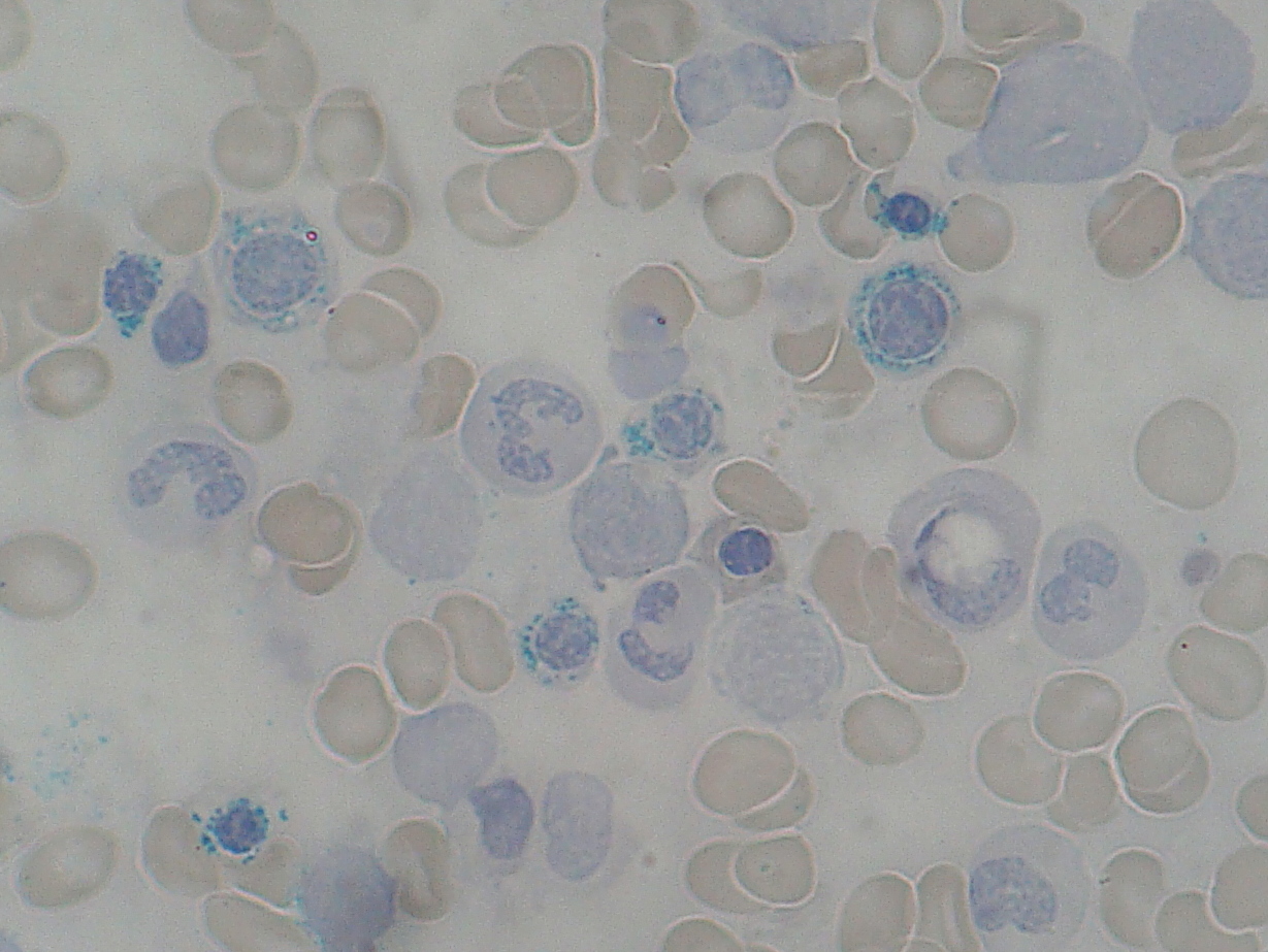
    The image below from Wiki Commons shows ring sideroblasts:


    Causes of sideroblastic anaemia include:
    • congenital causes
      • delta-aminolevulinate synthase deficiency
      • x linked recessive, rare
      • responds to pyridoxine
    • acquired causes
      • myelodysplasias
      • alcohol
      • lead
      • chemotherapy
      • anti-TB meds

    Treatment is to treat the cause, supportive care and pyridoxine.