Thursday, 7 October 2010

MRCP revision battle 26.1: Parkinson's disease

I'm back on the revision bandwagon and am ready for another 7 battles...


MRCP revision battle 26.1: Parkinson's disease
MRCP revision battle 26.2: Lewy Body Dementia
MRCP revision battle 26.3: Malignant hyperthermia
MRCP revision battle 26.4: Benign intracranial hypertension
MRCP revision battle 26.5: Coarctation of the aorta
MRCP revision battle 26.6: Heparin
MRCP revision battle 26.7: Essential tremor


MRCP revision battle 26.1: Parkinson's disease/Parkinsonism


Parkinsonism is a triad of:
  • resting tremor (3 to 5 Hz)
  • rigidity
  • bradykinesia
 There is also loss of postural reflexes.


It is Parkinson's disease if there is degeneration of the substantia nigra dopaminergic neurones with Lewy bodies.


There are a whole list of associated features that I'm sure you're familiar with, but if you fancy a brief recap read the list below...
  • expressionless, mask-like face
  • dribbling
  • festinant gait = short shuffling steps with flexed trunk)
  • micrographia


Management of Parkinsons is by a multidisciplinary team and the pharmacological management of it is notoriously tricky, with multiple drugs which often decrease in efficacy with use and may result in 'on-off' symptoms.


Even the NICE guidelines recognise the challenge in drug prescription, stating that "it is not possible to identify a universal first-line drug choice".  They single out the following as possible first line options:
  • levodopa
    • use lowest dose possible
    • give with carbidopa to prevent peripheral breakdown
    • is of no use in neuroleptic induced Parkinsons
  • dopamine agonists
    • bromocriptine*, cabergoline*, ropinole
    • if an argot-derived agonist is used (*) ESR, UEs and CXR should be done before starting and annually while on treatment
  • MAO-B inhibitors
    • selegiline
    • decreases dopamine breakdown


Second line treatment is combining the above options, or one of the above with:
  • COMT inhibitors
    • entacapone
    • inhibit dopamine breakdown


Other medications which may be useful for specific effects include:
  • amantidine
  • apomorphine
  • antimuscarinics
    • procyclidine
    • more useful for drug-induced Parkinsons


A couple of other key points from the NICE guidelines on Parkinsons are:
  • 'drug holidays' should not be undertaken due to risk of neuroleptic malignant syndrome on restarting
  • clinicians should be aware of dopamine dysregulation syndrome, which is an uncommon disorder in which misuse of dopaminergic medication is associated with behaviours such as hypersexuality, pathological gambling and sterotypic motor acts


For the really keen:


For everyone else, lets move on to Lewy body dementia.

MRCP revision battle 26.2: Lewy Body Dementia

Lewy Body Dementia accounts for around 20% of cases of dementia.


The key feature is Lewy Bodies (=intracytoplasmic neuronal inclusion bodies) in substantia nigra, paralimbic and neocortical areas.


Since this would obviously only be confirmed post-mortem diagnosis is made clinically:
  • Parkinsonian features
  • fluctuating cognitive loss
  • visual hallucinations

Neuroleptics must be avoided as their use would risk inducing irreversible Parkinsonism.


Treatments which may help include donepezil and rivastigmine.


Next up - malignant hyperthermia

MRCP revision battle 26.3: Malignant hyperthermia

Malignant hyperthermia is a rare, life-threatening condition in which there is a huge increase in the body's skeletal muscle oxidative metabolism, resulting in:
  • hypercapnia
  • increased oxygen consumption
  • hyperthermia (T>38C)


Secondary problems which may develop from malignant hyperthermia include:
  • renal failure
  • arrhythmias
  • DIC


Malignat hyperthermia may be caused by:
  • any inhaled anaesthetic agent
  • Succinylcholine  


It is often inherited in an autosomal dominant fashion.


Treatment is with dantrolene.


Onwards to benign intracranial hypertension...

MRCP revision battle 26.4: Benign intracranial hypertension

Benign intracranial hypertension classically affects overweight young females.

It presents like there should be a mass in the brain, but none can be found.


Features include:
  • headache
  • blurred vision
  • dizziness
  • horizontal diplopia
  • papilloedema


Benign intracranial hypertension is associated with:
  • COC
  • steroids
  • tetracyclines
  • vitamin A
  • nitrofurantoin
  • isontertinoin
  • danazol

Management is:
  • weight loss
  • acetazolamide
  • loop diuretics
  • prednisolone
  • therapeutic lumbar puncture
  • shunt

Note that up to 10% of patients have permanent significant vision loss - so its name of 'benign' is a little misleading!  Optic nerve sheath fenestration can help prevent this.


Onwards for a spot of coarctation of the aorta...

MRCP revision battle 26.5: Coarctation of the aorta

Coarctation of the aorta (=narrowing of the aorta) classically occurs just distal to the left subclavian artery.

It may present at birth with heart failure, or be discovered later in life, eg during investigation for hypertension.


Most adults with coarctation of the aorta are asymptomatic.  Occasionally they may complain of:
  • headache
  • recurrent epistaxis
  • claudication of the calf muscles


Clinical examination may reveal:
  • radio-femoral delay
  • midsystolic murmur


Complications of coarctation include:
  • hypertension
  • LVF
  • endocarditis

Conditions associated with coarctation include:
  • bicuspid aortic valve (10-20%)
  • PDA
  • VSD
  • Berry aneurysms
  • Turners syndrome
  • renal abnormalities


CXR may show rib notching due to formation of collaterals


Management is stenting/surgery.


Now for the penultimate battle of the day, heparin...

MRCP revision battle 26.6: Heparin

Heparin comes in 2 'flavours':
  1. low molecular weight heparin (AKA dalteparin/fragmin, enoxaparin/clexane, tinzaparin) 
  2. unfractionated (AKA 'heparin')

So what are the differences?
  • molecular weight
    • LMW heparin, as the name suggests, has a lower molecular weight - 5000 compared to unfractionated heparin's 13000
  •  half life
    • LMW heparin has a longer half life than unfractionated heparin
  •  mechanism of action
    • LMW heparin inhibits factor Xa but has little effect on antithrombin
    • unfractionated heparin binds antithrombin
  • affect on aPTT
    • LMW heparin has little effect on aPTT
    • unfractionated heparin prolongs aPTT
  • side effects
    • both can cause osteoporosis and thrombocytopenia but the risks are greatest with unfractionated heparin
  • reversibility
    • unfractionated heparin can be fully reversed by protamine
    • LMW heparin cannot be fully reversed by protamine

A side effect of protamine to be aware of is hypotension.


HIT (=heparin induced thrombocytopenia) is a potentially serious side effect of heparin use which will be covered tomorrow.


With that to look forward to lets shake on to the last battle of the day, essential tremor...

MRCP revision battle 26.7: Essential tremor

Essential tremor can be sporadic or inherited; if inherited its inheritance pattern is believed to be autosomal dominant with incomplete penetrance.

35% of patients with essential tremor will have no family history.


It tends to be worse when the arms are outstretched.


Essential tremor improves with alcohol (not advised as a treatment option!)


Treatment tends to be:
  • propranolol
  • primidone if propranolol contra-indicated (eg in asthma)


Congratulations on surviving another day's battles!

Wednesday, 6 October 2010

MRCP revision battle 25.1: Neuroleptic Malignant Syndrome

Having been blessed by a set of on-calls with some nice interesting cases I'm full of enthusiasm for today's battles, so lets get on with it!


MRCP revision battle 25.1: Neuroleptic malignant syndrome
MRCP revision battle 25.2: Behcets disease
MRCP revision battle 25.3: Subarachnoid haemorrhage
MRCP revision battle 25.4: Normal pressure hydrocephalus
MRCP revision battle 25.5: Gas gangrene
MRCP revision battle 25.6: Methaemoglobinaemia
MRCP revision battle 25.7: The weeverfish


MRCP revision battle 25.1: Neuroleptic malignant syndrome


A nice straightfoward battle to begin with...


Neuroleptic malignant syndrome is a life-threatening condition associated with use of antipsychotic drugs.


Symptoms and signs include:
  • hyperthermia
  • rigidity/muscle cramps
  • extrapyramidal signs
  • autonomic dysfunction (labile BP, sweating, urinary incontinence
  • confusion

Investigations show:
  • raised CK
  • raised WCC

Treatment is:
  • cooling
  • dantrolene (a muscle relaxant; note dantrolene is also used in malignant hyperthermia, which will be one of tomorrows battles)


Now on to a slightly less straightforward battle, Behcets disease...

MRCP revision battle 25.2: Behcets disease

Behcet's disease is a systemic vasculitis of unknown cause.


The classic triad of Behcet's disease is:
  • oral ulcers
  • genital ulcers
  • anterior uveitus
which I remember as GOA, like the place in India.


Other possible features include:
  • thrombophlebitis
  • DVT
  • erythema nodosum
  • pathergy reaction (=needle prick leads to pustule formation)
  • arthritis
  • aseptic meningitis
  • ataxia
  • diarrhoea


Males are more commonly affected than females.
It is commonest in Turkey and Japan.
30% of patients have a positive family history.


HLA B5 is associated with anterior uveitis and HLA B12 is associated with recurrent ulceration.


Severe disease requires steroids/ciclosporin/azathioprine.


Next up - SAH...

MRCP revision battle 25.3: Subarachnoid haemorrhage

Subarachnoid haemorrhage is bleeding into the space between the subarachnoid membrane and the pia mater.

It accounts for 5-10% of all strokes.


Causes:
  • ruptured berry aneurysm (80%)
  • malformations (15%)
  • post trauma


Common sites of berry aneurysms are:
  • junction of the posterior communicating artery with the internal carotid
  • junction of the anterior communicating artery with the anterior cerebral artery
  • the bifurcation of the middle cerebral artery

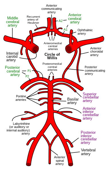
If (like me) your anatomy of the circle of willis is a little rusty the wiki commons image below might help refresh your memory:


15% of berry aneurysms are multiple.


Subarachnoid haemorrhages are associated with:
  • polycystic kidneys
  • coarctation of the aorta
  • Ehlers-Danlos syndrome
  • PAN


Classical presentation is a thunderclap occipital headache "as if kicked in head" with vomiting and neck stiffness.
Around 6% of patients will have had a sentinel headache before.


Investigation is:
  • CT - >90% of bleeds detected
  • LP - done >12hrs after onset looking for xanthochromia


Management:
  • neurosurgical referral
  • prompt angiography if surgery likely
  • nimodipine


On to another neuro topic - normal pressure hydrocephalus

MRCP revision battle 25.4: Normal pressure hydrocephalus

Normal pressure hydrocephalus is a triad of:
  1. dementia
  2. gait abnormality
  3. urinary incontinence
(remember as DUG!)


It is caused by a defect in the absorption of CSF.
Causes include: meningitis, head injury, subarachnoid haemorrhage.


Note that there is NO headache and NO papilloedema.


Investigations:
  • CT head (predictably) - enlarged ventricles
  • large volume lumbar puncture - ? high opening pressure, ? improvement of symptoms
  • lumbar infusion test

Treatment is a shunt.


Next up... gas gangrene!

MRCP revision battle 25.5: Gas gangrene

Gas gangrene is a life threatening bacterial infection.

It is characterised by muscle necrosis, gas production and sepsis.

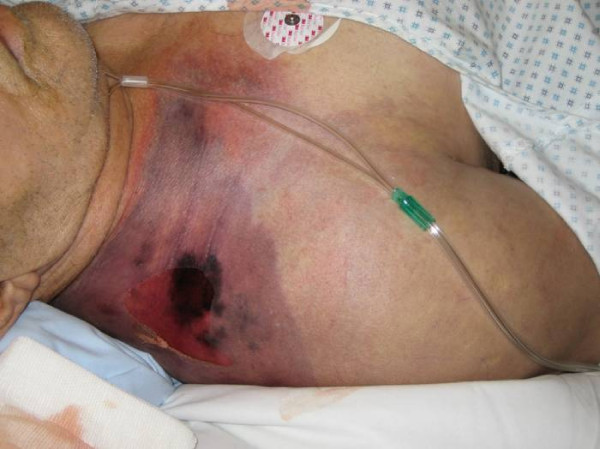
A typical appearance is shown below:

                                       From Wiki Commons, taken by Engelbert Schröpfer, Stephan Rauthe and Thomas Meyer.


Most cases are caused by trauma innoculation with bacteria, for example a farmer standing on a pitchfork.
Very rarely it may occur spontaneously, for example in the context of neutropenia.


The commonest causative organism is clostridium perfringens.


Management is:
  • urgent surgical debridement
  • IV Abx 
  • hyperbaric oxygen if available


Next up - methaemoglobinaemia

MRCP revision battle 25.6: Methaemoglobinaemia

Methaemoglobin (MetHb) is formed when the iron in haemoglobin is oxidised from the ferrous state (Fe2+) to the ferric state (Fe3+) 


Methaemoglobinaemia means raised MetHb levels, and as MetHb has a far higher affinity for oxygen than normal haemoglobin this results in tissue hypoxia as the oxygen being carried in the blood is not given up to the tissues.


Clinical features depend on the % of MetHb:
  • 15-20% - cyanosis
  • 20-45% - impaired consciousness
  • >55% - seizures, arrhythmias, coma
  • >70% - lethal


Pulse oximetry will show low oxygen sats, as will the printout from blood gas analysis - but the PaO2 will be high.


Causes of methaemoglobinaemia:
  • congenital
    • pyruvate kinase deficiency
    • G6PD deficiency
    • abnormal haemoglobin - HbH or HbM
    • diaphorase 1 deficiency
  • acquired - accelerated rate of oxidised formation due to meds
    • antibiotics - sulphonamides, trimethoprim, dapsone
    • drugs used in anaesthetics - lidocaine, procaine
    • others -primaquine


Management:
  • 1% solution of methylene blue
  • hyperbaric oxygen
  • exchange transfusion
  • dialysis
  • asorbic acid in patients with G6PD deficiency

So to the final battle of the day - weeverfish!

MRCP revision battle 25.7: The weeverfish

Just very briefly...

The weeverfish (AKA trachinus vipera) is found in shallow waters around the coast of the UK.

It is small and sandy-coloured, with sharp dorsal spines.


If you are unfortunate enough to stand on it you will get intense pain.

Happily the toxin produced by the weeverfish is denatured above 40C - so dunk the foot in hot water and the pain should stop.


On that most random of MRCP revision notes, lets end for the day!

Sunday, 3 October 2010

MRCP questions: War 24

 I now have a co-conspirator for MRCP revision on the go... lets call her Dr Biscuit and she is now writing all the wars!  How hard they are will probably reflect how bad her day has been... good luck!


As with previous 'wars' after 'battles' these are just a few quick questions to see if your brain cells have retained the information provided in battles 24:1 to 24:6.

The answers are available here


Question 1:
You are referred a patient who was diagnosed with Primary Biliary Cirrhosis 2 years ago. Name 3 signs you might find on clinical examination?

Question 2:
What blood test would you do to confirm the diagnosis of PBC?


Question 3:
A 22 year old female patient presents with DIC and hepatosplenomegaly.  Blood film shows cells which are strongly sudan black/peroxidase positive. What is the diagnosis and what is the gene translocation?


Question 4:
Name 3 drugs which can be used to treat AML?


Question 5:
Name 3 conditions associated with angioid retinal streaks?


Question 6:
List 3 drugs/groups of drugs which can cause SIADH?


Question 7:
Your patient is hyponatraemic with a sodium of 120.  On examination they appear clinically dehydrated. What investigation would you request next to help you elucidate the cause of the hyponatraemia?


Question 8:
Your patient is hyponatraemic with a sodium of 118.  Name four signs or symptoms they might display?


Question 9:
A patient is admitted with confusion. You are unable to obtain a history from her family but she takes no medications.  Her sodium is 119.  On examination she appears euvolaemic.  The admitting doctor has sent a urine sample which shows a urinary na of 34, and urine osmolality of 750. What is the likely diagnosis?


Question 10:
What are the 2 main effects of ADH?


The answers are available here

Friday, 1 October 2010

MRCP revision battle 24.1: Primary biliary cirrhosis

All this working and revising is leaving me with little internet-publishable material to put into my daily intros.  Looking for inspiration I ended up stumbling accross an utterly brilliant set of articles written back in the early 2000's by a (then) junior doctor called Michael Foxton in the Guardian.  Hopefully this link will take you to a list of his articles to soothe you after some revision.... but I warn you not to start looking before you've finished revising for the day because otherwise there is a real danger you will get no revision done!!


So today's battles are:

MRCP revision battle 24.1: Primary biliary cirrhosis
MRCP revision battle 24.2: Acute Myeloid Leukaemia
MRCP revision battle 24.3: Acute Promyelocytic Leukaemia
MRCP revision battle 24.4: Angioid retinal streaks
MRCP revision battle 24.5: SIADH
MRCP revision battle 24.6: Hyponatraemia
MRCP revision battle 24.7: Neutropenia and neutrophillia


MRCP revision battle 24.1: Primary biliary cirrhosis


I've never bonded well with this topic so this battle is a personal Waterloo for me...


Primary biliary cirrhosis is thought to be an autoimmune condition in which the interlobular bile ducts are damanged by chronic granulomatous inflammation. 


The result of this is:
  • progressive cholestasis
  • cirrhosis
  • portal hypertension


Females are more affected than males (9:1) and it tends to present in middle age.

It is associated with many other autoimmune conditions, including sjogrens, RA, systemic sclerosis, thyroid disease...


Primary biliary cirrhosis is often found incidentally by a raised alk phos on LFTs.Initially other LFTs may be normal but as the disease progresses bilirubin rises and PT may increase.


Associated signs (in late disease) include:
  • jaundice
  • xanthelasma
  • hepatosplenomegaly
  • clubbing


Complications include:
  • osteoporosis
  • portal hypertension
  • variceal haemorrhage


Diagnosis includes antibodies:
  • AMA M2 is highly specific and is positive in 98% of cases
  • SMA is positive in 30%
  • raised IgM


Treatment is:
  • cholestyramine for itching
  • osteoporosis prophylaxis
  • transplant


Once jaundice develops, without transplant the survival is < 2yrs.


On to AML...

MRCP revision battle 24.2: AML

Acute myeloid leukaemia is cancer of the myeloid line of blood cells.
Myeloid cells are essentially all blood cells that are not leukocytes.


It is the commonest leukaemia in adults.


Presentation may be by:
  • complications of marrow failure
    • anaemia
    • infection
    • bleeding
  • evidence of marrow infiltration
    • hepato/splenomegaly
    • gum hypertrophy
CNS involvement is rare in AML (contrasted with ALL, where it is common)


Diagnosis is by bone marrow biopsy.
Immunophenotyping/cytogenetic analysis is important to guide treatment and indicate prognosis.


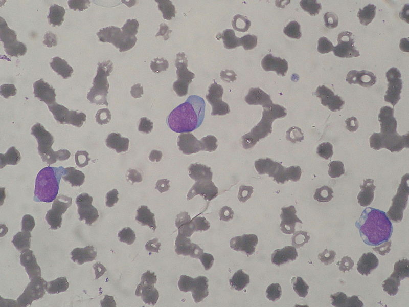
The key feature on microsopy is the auer rod - note the small rod in the cytoplasm of the central cell:


Classification was traditionally done by the FAB (French-American-British) classification, which ran from M0 to M7.  The important random facts I remember from this is that M2 is associated with t(8;21) and M3 (aka acute promyelocytic leukaemia, the topic of the next battle) is associated with t(15;17).  Both of these translocations are associated with a good prognosis.
Now the WHO classification is more commonly used.  It is rather complex and includes catagories such as 'AML with characteristic abnormalities' and 'AML therapy-related'  If you are interested the further reading link at the bottom will tell you more.


Treatment is supportive care and intensive chemotherapy, usually with cytosine arabinoside and daunorubicin, or bone marrow transplant.


Survival with no treatment is around 2 months; with chemo there is around 20% 3 yr survival.


For the really keen:


Now lets briefly look at acute promyelocytic leukaemia...

MRCP revision battle 24.3: Acute Promyelocytic Leukaemia

Acute Promyelocytic Leukaemia is the M3 subtype of AML.

It is associated with the t(15;17) translocation and the RARA gene.


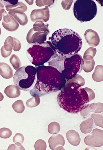
On microscopy you look for faggots (=collections of auer rods):

The cells are strongly sudan black/peroxidase positive.

Acute promyelocytic leukaemia tends to present in young adults (average approx 25 yrs) and the presentation is often DIC.


Despite this sounding bad, acute promyelocytic leukaemia actually has a good prognosis.


Remission can be induce with all-trans retinoic acid.


Thats enough heavy-going haematology for one day... lets move on to angioid retinal streaks...

MRCP revision battle 24.4: Angioid retinal streaks

Angioid retinal streaks are caused by breaks in Bruch's membrane


They look like dark red lines under the retina - almost like blood vessels.  I'd recommend using google to search for a selection of images to look at.


They are associated with SLAPPERS:
  • Sickle cell disease
  • Lead poisoning
  • Acromegaly
  • Pagets disease
  • Pseudoxanthoma elasticum
  • Ehlers-Danlos
  • Raised calicum/phosphate
  • Short people


 Onwards to SIADH...


MRCP revision battle 24.5: SIADH

SIADH = syndrome of inappropriate antidiuretic hormone secretion is a condition whose name really says it all: it is ADH being produced and released when it shouldn't be.


Quick bit of physiological background:
  • ADH is made in the magnocellular neurones in the supraoptic and paraventricular nuclei of the hypothalmus
  • ADH is stored in the posterior pituitary
  • it is released in response to raised plasma osmolality (osmoreceptors in hypothalamus) or decreased volume (baroreceptors in carotids, atria)
  • its effect is to increase the insertion of aquaporin 2 channels in the collecting ducts of the kidney, hence increasing water reabsorption.
  • it also increases peripheral vascular resistance


SIADH is diagnosed in the presence of:
  • concentrated urine ( sodium >20mmol/l and osmolality >500mosmol/kg) AND
  • hyponaturaemia (<125) or low plasma osmolality (<260mosmol/kg) AND
  • the absence of hypovolaemia, oedema or diuretics.


Causes of SIADH include:
  • malignancy: small cell lung cancer, pancreas, prostate, lymphoma
  • lung pathology: TB, pneumonia, aspergillosis
  • CNS pathology: injury, GBS, fits, subarachnoid/subdural haemorrhage, abscess, stroke
  • metabolic: porphyria
  • drugs: opiates, carbamazepine, SSRIs, TCA


So lets now finish todays battles by widening this topic out to hyponatraemia...