Sunday, 17 October 2010

MRCP revision battle 32.1: Deafness: Rinnes and Webers

Final day in the hat trick of eyes and neurology battles...


MRCP revision battle 32.1: Deafness: Rinnes and Webers
MRCP revision battle 32.2: Tinnitus
MRCP revision battle 32.3: TIA
MRCP revision battle 32.4: Epilepsy
MRCP revision battle 32.5: Macular degeneration
MRCP revision battle 32.6: Glaucoma
MRCP revision battle 32.7: Autonomic neuropathy


MRCP revision battle 32.1: Deafness: Rinnes and Webers

Hearing loss and Rinne and Weber's tests are MRCP favourites.


Remember there are 2 types of deafness:
  • conductive deafness
    • ear wax
    • otosclerosis
    • otitis media
    • glue ear
  • sensorineural deafness
    • acoustic neuroma
    • Pagets
    • MS
    • CVA
    • Menieres
    • head trauma
    • noise exposure
    • drugs
      • aminoglycosides (gentamycin)
      • furosemide
      • lead


Tests are done with a 256- 512 Hz tuning fork


Rinne's test:
  • hold vibrating tuning fork next to ear meatus, then place on mastoid
  • ask which is louder
  • air conduction (AC) > bone conduction (BC) = normal, or, if hearing is decreased, suggests sensorineural loss
  • BC>AC = conductive deafness = Rinne negative
  • Remember as: ALS (like the course) - air loudest: sensorineural.  


Weber's test:
  • hold vibrating tuning fork on forehead
  • ask which side it is heard loudest in
  • in sensorineural loss it laterals to the unaffected side
  • in conductive loss it laterals to the affected side
  • midline if normal, or bilateral sensorineural loss
  • Remember as: SUCA - sensorineural unaffected, conductive affected.


Next up - a spot of tinnitus...

MRCP revision battle 32.2: Tinnitus

Tinnitus is ringing/buzzing in ears.


Causes include:
  • eax
  • viral infections
  • presbyacusis
  • head injury
  • Menieres
  • drugs
    • aspirin
    • furosemide
    • gentamycin


Pulsatile tinnitus may indicate carotid artery stenosis or dissection


Now on to the meatier topic of TIAs...

MRCP revision battle 32.4: Epilepsy

Epilepsy is defined as a recurrent tendency to spontaneous, intermittent, abnormal electrical activity in part of the brain, manifest as seizures (OHCM definition)


Epileptic seizures are subdivided into:
  • partial seizures = features come from one part of the brain
    • simple partial = consciousness not impaired
    • complex partial = consciousness impaired - usually temporal lobe, aura may preceed and automatisms may feature
  • generalised seizures = features not localisable to one part of the brain
    • absence = petit mal = brief <10 second pauses - 3Hz spikes
    • atonic = becomes flaccid
    • tonic-clonic - classic stiffening and jerking
    • myoclonic


Treatment is usually started after the second seizure.   It would be started after the first if any of the following conditions apply:
  • neurological deficit
  • structural abnormality
  • EEG unequivocal
  • pt/family/carers keen for treatment


First line treatment depends on the seizure type:
  • generalised
    • 1st line: sodium valproate
    • 2nd line: lamotrigine
  • partial
    • 1st line: carbamezepine
    • 2nd line: sodium valproate
  • absence
    • sodium valproate or ethosuximide
    • note that carbamazepine may worsen absence seizures

Remember that both sodium valproate and lamotrigine are associated with Steven Johnson syndrome.


Cannot drive until seizure-free for 1 yr, or 3 yrs of night time only seizures.
HGV drivers need to be off meds and seizure-free for 10 yrs.


If a pt has no seizures for 2 yrs, you may consider stopping meds over 2-3 months.


On to battle 32.5...

MRCP revision battle 32.3: TIA

TIA (=transient ischaemic attack) = sudden onset of focal CNS phenomena which last <24 hours.  Caused by temporary occlusion of part of the cerebral circulation.


Causes of TIAs:
  • thromboembolism
    • chiefly from carotids
    • may be from heart - AF, mural thrombus etc)
  • hyperviscosity
    • myeloma
    • polycythaemia
    • sickle cells
    • very high white cells


Management depends on the ABCD2 score, which is calculated as shown below:
  • age >60 : 1 point
  • BP greater or equal to 140/90 : 1 point
  • clinically:
    • unilateral weakness : 2 points
    • speech disturbance without weakness : 1 point
  • duration
    • >60 mins : 2 points
    • 10 - 59 mins : 1 point
  • diabetes : 1 point


ABCD2 score of 4 or more, or crescendo TIAs (=2 or more in one week):
  • specialist review within 24 hrs
  • start 300mg aspirin OD


ABCD2 score of 3 or below, or symptoms >1 week ago:
  • specialist review within a week
  • start 300mg aspirin od

After specialist review, usually 75mg aspirin OD and dipyridamol OD for 2 yrs.
If pt is aspirin intolerant, prescribe monotherapy of clopidogrel.


If assessed to be a candidate for carotid endarterectomy, imaging should be performed within 1 week of symptom onset and if carotid stenosis of:
  • 50–99% according to NASCET criteria, or 
  • 70–99% according to ECST criteria 
carotid endarterectomy should be performed within 2 weeks of symptom onset.


Remember pt must inform DVLA and no driving for 1 month (car) or 1 yr (lorry).  This increases to 3 months for car if multiple TIAs.


Of course other risk factors (hypertension, alcohol, smoking, etc etc etc) should also be addressed.


Differentials for TIA may include:
  • migraine
  • epilepsy
  • hypoglycaemia
  • malignant hypertension 
  • MS


Lets move on to tackle epilepsy...

MRCP revision battle 32.5: Macular degeneration

Macular degeneration is the commonest cause of blindness in the UK.


There are 2 types:
  • dry 
    • drusen - yellow spots in Bruchs membrane
  • wet
    • choroidal neovascularisation
    • worse prognosis

Below is an image of dry macular degeneration showing drusen.  To link to an image of wet macular degeneration click here


Risk factors for macular degeneration are:
  • age >60
  • female
  • smoking
  • caucasian
  • family history


Treatment - there is no cure:
  • high dose beta caratene - but may increase risk of lung cancer in smokers
  • vitamin C and E - but vit E may increase risk of heart failure
  • zinc
  • for wet:
    • photocoagulation
    • anti VEGF
    • Ranibizumab
    • photodynamic therapy


Keeping on eyes, next up is glaucoma...

MRCP revision battle 32.6: Glaucoma

Glaucoma is damage to the optic nerve, generally due to increase in intra-ocular pressure.


There are 2 types of glaucoma:
  1. acute = closed angle glaucoma
  2. chronic = open angle glaucoma


Acute closed angle glaucoma

Acute closed angle glaucoma presents as:
  • pain
  • decreased visual acuity
  • worsening of pain with mydriasis
  • hard, red eye
  • halos around lights
  • semi-dilated, non-reactive pupil
  • pt systemically unwell

Risk factors for closed angle glaucoma include hypermetropia and pupillary dilation.


Management is:
  • urgent opthalmic review
  • IV acetazolamide (a carbonic anhydrase inhibitor; decreases aqueous secretions)
  • topical pilocarpine (constricts pupil)


Open angle glaucoma


This affects 2% of over 40s.


Risk factors include:
  • family history
  • black
  • myopia
  • hypertension
  • diabetes


Features include:
  • peripheral field loss
  • decreased visual acuity
  • optic disc cupping


Treatments include:
  • eye drops (beta blockers or prostaglandins)
  • possibly surgery.


Now to our final battle of the day, autonomic neuropathy

MRCP revision battle 32.7: Autonomic neuropathy

Autonomic neuropathy is characterised by:
  • postural hypotension
  • erectile dysfunction
  • constipation or diarrhoea
  • urinary retention
  • inability to sweat
  • loss of decreased heart rate on deep breathing
  • Horners
  • Holmes-Adie pupil


Causes:
  • endocrine
    • diabetes
  • infective
    • HIV
    • syphilis
    • leprosy
  • autoimmune
    • SLE
  • other
    • GBS
    • MSA
    • parkinson's disease
    • liver/renal failure.


Treatment is symptomatic; remember fludrocortisone as an option for severe postural hypotension.


On that list-intensive battle lets retire for the day.

Saturday, 16 October 2010

MRCP revision battle 31.1: Wernicke's Encephalopathy

Day 2 of 3 for lots of neuro...


MRCP revision battle 31.1: Wernicke's Encephalopathy
MRCP revision battle 31.2: Korsakoff's Syndrome
MRCP revision battle 31.3: Multiple Sclerosis
MRCP revision battle 31.4: Menieres Disease
MRCP revision battle 31.5: CJD
MRCP revision battle 31.6: Herpes Zoster Opthalmicus
MRCP revision battle 31.7: Ramsey Hunt Syndrome


MRCP revision battle 31.1: Wernicke's Encephalopathy


Wernicke's encephalopathy is caused by thiamine (=vitamin B1) deficiency.


The classic triad associated with Wernicke's encephalopathy is:
  • opthalmoplegia
  • ataxia
  • confusion

There may also be hypothermia and hypotension.


Thiamine deficiency may result from:
  • alcoholism
  • carcinoma of stomach
  • anorexia
  • vomiting


CT brain may show petichial haemorrhages and a low red cell transketolase blood test could prove thiamine deficiency (rarely performed)


Treatment is parenteral thiamine (pabrinex)

If untreated, 20% of patients will die and 85% will develop Korsakoffs.


Which leads us nicely into revision battle 31.2: Korsakoffs syndrome....

MRCP revision battle 31.2: Korsakoff's Syndrome

Korsakoff's Syndrome is a condition in which there is an inability to build new memories but preservation of other cognitive functions.


It is a chronic disorder, usually following Wernicke's encephalopathy.


The patient has an inability to consolidate new info, retrograde amnesia, patchy preservation of long term memory, a lack of insight and confabulates (=fabricates information to make up for memory loss)


Treatment is with thiamine but under 20% of patients respond.


On to 31.3... MS...

MRCP revision battle 31.3: Multiple Sclerosis

Multiple sclerosis is a cell-mediated autoimmune disorder characterised by plaques of demyelination at sites throughout the CNS but not in the peripheral nerves.


4 main types:
  1. relapsing-remitting
    • accounts for 80% of cases
  2. secondary progressive
    • up to 50% of relapsing-remitting patients eventually develop this
  3. primary progressive
    • 10-15% of cases
    • older age of onset
  4. progressive-relapsing


Initial presentation tends to be a single symptom, such as:
  • optic neuritis 
    • pain on eye movement and rapid deterioration in central vision, loss of colour vision
    • treatment is methylpred
    • single episode gives 40-60% chance of subsquent MS
  • numbness or tingling in limbs
  • leg weakness
  • ataxia


Diagnosis is by (NB none is specific):
  • lesions disseminated in time and location
  • T2 weighted MRI showing demyelinating plaques
  • delayed visual evoked response potentials
  • oligoclonal bands in CSF but not serum


Good prognostic factors are:
  • relapsing-remitting course
  • female
  • young onset
  • sensory symptoms

Risk of MS is increased 20-40% in siblings and there is 25% concordance in monozygotic twins.


Treatment is:
  • IV methylprednisolone
    • shortens relapses
    • no effect on incidence of attacks
  • beta interferon
    • decreases relapses by 30%
    • given if
      • relapsing-remitting course and 2 relapses in 2 yrs and can walk 100m unaided
      • secondary progressive and 2 relapses in 2 yrs and 100m aided
    • side effect: flu-like
  • baclofen
    • decreases spascity

Now for the less heavy-going Menieres disease...

MRCP revision battle 31.4: Meniere's Disease

Meniere's Disease is a disorder of the inner ear causing vertigo and hearing loss.


Features include:
  • vertigo
  • tinnitus
  • sesorineural hearing loss
  • aural fullness

There may be nystagmus and a positive romberg's test


It is usually unilateral and is believed to be associated with endolymphatic hydrops (=increased fluid in ear)


Rarely it may be associated with drop attacks - no LOC or vertigo, but suddenly falling to one side.


Management:
  • prochlorperazine
  • inform DVLA and stop driving until controlled.


 Next up: that formally very topical topic CJD...

MRCP revision battle 31.5: CJD

Creutzfeldt-Jakob Disease (=CJD) is a prion disease which causes a rapidly progressive dementia with myoclonus.


The majority of cases are sporadic.  There is also an inherited form and an acquired form (=varient CJD, from meat infected with BSE)


CSF examination is usually normal but EEG shows characteristic biphasic high-amplitude sharp waves.


Death is usually within 6 months.


On a lighter note lets look at herpes zoster opthalmicus...

MRCP revision battle 31.6: Herpes Zoster Opthalmicus

Herpes zoster opthalmicus is the reactivation of varicella zoster in the area supplied by the opthalmic division of the trigeminal nerve.


It occurs in roughly 10% of cases of shingles.


There may be Hutchinson's sign = rash on the tip of nose, which indicates nasocillary involvement and is a strong risk factor for ocular involvement.


Treatment is oral antivirals for 7 to 10 days and urgent opthalmic review if there is ocular involvement.


On to the final revision battle of the day...

MRCP revision battle 31.7: Ramsey Hunt Syndrome

Ramsey Hunt Syndrome is the reactivation of varicella zoster in the geniculate ganglion of the facial nerve.  


It is characterised by:
  • herpes zoster oticus (=herpetic eruption in external auditory meatus)
  • facial nerve palsy
  • +/- deafness, tinnitus and vertigo


Treatment is with oral antivirals

Friday, 15 October 2010

MRCP revision battle 30.1: Retinitis Pigmentosa

Today is the start of a 3 day neuro/opthalmology blitz, so hold on to your hats....


MRCP revision battle 30.1: Retinitis Pigmentosa
MRCP revision battle 30.2: Oculogyric Crisis
MRCP revision battle 30.3: Myotonic Dystrophy
MRCP revision battle 30.4: Trinucleotide repeat disorders
MRCP revision battle 30.5: Huntington's Chorea
MRCP revision battle 30.6: Friedreich's ataxia
MRCP revision battle 30.7: Alzheimer's Disease


MRCP revision battle 30.1: Retinitis Pigmentosa

Retinitis pigmentosa is a type of progressive retinal dystrophy which eventually leads to blindness.

It has a very characteristic fundoscopic appearence with black mottling of the retina and a pale optic disc, as illustrated below in an image by Christian Hammel (from wiki commons):


It can be inherited in an autosomal dominant, autosomal recessive or X linked recessive fashion.


Night blindness is often the first sign.
Later comes funnel/tunnel vision.


It is associated with many conditions, including:
  • Kearns Sayre (remember battle 15.1?)
  • Ushers syndrome = retinitis pigmentosa with sensorineural deafness
  • congenital toxoplasmosis

There is no cure for retinitis pigmentosa but progression of the disease can be slowed by vitamin A.


On to the second battle of the day...

MRCP revision battle 30.2: Oculogyric Crisis

The key features of an oculogyric crisis are restlessness and upward deviation of the eyes (although this only occurs in severe cases).


Oculogyric crises may be precipitated by a range of drugs including:
  • neuroleptics
  • metoclopramide/domperidone
  • nifedipine
  • TCA
  • carbamazepine

Several clinical conditions can also cause oculogyric crises, for example:
  • parkinsons disease
  • post encephalitis
  • bilateral thalmic infarction
  • MS


Treatment is with procyclidine, an anticholinergic.


On to the next battle...

MRCP revision battle 30.3: Myotonic Dystrophy

Myotonic dystrophy is an autosomal dominant condition in which there is muscle weakness and myotonia.  It is a trinucleotide repeat disorder.

Onset tends to be in the 30s.


There are 2 types:


Dystrophia myotonica (=DM) 1
  • 98% of cases
  • more severe
  • distal muscle weakness
  • chromosome 19
Dystrophia myotonica 2:
  • 2% of cases
  • less severe
  • proximal muscle weakness
  • chromosome 3

Features associated with myotonic dystrophy include:
  • long faces
  • frontal baldness
  • myotonia and weakness
  • testicular/ovarian atrophy
  • bilateral ptosis
  • cateract
  • cardiomyopathy
  • diabetes
  • miotic pupils 
  • mildly decreased IQ

It is incurable but phenytoin can improve the myotonia.


Lets move on to consider an overview of trinucleotide repeat disorders

MRCP revision battle 30.4: Trinucleotide repeat disorders

Trinucleotide repeat disorders are conditions in which 3 nucleotides in the DNA are expanded.

The most 'famous' are:
  • Huntingtons chorea (CAG) - covered in next battle
  • Fragile X (CGG)
  • Myotonic dystrophy (CTG) - covered in previous battle
  • Friedreichs ataxia (GAA) - covered in battle 30.6


Two concepts to be born in mind for trinucleotide repeat disorders are:

Anticipation
= the disease presents earlier and is worse in successive generations

Somatic instability = expansion increases as patient gets older


So lets briefly cover the most famous, Huntingtons...

MRCP revision battle 30.5: Huntington's Chorea

Huntington's chorea is an autosomal dominant condition due to CAG repeats.

It is carried on chromosome 4.


Symptoms tend to start in the 40s with a progression from chorea to irritability to dementia and death.


There is no cure.  The chorea may be treated with a dopamine antagonist such as tetrabenazine.


Note that Hunington's has complete penetrance so a child of a sufferer has a 50% chance of being affected.  Due to anticipation if they are affected they are also likely to develop symptoms earlier.


Next up: Friedreich's ataxia...

MRCP revision battle 30.6: Friedreich's ataxia

Friedreichs ataxia is an autosomal recessive trinucleotide repeat disorder (GAA)


It causes degeneration of nerve tracts resulting in cerebellar ataxia, dysarthria, nystagmus and dysdiadocholokinesis.


Muscles are weak, reflexes are depressed but plantars are upgoing.


There is often scoliosis and high arches of the feet.

Cardiomyopathy is another complication.


There is no cure.


Lets now try and forget all these incurable diseases with a touch of Alzheimer's...

MRCP revision battle 30.7: Alzheimer's Disease

Alzheimer's disease is the leading cause of dementia.  This battle aims to pick out a few juicy facts for MRCP; http://alzheimers.org.uk/ can provide more in-depth information if you are interested.


The earliest symptom of AD is typically forgetfulness.


5% of cases are autosomal dominant, with chromosomes 1, 14, 19 and 21 implimented.

After age, apoE4 (chromosome 19) is the most significant risk factor for AD.


Beta amyloid plaques and hyperphosphorylated tau protein tangles are found within neurones post-mortem.
The density of the tangles can correlate with the severity of the dementia.


Hippocampal atrophy is prominant.


Cholinesterase inhibitors such as donepezil, rivastigmine and galantamine are licensed for patients with a MMSE of between 10 and 20.

Memantine (an antiglutamatergic) may be useful in severe AD.


As Bugs Bunny would say, thhhhatttttts alll folllkkkksss.... until tomorrow!

Thursday, 14 October 2010

MRCP revision battle 29.1: Hypoglycaemia

After nearly a week away I'm back with a bumper set of battles, the first of which is hypoglycaemia, which has been a recurring theme in both my patients and myself this week!  Enjoy!


MRCP revision battle 29.1: Hypoglycaemia
MRCP revision battle 29.2: Thyroid eye disease
MRCP revision battle 29.3: Hyperlipidaemia
MRCP revision battle 29.4: Lipid-lowering treatment
MRCP revision battle 29.5: Restless legs syndrome
MRCP revision battle 29.6: Histocytosis X
MRCP revision battle 29.7: Bartter's Syndrome
MRCP revision battle 29.8: Gitelman Syndrome
MRCP revision battle 29.9: Liddle's Syndrome


MRCP revision battle 29.1: Hypoglycaemia


Hypoglycaemia is defined as plasma glucose <3mmol/l.


In general in diabetics autonomic symptoms of hypoglycaemia (sweating, anxiety, tremor, palpitations) occur below 3.5mmol and neurological symptoms (confusion, drowsiness, seizures, coma) occur below 2.5mmol.

If a diabetic has frequent hypos they may become 'insensitive' and no longer experience symptoms before becoming unresponsive.  Symptoms may be 'restored' by carefully avoiding hypos for 3 months.


Causes of hypoglycaemia are easy to remember as it "IS PLAIN" to see...
  • Insulin
  • Sulphonyureas/other drugs
  • Pituitary insufficiency
  • Liver failure
  • Addisons disease or alcohol
  • Insulinoma
  • Neoplasms eg retroperitoneal fibrosarcomas than secrete IGF

Take bloods for glucose, insulin, c-peptide and plasma ketones.
Normal/high insulin, no ketones: insulinoma, drugs
Insulin low, no ketones: non-pancreatic neoplasm
Insulin low, ketones high: alcohol, addissons, pituitary insufficiency.


Post-pradial hypoglycaemia can occur post-gastrectomy


Whipples Triad is a set of criteria which if fulfilled suggest a patient's symptoms are due to hypoglycaemia:
  • symptoms suggestive of hypoglycaemia
  • BM < or equal to 2.5
  • symptoms relieved by food.

Treatment

I'm sure this is all old-hat to you all... get the pt to eat if they can, 1mg IM glucagon if they can't and no IV access (remember glucagon effects only last for 20 mins and may not work at all in alcoholics) and if IV is an option the 50mls of 50% glucose or the more modern 200mls of 10% (less abrasive to the veins)


Now for some TED time...

MRCP revision battle 29.2: Thyroid eye disease

Thyroid eye disease, referred to by its friends as 'TED', can occur in people who are hyperthyroid, hypothyroid or euthyroid.


Thyrotoxicosis from any cause can cause lid lag and lid retraction.


However, only Graves disease causes:
  • periorbital oedema
  • conjunctivial injection
  • proptosis/exophthalmos
  • opthalmoplegia/diplopia
  • papilloedema

(as an aside, I've always been mystified by the difference between proptosis and exophthalmos.  Unfortunately it appears I'm not the only one as some sources suggest the difference is related to the degree of protrusion whilst others say exophthalmos is used if the aetiology is endocrine and proptosis is used if the aetiology is not endocrine...)


25-50% of patients with Grave's disease have TED.


Risk of TED is increased in smokers.


The danger of TED is optic nerve compression.  Symptoms/signs of optic nerve compression include:
  • blurred vision/decrease VA
  • decreased colour vision
  • a relative afferent papillary defect


Treatment for TED is topical lubricants, steroids if severe and possibly even surgery.


Now for a horrid battle with hyperlipidaemia...

MRCP revision battle 29.3: Hyperlipidaemia

This battle is quickly going to get complex and aims only to be an MRCP-focused overview rather than a comprehensive discussion.  It is divided into 5 stages:
  1. The basics
  2. Hypercholesterolaemia
  3. HDL - the good cholesterol
  4. Hypertriglyceridaemia
  5. Mixed


1. The Basics

Hyperlipidaemia = raised lipids.  Lipids come in 4 main 'flavours':

  1. chylomicrons = carry triglyceride
  2. LDL = mainly cholesterol (50%, 10% TG) = the 'bad' flavour
  3. HDL = mainly phospholipid = the 'good' flavour, carry cholesterol back to the liver
  4. VLDL = mainly triglyceride (60%, 20% cholesterol)

Signs:
  • corneal arcus = grey-white ring around cornea
  • xanthelasma = yellow collection of cholesterol under skin
  • xanthomata = 'lumps' of cholesterol
  • eruptive xanthomata = small yellow-orange papules that appear all over body
  • lipaemia retinalis = 'creamy' appearence of blood vessels on fundoscopy

So lets look first at cholesterol and hypercholesterolaemia.


2. Hypercholesterolamia

a. Familial
  • LDL receptor dysfunction
    • cholesterol 7.5-16
    • raised LDL
    • tendon xanthomata, corneal arcus and xanthelasma
    • heterozygous prevalance 1/500 --> MI in 40s
    • homozygous --> MI in 20s
  • Polygenic hypercholesterolaemia
    • cholesterol 6.5-9
    • raised LDL
    • xanthelasma and corneal arcus

 b. Acquired
  • nephrotic syndrome
  • renal transplant
  • cholestasis
  • hypothyroidism


3.  HDL

HDL is 'good' cholesterol

It is higher in:
  • thin people
  • exercise
  • oestrogens
  • alcohol
  • low triglycerides

It is lower in:
  • obesity
  • sedentary states
  • post-puberty males
  • smoking


4. Hypertriglyceridaemia

a. Familial
  • failure to metabolise chylomicrcons
  • features include:
    • eruptive xanthomata
    • lipaemia retinalis
    • retinal vein thrombosis
    • pancreatitis
    • hepatosplenomegaly

b. Secondary
  • diabetes
  • obestiy
  • alcohol
  • chronic renal failure
  • liver disease
  • drugs such as thiazides, beta blockers or oestrogens


5. Mixed hyperlipidaemia
  • Look for palar xanthomas and tuberous xanthomas


After that wordy and depressing battle, lets move on to treatment of hyperlipidaemia...

MRCP revision battle 29.4: Lipid-lowering treatment

NICE recommends that all those with a 10 yr cardiovascular risk of >20% should be offered lipid-lowering therapy.

1st line: 40mg simvastatin
Measure LFTs when starting, at 3 months and at 12 months.


In primary prevention there is no 'target' cholesterol
In secondary prevention the target is less than 4mmol/l cholesterol and less than 2mmol/l LDL


If statins are not tolerated consider:
  • fibrates
  • ezetimibe
  • nicotinic acid


Mechanisms of action


Statins are HMG CoA reductase inhibitors.  They therefore work by decreasing the production of cholesterol in the liver.  The decreased production of cholesterol in the liver also results in the liver absorbing and processing more LDL, hence further reducing cholesterol.


Grapefruit juice decreases the metabolism of statins.  Patients on statins are therefore advised to avoid grapefruit juice as it would increase the risk of the rare side effect of rhabdomyolysis


Ezetimibe works by preventing intestinal absorption of cholesterol.


Fibrates are PPAR alpha receptor agonists.  They therefore increase clearance of VLDL and remnant particles and decrease TG secreation.

Fibrates and statins tend not to be used together due to increased risk of rhabdomyolysis.


Nicotinic acid (=vitamin B3) blocks breakdown of fats in adipose tissue. 
It can cause facial flushing.


For the really keen:
NICE lipid modification guide

After those heavy-going battles lets tackle something light - restless legs syndrome...                             

MRCP revision battle 29.5: Restless legs syndrome

Restless legs syndrome, sometimes known as Ekbom's syndrome, is a condition in which a person has unpleasant sensations in their legs and a desire to move them.


Restless legs may be idiopathic or secondary to:
  • pregnancy
  • iron deficiency
  • ureamia
  • diabetes
  • RA

1st line treatments are dopamine agonists such as pramipexole
2nd line treatments include benzodiazepines or gabapentin.


To keep the neurones jumping lets move on to a completely different topic again - histiocytosis X...

MRCP revision battle 29.6: Histiocytosis X

Histocytosis X, aka Langerhans-cell histiocytosis, is a group of disorders in which there is organ infiltration by granulomatous lesions containing clonally proliferated dendrintic (=Langerhams) cells.


The most commonly affected organs are lungs and bones.


Lung histiocytosis tends to affect young adults and is commonest in smokers.

The CXR shows multiple ring shadows with diffuse reticulo-nodular opacities affecting mainly the upper and mid zones.


Biopsy would show characteristic Birbeck granules.


Treatment is ?local excision, ?steroids.


Now onwards for a run of the last 3 short battles of the day, starting with Bartter's Syndrome...

MRCP revision battle 29.7: Bartter's Syndrome

Bartter's Syndrome is an autosomal recessive condition in which there is defective chloride reabsorption in the Na-K-2Cl channels in the loop of Henle resulting in severe hypokalaemia.


It presents in childhood with failure to thrive and polyuria/polydipsia


Blood pressure is normal/low.

There is hyperreninaemia and hypertrophy of the JGA.


Treatment is with potassium replacement and NSAIDs.


Next... Gitelman Syndrome....

MRCP revision battle 29.8: Gitelman Syndrome

Gitelman Syndrome is an autosomal recessive disorder in which there is decreased salt reabsorption in the distal tubule resulting in hypokalaemia due to secondary hyperaldosteronism.


Patients with Gitelman Syndrome have:
  • metabolic alkalosis
  • low potassium
  • low magnesium
  • usually normal blood pressure

Treatment is magnesium and potassium replacement.


Finally - Liddles!

MRCP revision battle 29.9: Liddle's Syndrome

Liddle's syndrome is an autosomal dominant condition caused by enhanced sodium reabsorption in the DCT.

It results in:
  • hypertension
  • hypokalaemia
  • metabolic alkalosis

Treatment is salt restriction, potassium, amiloride.

Saturday, 9 October 2010

MRCP revision battle 28.1: Haemochromatosis

I feel like I'm possibly starting to make some inroads into this MRCP revision thing, and my chocolate dependency level has decreased a little.  Today is another random assortment of gems:


MRCP revision battle 28.1: Haemochromatosis
MRCP revision battle 28.2: Syphilis
MRCP revision battle 28.3: Third nerve palsy
MRCP revision battle 28.4: G6PD deficiency
MRCP revision battle 28.5: Kaposi's sarcoma
MRCP revision battle 28.6: Chronic Myeloid Leukaemia
MRCP revision battle 28.7: Gerstmann's Syndrome


MRCP revision battle 28.1: Haemochromatosis


Haemochromatosis is an excessive accumulation of iron.
There is increased intestinal absorption leading to deposition in joints, skin, the heart, liver, pancreas, adrenals and pituitary.


It is an autosomal recessive disorder, inherited on chromosome 6.


Presentation is initially with tiredness and arthralgia

Later problems include:
  • diabetes mellitus (bronze diabetes)
  • slate-grey skin
  • liver disease/cirrhosis
  • cardiac failure/cardiomegaly
  • pseudogout
  • hypopituitism
  • >30% develop hepatocellular carcinoma


Males are affected earlier and more severely than females.


Diagnosis is by:
  • transferrin saturation >50%
  • ferritin >300-500micrograms/l (depending on guidelines)
  • liver biopsy  - Perls stain to show hepatic iron >180micromol/g


Treatment is regular venesection and ?chelation with desferrioxamine


Haemosiderosis is secondary haemochromatosis.  


Causes of haemosiderosis include:
  • beta thalassemia
  • sideroblastic anaemia
  • aplastic anaemia
  • transfusions
  • alcoholic cirrhosis
  • chronic viral hepatitis
  • porphyria cutanea tarda


Thats enough iron for one day... onwards to some syphilis...

MRCP revision battle 28.2: Syphillis

Syphilis is another of those conditions which is a common differential in MRCP questions.


Syphilis is caused by the spirochaete treponema pallidum.


There are 4 stages of syphilis:


1) Primary
  • painless genital ulcer
  • known as a chancre
  • highly infectious


2) Secondary
  • occurs 4-8 weeks after the chancre
  • rash, malaise, lymphadenopathy, temperature
  • condylomata lata


3) Tertiary syphilis
  • >2yrs after secondary
  • gummas (=granulomas) form in skin, mucosa and viscera


4) Quaternary syphilis
  • cardiovascular
    • aortic aneurysm
    • aortic regurgitation
  • neurosyphilis
    • cranial nerve palsies
    • general paralysis of the insane
    • tabes dorsalis
      • sensory ataxia
      • numb legs
      • lightening pains
      • upgoing plantars
      • argyll robertson pupil


Tests:
  • cardiolipin antibody = VDRL, RPR
    • not syphilis specific
    • insensitive in late syphillis
    • becomes negative after treatment
    • false +ives in pregnancy, SLE, TB, leprosy, malaria, HIV
  • treponeme-specific antibody = TPHA
    • remains positive after treatment
  • dark ground microscopy


Treatment:
  • IM penicillin or PO doxcycline


Random eponymous warning - Jarisch-Herxheimer reaction:
  • raised pulse, temp and vasodilation
  • occurs hours after 1st dose of antibiotics
  • due to release of endotoxin
Occurs in around 90% of patients with secondary syphilis.


Now on for a bit of third nerve palsy....

MRCP revision battle 28.3: Third nerve palsy

Cranial nerve III = oculomotor nerve arises in the rostral midbrain at the level of the superior colliculus.


It has 2 adjacent nuclei:
  • oculomotor nucleus - somatic fibres (eye movements)
  • edinger-westphal nucleus - visceral fibres (pupillary constriction)

The oculomotor nerve passes between the posterior cerebral and superior cerebellar arteries, then on through the cavernous sinus and out through the superior orbital fissure.


In the orbit it splits into:
  • superior branch - supplies levator palpebrae
  • inferior branch - supplies medial rectus, inferior rectus and inferior oblique muscles and carries the visceral fibres

Complete 3rd nerve palsies tend to be peripheral rather than central in origin as the nucleus is big.


Classic 3rd nerve palsy:
  • ptosis
  • 'down and out' pupil
  • dilated pupil

If the pupil is spared it is sometimes referred to as a 'medical' third nerve palsy, whereas if it is fixed and dilated it is a 'surgical' third nerve palsy.

The reasoning behind this is that the visceral constrictive fibres run on the outside of the nerve so are spared in vascular aetiologies.


Causes of third nerve palsy:
  • diabetes (75% pupil-sparing)
  • temporal arteritis
  • SLE
  • MS
  • cavernous sinus thrombosis
  • amyloid
  • posterior communicating artery aneurysm (usually painful)
  • tumour


Webers syndrome: ipsilateral third nerve palsy with contralateral hemiplegia --> signifies a midbrain stroke.


After that pleasant forray into the world of neurology on to something completely different - G6PD deficiency...

MRCP revision battle 28.4: G6PD deficiency

Glucose-6-phosphate dehydrogenase deficiency is the commonest RBC enzyme defect.

It is x-linked recessive.


It causes intravascular haemolysis.


Generally patients are asymptomatic unless a crisis is precipitated, in which case they become anaemic and jaundiced.

Precipitants of crisis include:
  • drugs
    • primaquine
    • sulfonamides
    • ciprofloxacin
    • aspirin
  • broad/fava beans
  • illness


During a crisis a blood film will show:
  • bite cells
  • heinz bodies (=inclusions within RBCs of denatured haemoglobin)
 click here for a whole page of images of Heinz bodies


 Management is to avoid precipitants and transfuse if necessary.


On to Kaposi's sarcoma...

MRCP revision battle 28.5: Kaposi's sarcoma

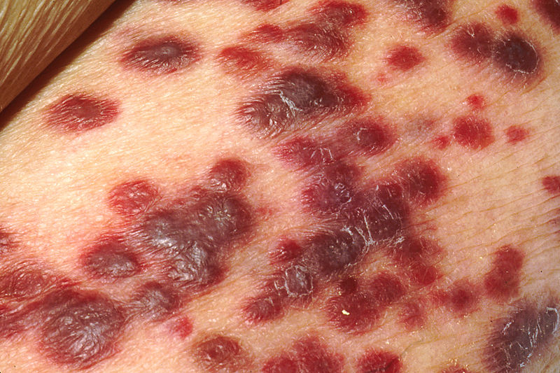
Kaposi's sarcoma is a red, brown, purple or blue plaque on the skin or mucosa


Kaposi's sarcoma is derived from capillary endothelial cells or fibrous tissue.


It is associated with HHV-8.


It may be secondary to immunosupression, or a diagnosing feature of HIV infection.


Next up - CML....

MRCP revision battle 28.6: Chronic Myeloid leukaemia

Chronic myeloid leukaemia is an uncontrolled clonal proliferation of myeloid cells.

It tends to affect middle aged people (40 to 60 years)


Classical presentation is with:
  • decreased weight
  • tiredness
  • sweating

Possible additional presentations are:
  • gout due to increased purine breakdown
  • abdo pain due to splenomegaly
  • bleeding due to platelet dysfunction

Approximately 30% are detected by chance.


Features include:
  • elevated WCC - often 100-500
  • massive neutrophilia with left shift
  • low neutrophil alkaline phosphatase
  • splenomegaly in >75%


The philadelphia chromosome is present in >80% of cases

The philadelphia chromosome:
  • is a hybrid chromosome formed by t(9;22)
  • forms a gene BCR-ABL which results in excess tyrosine kinase activity
  • found in 80% CML, 5% children with ALL, 25% of adult ALL and 1% adult AML.
  • is associated with a good prognosis in CML but a poor prognosis in all other leukaemias


Treatment for CML is:
  • hydroxyurea
  • imatimib = glivec = specific BCR-ABL tyrosine kinase inhibitor
  • bone marrow transplant


CML transforms to AML in 80% of cases and ALL in 20%


Last battle of the day is Gertmann's Syndrome...

MRCP revision battle 28.7: Gerstmann's Syndrome

Gerstmann's Syndrome is a condition characteristed by:
  1. dysgraphia
  2. dyscalculia
  3. finger agnosia
  4. left-right disorientation

Gerstmann's Syndrome is associated with lesions in the dominant brain hemisphere in the region of the angular gyrus of the parietal lobe.


On that esoteric note, see you tomorrow!

Friday, 8 October 2010

MRCP revision battle 27.1: Heparin induced thrombocytopenia (HIT)

Today we have an epic set of battles with some real hard-hitters hidden amongst them, including sarcoidosis which is a differential for around 110% of MRCP questions (or at least thats what it feels like)   But if you get to the last battle you will be rewarded with lots of pictures (the joys of poikilocytosis) and then a link to look at some interesting non-medical pictures...


So the carrot is dangled, and here are the sticks:

MRCP revision battle 27.1: Heparin induced thrombocytopenia (HIT)
MRCP revision battle 27.2: Sarcoidosis
MRCP revision battle 27.3: Lofgren's syndrome
MRCP revision battle 27.4: Pancreatitis
MRCP revision battle 27.5: Visual field defects
MRCP revision battle 27.6: Paroxysmal nocturnal haemoglobinuria
MRCP revision battle 27.7: Poikilocytosis


MRCP revision battle 27.1: Heparin induced thrombocytopenia (HIT)

Heparin induced thrombocytopenia is one of those fabulous conditions whose name explains it all: low platelets, caused by heparin.  It is often abbreviated to HIT, which confused me greatly when I first had a patient affected by it and my consultant declared "ah, he's got HIT" which made me wonder who he was suspecting of abuse on the ward...


Technically there are 2 types of HIT:
  • Type 1 - occurs in first 2 days, is non-immune and the platelet count spontaneously recovers 
  • Type 2 - occurs 4 to 10 days after starting heparin, is immune-mediated and potentially life-threatening.

However, when HIT is mentioned, people generally mean type 2.


HIT is caused by antibodies against the heparin-platelet factor 4 complex.  These antibodies can be found in 90% of patients with HIT... but may also be present in unaffected patients taking heparin.


Ironically, although HIT is characterised by low platelets it is an incredibly pro-thrombotic condition.  As a result patients are likely to get PEs/DVTs and warfarin must absolutely not be started (since warfarin is initially prothrombotic too).  As you can see HIT causes something of a management problem - thrombosis due to the treatment you would give for thrombosis...


Management of HIT will involve specialist haematological input but in terms of MRCP question answers think:
  • stop heparin
  • stop/reverse warfarin
  • give lepirudin (=highly specific direct inhibitor of thrombin)


Note that HIT is more likely with unfractionated than LMW heparin.


Onwards to sarcoidosis...

MRCP revision battle 27.2: Sarcoidosis

Sarcoidosis is a multi-system granulomatous disorder of unknown cause.


It is asymptomatic in up to 40% of cases, being discovered incidentally on a routine CXR.


Features of sarcoidosis may be divided into pulmonary and non-pulmonary:

Pulmonary features of sarcoidosis:
  • dry cough
  • progressive dyspnoea
  • chest pain

Non-pulmonary features of sarcoidosis include:
  • lymphadenopathy (commonest in non-whites)
  • erythema nodosum (commonest in white females)
  • polyarthralgia
  • hepatomegaly
  • splenomegaly (around 25%)
  • anterior uveitis (around 25%)
  • glaucoma
  • Bells palsy
  • lupus pernio
  • hypercalcaemia
  • renal stones
  • pituitary dysfunction
  • cardiomyopathy...


90% of patients with sarcoid will have an abnormal CXR.
Staging of CXR in sarcoid is:
  • 0 = clear CXR
  • 1 = bilateral hilar lymphadenopathy
  • 2 = bilateral hilar lymphadenopathy plus pulmonary infiltration
  • 3 = pulmonary infiltration only
  • 4 = fibrosis, honeycombing, pleural involvement


Investigations:
  • raised ESR
  • lymphopenia
  • raised LFTs
  • raised serum ACE
  • raised calcium (5%)
  • tuberculin skin test is positive in around a third

Management:
  • BHL alone doesn't need treatment
  • acute sarcoidosis : NSAIDs and bed rest
  • prednisolone ( 40mg OD for 4-6 weeks then reducing dose over one year) is indicated if:
    • parachymal lung disease
    • uveitis
    • hypercalcaemia
    • neurological/cardiac involvement

Lets move on to briefly consider lofgrens syndrome...

MRCP revision battle 27.3: Lofgren's syndrome

Lofgrens syndrome is a subtype of sarcoidosis, characterised by:
  • erythema nodosum
  • bilateral hilar lymphadenopathy
  • fever
  • athralgia

It affects females more than males (85:15)


It carries a good prognosis - unlike sarcoidosis is unlikely to become chronic and most cases resolve in 6 months to 2 years.


On to something which often doesn't have a good prognosis... pancreatitis...

MRCP revision battle 27.4: Pancreatitis

Acute pancreatitis tends to present as epigastric/central abdominal pain radiating to the back with vomiting.  The pain may be improved by sitting forward.

Signs may be few; periumbilical discoloration (Cullen's sign) or flank discoloration (Grey Turner's sign) are rare.


Causes of pancreatitis can be remembered by I GET SMASHED:
  • idiopathic
  • gallstones
  • ethanol
  • trauma
  • steroids
  • mump
  • autoimmune
  • scorpion venom
  • hyperlipidaemia, hypercalcaemia, hypothermia
  • ERCP
  • drugs (steroids, ocreotide, sulphonamides, tetracyclines, azathioprine, sodium valproate)

Investigations:
  • bloods including amylase 
    • note amylase normalises in 24-48hrs; serum lipase is more sensitive and specific
  • AXR: ?sentinel loop ?loss of psoas shadow
  • CT abdo is the investigation of choice
  • score using the Modified Glasgow criteria.

Modified Glasgow criteria (helpfully spells out 'pancreas'):
  • Pa O2 <8
  • Age >55
  • Neurophils: WCC >15
  • Calcium: <2
  • Renal function: urea >16
  • enzymes: LDH >600; AST >200
  • Albumin: <32
  • Sugars: glucose >10
3 or more suggests severe pancreatitis.


Management of acute pancreatitis is:
  • lots of IV fluid, catheter for fluid balance
  • analgesia
  • close observation
  • treat cause

Complications of pancreatitis include:
  • early
    • sepsis
    • shock
    • ARDS
    • renal failure
    • DIC
    • hypocalcaemia
    • hyperglycaemia - 5% need insulin
  • late
    • necrosis and pseudocyst (=fluid in lesser sac)
    • abscess
    • fistulae
    • chronic pancreatitis


Now for something completely different, visual field defects...

MRCP revision battle 27.5: Visual field defects

Visual fields pop up frequently in MRCP questions so it pays to learn this well!

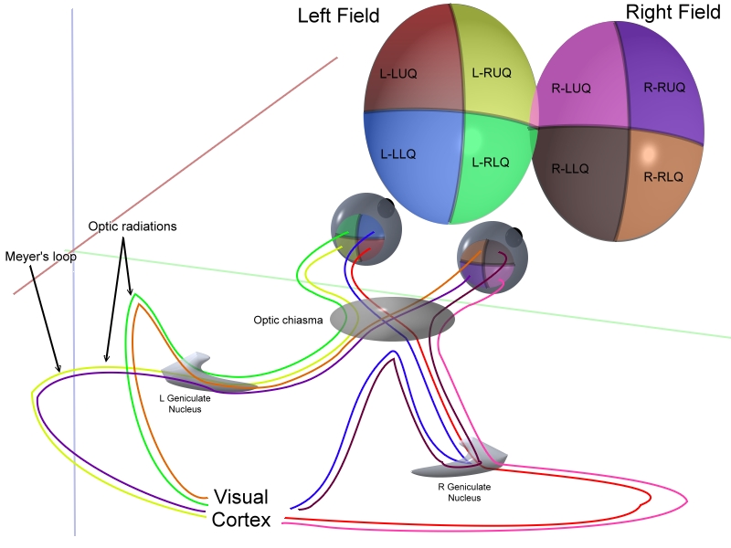
The basic anatomy is illustrated below:


Armed this this anatomy lets look at the various patterns of field defect...


1: Bitemporal hemianopia
  • caused by compression of the optic chiasm
  • upper affected more than lower = due to pituitary tumour
  • lower affected more than upper = due to craniopharyngioma
  • I personally remember this as UP  London City


2: Homonymous quadrantanopia

  • superior homonymous quadrantanopia is a lesion in the temporal lobe
  • inferior homonymous quadrantanopia is a lesion in the parietal lobe
  • this can be remembered by thinking PITS - parietal inferior temporal superior
  • if the defect is incongruous it is in the optic tract
  • if the defect is congruous it is in the optic radiation/cortex


3: Homonymous hemianopia
  • injury to the brain (eg bleed, tumour) on the opposite side to the field defect


4: Central scotoma

  • usually caused by optic neuritis


5: Binasal hemianopia
  • rare
  • ?calcification of carotids


Note that the fovea is supplied by both the PCA and MCA so may be spared in a PCA CVA.


In the calcarine sulcus peripheral regions are processed anteriorly while central regions are processed posteriorly.


Finally remember cortical blindness:
  • caused by bilateral occipital infarcts
  • pupillary responses are preserved
  • possibly small macular sparing
  • patient may deny they have vision loss (=Anton's syndrome)


Wow, that was lots for one battle... lets move on to some urine for light relief...

MRCP revision battle 27.6: Paroxysmal nocturnal haemoglobinuria

Paroxysmal nocturnal haemoglobinuria is a condition in which blood cells are sensitive to complement-mediated lysis due to an inherited lack of surface glucosylphosphatidylinositol (GPI)


Features of paroxysmal nocturnal haemoglobinuria include:
  • cola coloured/red urine in morning due to increase in RBC lysis overnight
  • intravascular haemolysis
  • pancytopenia
  • thrombosis


Diagnosis is by urinary haemosiderin or + Ham's test (rarely done now)


Treatment is anticoagulation, transfusions when needed and possibly the smart new monoclonal antibody called eculizimab.


Now on to the last battle and some pretty pictures...

MRCP revision battle 27.7: Poikilocytosis

Poikilocytosis = abnormality of red blood cell shape.

Here's a quick crash course of the various shapes and their causes.  Blood slides are all from wiki commons.


1. Teardrop cells - myelofibrosis


2. Helmet cells - microangiopathic haemolytic anaemias

click here to link to an image covered by copyright


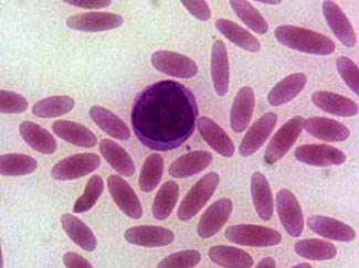
3. Elliptocytes = Pencil cells - seen in hereditary elliptocytosis and iron deficiency


4. Sickle cells - seen in sickle cell disease


5. Spherocytes - seen in any haemolysis and in hereditary spherocytosis


6. Target cells - seen in liver disease, post splenectomy, iron deficieny, thalassaemia, haemoglobinopathies


click here to link to an image covered by copyright


7. Acanthocyte/burr cell - acanthocytes are seen in liver disease.  Burr cells (=slightly less spikey than acanthocytes) appear in uraemia.


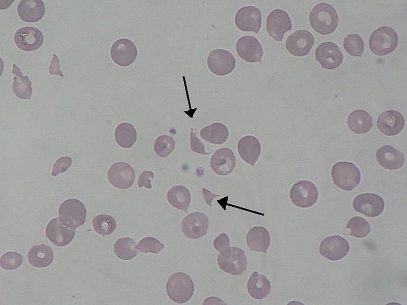
8. Schistocyte  = shredded red blood cells, seen in DIC, mechanical heart valves, microangiopathic diseases, haemolytic anaemias


Thats all the battles for today; as promised at the beginning  click here if you want to see some interesting pictures of what your workplace might look like if abandoned for a few years...

Thursday, 7 October 2010

MRCP revision battle 26.1: Parkinson's disease

I'm back on the revision bandwagon and am ready for another 7 battles...


MRCP revision battle 26.1: Parkinson's disease
MRCP revision battle 26.2: Lewy Body Dementia
MRCP revision battle 26.3: Malignant hyperthermia
MRCP revision battle 26.4: Benign intracranial hypertension
MRCP revision battle 26.5: Coarctation of the aorta
MRCP revision battle 26.6: Heparin
MRCP revision battle 26.7: Essential tremor


MRCP revision battle 26.1: Parkinson's disease/Parkinsonism


Parkinsonism is a triad of:
  • resting tremor (3 to 5 Hz)
  • rigidity
  • bradykinesia
 There is also loss of postural reflexes.


It is Parkinson's disease if there is degeneration of the substantia nigra dopaminergic neurones with Lewy bodies.


There are a whole list of associated features that I'm sure you're familiar with, but if you fancy a brief recap read the list below...
  • expressionless, mask-like face
  • dribbling
  • festinant gait = short shuffling steps with flexed trunk)
  • micrographia


Management of Parkinsons is by a multidisciplinary team and the pharmacological management of it is notoriously tricky, with multiple drugs which often decrease in efficacy with use and may result in 'on-off' symptoms.


Even the NICE guidelines recognise the challenge in drug prescription, stating that "it is not possible to identify a universal first-line drug choice".  They single out the following as possible first line options:
  • levodopa
    • use lowest dose possible
    • give with carbidopa to prevent peripheral breakdown
    • is of no use in neuroleptic induced Parkinsons
  • dopamine agonists
    • bromocriptine*, cabergoline*, ropinole
    • if an argot-derived agonist is used (*) ESR, UEs and CXR should be done before starting and annually while on treatment
  • MAO-B inhibitors
    • selegiline
    • decreases dopamine breakdown


Second line treatment is combining the above options, or one of the above with:
  • COMT inhibitors
    • entacapone
    • inhibit dopamine breakdown


Other medications which may be useful for specific effects include:
  • amantidine
  • apomorphine
  • antimuscarinics
    • procyclidine
    • more useful for drug-induced Parkinsons


A couple of other key points from the NICE guidelines on Parkinsons are:
  • 'drug holidays' should not be undertaken due to risk of neuroleptic malignant syndrome on restarting
  • clinicians should be aware of dopamine dysregulation syndrome, which is an uncommon disorder in which misuse of dopaminergic medication is associated with behaviours such as hypersexuality, pathological gambling and sterotypic motor acts


For the really keen:


For everyone else, lets move on to Lewy body dementia.

MRCP revision battle 26.2: Lewy Body Dementia

Lewy Body Dementia accounts for around 20% of cases of dementia.


The key feature is Lewy Bodies (=intracytoplasmic neuronal inclusion bodies) in substantia nigra, paralimbic and neocortical areas.


Since this would obviously only be confirmed post-mortem diagnosis is made clinically:
  • Parkinsonian features
  • fluctuating cognitive loss
  • visual hallucinations

Neuroleptics must be avoided as their use would risk inducing irreversible Parkinsonism.


Treatments which may help include donepezil and rivastigmine.


Next up - malignant hyperthermia

MRCP revision battle 26.3: Malignant hyperthermia

Malignant hyperthermia is a rare, life-threatening condition in which there is a huge increase in the body's skeletal muscle oxidative metabolism, resulting in:
  • hypercapnia
  • increased oxygen consumption
  • hyperthermia (T>38C)


Secondary problems which may develop from malignant hyperthermia include:
  • renal failure
  • arrhythmias
  • DIC


Malignat hyperthermia may be caused by:
  • any inhaled anaesthetic agent
  • Succinylcholine  


It is often inherited in an autosomal dominant fashion.


Treatment is with dantrolene.


Onwards to benign intracranial hypertension...

MRCP revision battle 26.4: Benign intracranial hypertension

Benign intracranial hypertension classically affects overweight young females.

It presents like there should be a mass in the brain, but none can be found.


Features include:
  • headache
  • blurred vision
  • dizziness
  • horizontal diplopia
  • papilloedema


Benign intracranial hypertension is associated with:
  • COC
  • steroids
  • tetracyclines
  • vitamin A
  • nitrofurantoin
  • isontertinoin
  • danazol

Management is:
  • weight loss
  • acetazolamide
  • loop diuretics
  • prednisolone
  • therapeutic lumbar puncture
  • shunt

Note that up to 10% of patients have permanent significant vision loss - so its name of 'benign' is a little misleading!  Optic nerve sheath fenestration can help prevent this.


Onwards for a spot of coarctation of the aorta...

MRCP revision battle 26.5: Coarctation of the aorta

Coarctation of the aorta (=narrowing of the aorta) classically occurs just distal to the left subclavian artery.

It may present at birth with heart failure, or be discovered later in life, eg during investigation for hypertension.


Most adults with coarctation of the aorta are asymptomatic.  Occasionally they may complain of:
  • headache
  • recurrent epistaxis
  • claudication of the calf muscles


Clinical examination may reveal:
  • radio-femoral delay
  • midsystolic murmur


Complications of coarctation include:
  • hypertension
  • LVF
  • endocarditis

Conditions associated with coarctation include:
  • bicuspid aortic valve (10-20%)
  • PDA
  • VSD
  • Berry aneurysms
  • Turners syndrome
  • renal abnormalities


CXR may show rib notching due to formation of collaterals


Management is stenting/surgery.


Now for the penultimate battle of the day, heparin...

MRCP revision battle 26.6: Heparin

Heparin comes in 2 'flavours':
  1. low molecular weight heparin (AKA dalteparin/fragmin, enoxaparin/clexane, tinzaparin) 
  2. unfractionated (AKA 'heparin')

So what are the differences?
  • molecular weight
    • LMW heparin, as the name suggests, has a lower molecular weight - 5000 compared to unfractionated heparin's 13000
  •  half life
    • LMW heparin has a longer half life than unfractionated heparin
  •  mechanism of action
    • LMW heparin inhibits factor Xa but has little effect on antithrombin
    • unfractionated heparin binds antithrombin
  • affect on aPTT
    • LMW heparin has little effect on aPTT
    • unfractionated heparin prolongs aPTT
  • side effects
    • both can cause osteoporosis and thrombocytopenia but the risks are greatest with unfractionated heparin
  • reversibility
    • unfractionated heparin can be fully reversed by protamine
    • LMW heparin cannot be fully reversed by protamine

A side effect of protamine to be aware of is hypotension.


HIT (=heparin induced thrombocytopenia) is a potentially serious side effect of heparin use which will be covered tomorrow.


With that to look forward to lets shake on to the last battle of the day, essential tremor...

MRCP revision battle 26.7: Essential tremor

Essential tremor can be sporadic or inherited; if inherited its inheritance pattern is believed to be autosomal dominant with incomplete penetrance.

35% of patients with essential tremor will have no family history.


It tends to be worse when the arms are outstretched.


Essential tremor improves with alcohol (not advised as a treatment option!)


Treatment tends to be:
  • propranolol
  • primidone if propranolol contra-indicated (eg in asthma)


Congratulations on surviving another day's battles!

Wednesday, 6 October 2010

MRCP revision battle 25.1: Neuroleptic Malignant Syndrome

Having been blessed by a set of on-calls with some nice interesting cases I'm full of enthusiasm for today's battles, so lets get on with it!


MRCP revision battle 25.1: Neuroleptic malignant syndrome
MRCP revision battle 25.2: Behcets disease
MRCP revision battle 25.3: Subarachnoid haemorrhage
MRCP revision battle 25.4: Normal pressure hydrocephalus
MRCP revision battle 25.5: Gas gangrene
MRCP revision battle 25.6: Methaemoglobinaemia
MRCP revision battle 25.7: The weeverfish


MRCP revision battle 25.1: Neuroleptic malignant syndrome


A nice straightfoward battle to begin with...


Neuroleptic malignant syndrome is a life-threatening condition associated with use of antipsychotic drugs.


Symptoms and signs include:
  • hyperthermia
  • rigidity/muscle cramps
  • extrapyramidal signs
  • autonomic dysfunction (labile BP, sweating, urinary incontinence
  • confusion

Investigations show:
  • raised CK
  • raised WCC

Treatment is:
  • cooling
  • dantrolene (a muscle relaxant; note dantrolene is also used in malignant hyperthermia, which will be one of tomorrows battles)


Now on to a slightly less straightforward battle, Behcets disease...

MRCP revision battle 25.2: Behcets disease

Behcet's disease is a systemic vasculitis of unknown cause.


The classic triad of Behcet's disease is:
  • oral ulcers
  • genital ulcers
  • anterior uveitus
which I remember as GOA, like the place in India.


Other possible features include:
  • thrombophlebitis
  • DVT
  • erythema nodosum
  • pathergy reaction (=needle prick leads to pustule formation)
  • arthritis
  • aseptic meningitis
  • ataxia
  • diarrhoea


Males are more commonly affected than females.
It is commonest in Turkey and Japan.
30% of patients have a positive family history.


HLA B5 is associated with anterior uveitis and HLA B12 is associated with recurrent ulceration.


Severe disease requires steroids/ciclosporin/azathioprine.


Next up - SAH...

MRCP revision battle 25.3: Subarachnoid haemorrhage

Subarachnoid haemorrhage is bleeding into the space between the subarachnoid membrane and the pia mater.

It accounts for 5-10% of all strokes.


Causes:
  • ruptured berry aneurysm (80%)
  • malformations (15%)
  • post trauma


Common sites of berry aneurysms are:
  • junction of the posterior communicating artery with the internal carotid
  • junction of the anterior communicating artery with the anterior cerebral artery
  • the bifurcation of the middle cerebral artery

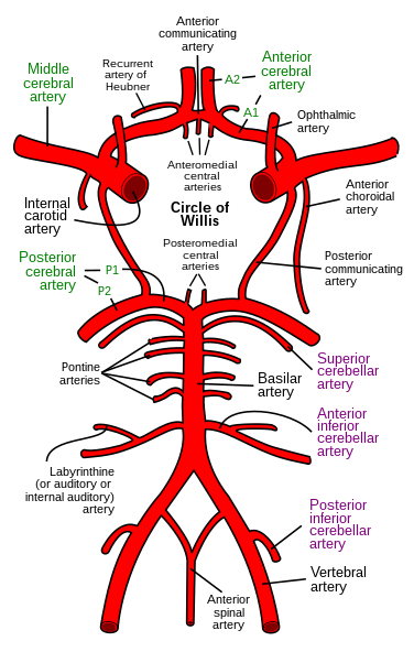
If (like me) your anatomy of the circle of willis is a little rusty the wiki commons image below might help refresh your memory:


15% of berry aneurysms are multiple.


Subarachnoid haemorrhages are associated with:
  • polycystic kidneys
  • coarctation of the aorta
  • Ehlers-Danlos syndrome
  • PAN


Classical presentation is a thunderclap occipital headache "as if kicked in head" with vomiting and neck stiffness.
Around 6% of patients will have had a sentinel headache before.


Investigation is:
  • CT - >90% of bleeds detected
  • LP - done >12hrs after onset looking for xanthochromia


Management:
  • neurosurgical referral
  • prompt angiography if surgery likely
  • nimodipine


On to another neuro topic - normal pressure hydrocephalus

MRCP revision battle 25.4: Normal pressure hydrocephalus

Normal pressure hydrocephalus is a triad of:
  1. dementia
  2. gait abnormality
  3. urinary incontinence
(remember as DUG!)


It is caused by a defect in the absorption of CSF.
Causes include: meningitis, head injury, subarachnoid haemorrhage.


Note that there is NO headache and NO papilloedema.


Investigations:
  • CT head (predictably) - enlarged ventricles
  • large volume lumbar puncture - ? high opening pressure, ? improvement of symptoms
  • lumbar infusion test

Treatment is a shunt.


Next up... gas gangrene!

MRCP revision battle 25.5: Gas gangrene

Gas gangrene is a life threatening bacterial infection.

It is characterised by muscle necrosis, gas production and sepsis.

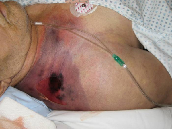
A typical appearance is shown below:

                                       From Wiki Commons, taken by Engelbert Schröpfer, Stephan Rauthe and Thomas Meyer.


Most cases are caused by trauma innoculation with bacteria, for example a farmer standing on a pitchfork.
Very rarely it may occur spontaneously, for example in the context of neutropenia.


The commonest causative organism is clostridium perfringens.


Management is:
  • urgent surgical debridement
  • IV Abx 
  • hyperbaric oxygen if available


Next up - methaemoglobinaemia

MRCP revision battle 25.6: Methaemoglobinaemia

Methaemoglobin (MetHb) is formed when the iron in haemoglobin is oxidised from the ferrous state (Fe2+) to the ferric state (Fe3+) 


Methaemoglobinaemia means raised MetHb levels, and as MetHb has a far higher affinity for oxygen than normal haemoglobin this results in tissue hypoxia as the oxygen being carried in the blood is not given up to the tissues.


Clinical features depend on the % of MetHb:
  • 15-20% - cyanosis
  • 20-45% - impaired consciousness
  • >55% - seizures, arrhythmias, coma
  • >70% - lethal


Pulse oximetry will show low oxygen sats, as will the printout from blood gas analysis - but the PaO2 will be high.


Causes of methaemoglobinaemia:
  • congenital
    • pyruvate kinase deficiency
    • G6PD deficiency
    • abnormal haemoglobin - HbH or HbM
    • diaphorase 1 deficiency
  • acquired - accelerated rate of oxidised formation due to meds
    • antibiotics - sulphonamides, trimethoprim, dapsone
    • drugs used in anaesthetics - lidocaine, procaine
    • others -primaquine


Management:
  • 1% solution of methylene blue
  • hyperbaric oxygen
  • exchange transfusion
  • dialysis
  • asorbic acid in patients with G6PD deficiency

So to the final battle of the day - weeverfish!

MRCP revision battle 25.7: The weeverfish

Just very briefly...

The weeverfish (AKA trachinus vipera) is found in shallow waters around the coast of the UK.

It is small and sandy-coloured, with sharp dorsal spines.


If you are unfortunate enough to stand on it you will get intense pain.

Happily the toxin produced by the weeverfish is denatured above 40C - so dunk the foot in hot water and the pain should stop.


On that most random of MRCP revision notes, lets end for the day!

Sunday, 3 October 2010

MRCP questions: War 24

 I now have a co-conspirator for MRCP revision on the go... lets call her Dr Biscuit and she is now writing all the wars!  How hard they are will probably reflect how bad her day has been... good luck!


As with previous 'wars' after 'battles' these are just a few quick questions to see if your brain cells have retained the information provided in battles 24:1 to 24:6.

The answers are available here


Question 1:
You are referred a patient who was diagnosed with Primary Biliary Cirrhosis 2 years ago. Name 3 signs you might find on clinical examination?

Question 2:
What blood test would you do to confirm the diagnosis of PBC?


Question 3:
A 22 year old female patient presents with DIC and hepatosplenomegaly.  Blood film shows cells which are strongly sudan black/peroxidase positive. What is the diagnosis and what is the gene translocation?


Question 4:
Name 3 drugs which can be used to treat AML?


Question 5:
Name 3 conditions associated with angioid retinal streaks?


Question 6:
List 3 drugs/groups of drugs which can cause SIADH?


Question 7:
Your patient is hyponatraemic with a sodium of 120.  On examination they appear clinically dehydrated. What investigation would you request next to help you elucidate the cause of the hyponatraemia?


Question 8:
Your patient is hyponatraemic with a sodium of 118.  Name four signs or symptoms they might display?


Question 9:
A patient is admitted with confusion. You are unable to obtain a history from her family but she takes no medications.  Her sodium is 119.  On examination she appears euvolaemic.  The admitting doctor has sent a urine sample which shows a urinary na of 34, and urine osmolality of 750. What is the likely diagnosis?


Question 10:
What are the 2 main effects of ADH?


The answers are available here

Friday, 1 October 2010

MRCP revision battle 24.1: Primary biliary cirrhosis

All this working and revising is leaving me with little internet-publishable material to put into my daily intros.  Looking for inspiration I ended up stumbling accross an utterly brilliant set of articles written back in the early 2000's by a (then) junior doctor called Michael Foxton in the Guardian.  Hopefully this link will take you to a list of his articles to soothe you after some revision.... but I warn you not to start looking before you've finished revising for the day because otherwise there is a real danger you will get no revision done!!


So today's battles are:

MRCP revision battle 24.1: Primary biliary cirrhosis
MRCP revision battle 24.2: Acute Myeloid Leukaemia
MRCP revision battle 24.3: Acute Promyelocytic Leukaemia
MRCP revision battle 24.4: Angioid retinal streaks
MRCP revision battle 24.5: SIADH
MRCP revision battle 24.6: Hyponatraemia
MRCP revision battle 24.7: Neutropenia and neutrophillia


MRCP revision battle 24.1: Primary biliary cirrhosis


I've never bonded well with this topic so this battle is a personal Waterloo for me...


Primary biliary cirrhosis is thought to be an autoimmune condition in which the interlobular bile ducts are damanged by chronic granulomatous inflammation. 


The result of this is:
  • progressive cholestasis
  • cirrhosis
  • portal hypertension


Females are more affected than males (9:1) and it tends to present in middle age.

It is associated with many other autoimmune conditions, including sjogrens, RA, systemic sclerosis, thyroid disease...


Primary biliary cirrhosis is often found incidentally by a raised alk phos on LFTs.Initially other LFTs may be normal but as the disease progresses bilirubin rises and PT may increase.


Associated signs (in late disease) include:
  • jaundice
  • xanthelasma
  • hepatosplenomegaly
  • clubbing


Complications include:
  • osteoporosis
  • portal hypertension
  • variceal haemorrhage


Diagnosis includes antibodies:
  • AMA M2 is highly specific and is positive in 98% of cases
  • SMA is positive in 30%
  • raised IgM


Treatment is:
  • cholestyramine for itching
  • osteoporosis prophylaxis
  • transplant


Once jaundice develops, without transplant the survival is < 2yrs.


On to AML...