Wednesday, 15 September 2010

MRCP revision battle 12.2: CLL

I keep clerking patients who say they've got 'CLL, doctor' and I nod sagely and scribble it down, while desperately trying to recall any of my med school haematology lectures.  My brief summary for MRCP means that now hopefully my sage nod is slightly more appropriate!!


CLL (=chronic lymphocytci leukaemia) is a monoclonal proliferation of mature lymphocytes.

99% of cases of CLL involve B cells.

CLL is important clinically as it is the commonest cause of lymphocytosis in the elderly.
Males are more often affected than females, 2:1


Presentation:
  • no symptoms in 25%
  • recurrent infections (due to hypogammaglobulinaemia)
  • anaemia (warm AIHA in 10-15%)
  • decreased weight
  • sweats
  • anorexia


Signs includer:
  • enlarged rubbery, non-tender nodes
  • hepatomegaly
  • splenomegaly


Blood tests will show:
  • raised lymphocytes
  • low Hb, neutrophils and platelets
  • smear/smudge cells on blood film


Staging is done by the Rai classification, which can also be used to give the prognosis:
  • Stage 0 - lymphocytosis alone - mean survival >13 yrs
  • Stage 1 - lymphocytosis plus lymphadenopathy - mean survival 8 yrs
  • Stage 2 - lymphocytosis plus spleno/hepato megaly - mean survival 5 yrs
  • Stage 3 - lymphocytosis plus anaemia (defined as Hb <11) - mean survival 2 yrs
  • Stage 4 - lymphocytosis plus platelets <100 - mean survival 1 yr


Indicators of poorer prognosis include:
  • being male
  • age >70
  • lymphocytes >50
  • prolymphocytes >10%
  • doubling rate <12 months
  • raised LDH
  • CD 38 positive
  • ZAP 70 + (if ZAP 70 -ive, mean survival 25 years)


Management of CLL is:
  1. monitoring only if asymptomatic
  2. chlorambucil to decrease lymphocyte count, improve marrow function and decrease node size
  3. fludarabine (a chemo agent, purine analog that works by inhibiting DNA synthesis) if:
    • bulky disease
    • cytopenias due to marrow failure
    • short lymphocyte doubling time


CLL may transform to Richters syndrome.
Richters syndrome is the transformation of CLL to agressive B cell lymphoma.  LDH is raised.


Now onwards to a subtype of CLL, hairy-cell leukaemia.

MRCP revision battle 12.3: Hairy Cell Leukaemia

Hairy cell leukaemia is a subtype of CLL characterised by (suprise, suprise) cells that appear 'hairy' on microscopy.


Hairy cell leukaemia is 4 times more common in men than women.


Features of hairy cell leukaemia include:
  • pancytopenia
  • splenomegaly
  • skin vasculitis in 1/3 cases
  • 'dry tap' 


A key phrase to look out for in MRCP is 'tartrate resistant acid phosphatase positive'


Treatment of hairy cell leukaemia is chemotherapy.  Second line options include alpha interferon and rituximub.


Thats quite enough haematology for one day... lets move on for a 'rippingly' good time with aortic dissection...

MRCP revision battle 12.4: Aortic dissection

Aortic dissection is a scary condition with a mortality rate of 1% per hour.  But before we jump into it, lets begin with a quick recap of some basic anatomy.

The layers of the aorta are, from inside out:
  • tunica intima = epithelial cells
  • tunica media = smooth muscle and elastic fibres
  • tunica adventitia = connective tissue

Aortic dissection usually involves blood collecting in the media.


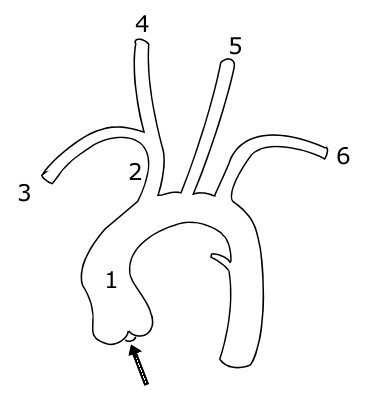
The aorta itself leaves the heart, goes up into an arch and then down again, giving off branches as it goes.  The picture below shows the vessels coming off the aorta:

1 indicates the ascending aorta, 2 the brachiocephalic trunk, 3 the right subclavian artery, 4 the right common carotid, 4 the right common carotid, 5 the left common carotid and 6 the left subclavian artery.

A knowledge of this anatomy aides understanding of the signs and symptoms of aortic dissection.
So what are the features of an aortic dissection?
  • sudden onset of 'tearing' or 'ripping' pain
  • if pain intrascapular, likely a descending dissection
  • MI
  • stroke
  • parathesia of arms
  • hemiparesis
  • many others, depending on the part of the aorta affected

Signs to look out for include:
  • aortic regurgitation - present in 1/3 ascending dissections
  • pulse deficit in an arm - present in 15%
  • BP difference in arms >20mmHg (but note 'normal' individuals may have a BP difference)


Aortic dissections may be classified using either the Stanford or DeBakey systems:
  • Stanford A = ascending dissection - accounts for 2/3 of dissections
    • this covers DeBakey I - ascending and descending aorta involved and
    • DeBakey II - ascending aorta only involved
  • Stanford B = descending aorta only dissected (distal to left subclavian artery) - accounts for 1/3 of dissections
    • = DeBakey III


 Management of aortic dissection:
  • if type A - IV labetalol and urgent surgery
  • if type B - IV labetalol only


Complications associated with type A dissections include:
  • aortic regurgitation
  • inferior MI
  • pericardial effusion
  • carotid dissection
  • loss of pulses


Investigations for dissection:
  • CXR 
    • shows widened mediastinum in 70% of cases
    •  Ring sign - calcification of aorta with wall displaced >5mm
  • CT
  • TOE

Risk factors for dissection include:
  • hypertension (80%)
  • Marfans
  • Noonans
  • Turners
  • trauma
  • pregnancy
  • coarctation
  • congenital bicuspid valve
  • giant cell artertitis
  • cocaine use

If you feel motivated to read more, I'd recommend this webpage: http://www.aorticdissection.com/Aortic%20Diagnosis.htm


A 'war' for yesterday's questions is here


MRCP questions: War 11

As with previous 'wars' after 'battles' these are just a few quick questions to see if your brain cells have retained the information provided in battles 10.1 to 10.6.

Grab a piece of paper, jot down your answers then compare them to my answers here

Question 1
List 4 factors that lead to a worse prognosis in GBS.


Question 2
What percentage of patients with GBS will make a complete or near complete recovery?


Question 3
What regular investigation do patients with GBS require?


Question 4
What is the classic triad of Miller-Fisher syndrome?


Question 5
What antibodies are usually associated with Miller-Fisher syndrome?


Question 6
What is syringomyelia?


Question 7
What are the cardinal symptoms of syrinomyelia?


Question 8
What is the inheritance of Kallmans syndrome?


Question 9
List 5 symptoms/signs of carcinoid syndrome


Question 10
What is the treatment of carcinoid syndrome?


answers here

Tuesday, 14 September 2010

MRCP revision battle 11.1: Guillain Barre Syndrome

When you're working 12 hr days (plus commute) fitting in revision can feel a bit like trying to squeeze that extra towel into the suitcase - you may manage it, but the suitcase is then stretched and prone to snap open at unfortunate times.  If you extend this metaphor to the suitcase being your sanity it explains why junior doctors can often become more emotionally incontinent/irrational as MRCP exams get nearer.  So whats the solution?  Well, in the suitcase scenario you'd add some of those rubber bands round to keep it closed.  In real life, your friends, family and managing to keep some form of hobby/interest going are your rubber bands.

Since I don't want to overpressure my 'rubber bands' I'm going to limit today's battles to

MRCP revision battle 11.1: Guillain Barre Syndrome
MRCP revision battle 11.2: Miller Fisher Syndrome
MRCP revision battle 11.3: Syringomyelia
MRCP revision battle 11.4: Kallmans
MRCP revision battle 11.5: Carcinoid Syndrome


MRCP revision battle 11.1: Guillain Barre Syndrome


Guillain Barre Syndrome is a rare, post-infective demyelinating polyneuropathy.


It tends to occur a few weeks after 'the flu' or a minor illness.  The most commonly associated pathogens are:
  • campylobacter jejuni
  • mycoplasma
  • CMV
  • EBV
  • HIV
  • zoster

Guillain Barre Syndrome is characterised by an ascending, symmetric muscle weakness.  Proximal muscles are more affected than distal ones.  Cranial nerves may also be involved, with CN VII being the most common.


There is usually a 'progressive' phrase for around 4 weeks, after which the patient starts to improve.


Features associated with GBS that appear in MRCP questions include:
  • papilloedema
  • urinary retention
  • arrhythmias

Diagnostically GBS is characterised by:
  • very high protein in CSF
  • decreased nerve velocity on EMG
  • areflexia

The prognosis for patients with GBS is worse if:
  • rapid onset of symptoms
  • older age
  • axomal neuropathoes
  • preceeded by campylobacter infection

Management is:
  • 4 hourly FVC, to check the patient's respiratory muscles aren't affected
  • ventilation if needed
  • plasma exchange
  • IV IG

Overall, 85% of patients make a complete/new complete recovery.
10% will relapse
Less than 5% will die.


Now to to meet a close relative of GBS, Miller Fisher Syndrome.

MRCP revision battle 11.2: Miller Fisher Syndrome

Miller Fisher Syndrome is essentially a rarer variant of Guillain Barre Syndrome.


It's classical triad is:
  • areflexia
  • ataxia
  • opthalmoplegia

Anti-ganglioside antibodies, especially to GQ1b, will be positive in 90% of cases.


On to syringomyelia...

MRCP revision battle 11.3: Syringomyelia and Syringobulbia

Syringomyelia and syringobulbia have always confused me, partly because for some reason I even have problems pronouncing their names!  However, in terms of MRCP there isn't too much you need to know...


Syringomyelia is a cyst/cavity forming in or close to the central canal of the spinal cord.


The cause may be anything that is hindering CSF circulation, but the classical cause is an Arnold Chiari malformation (=condition where the cerebellum herniates through the foramen magnum.)


The essential features are:
  • wasting and weakness of muscles of the hands (possibly 'claw hand')
  • loss of pain and temperature sensation in hands

Other possible signs/symptoms are varied and include Horner's syndrome, eye movement abnormalities and UMN signs in the legs.


Treatment is surgery.


If the cavity/cyst extends from the cord into the brain stem it is known as syringobulbia and causes bulbar palsy type symptoms, facial pain and dizziness.


After that neurology binge onto a slightly lighter topic, Kallmans...

MRCP revision battle 11.4: Kallmans

Kallmans syndrome is an x-linked recessive condition caused by failure of GnRH secreting neurones to migrate to the hypothalamus.


Its features are:
  • anosia
  • 'delayed puberty'
  • hypogonadism, crytoorchidism
  • may be tall or of normal height

Investigations: blood LH/FSH/testosterone and all either normal or low


The most important complication to remember for MRCP is the increased risk of osteoporosis

Next up: Carcinoid syndrome

MRCP revision battle 11.5: Carcinoid Syndrome

Carcinoid tumours are common - 1% of the population will have one at post mortem.

They primarily secrete serotonin (5HT) but may also produce histamine/adrenal hormones/insulin...


45% of carcinoid tumours are in the appendix and a further 30% in the ileum.
They can cause complications such as appendicitis, obstruction or intussception.


Having said carcinoid tumours are common, it's important to understand that carcinoid syndrome is rare - it only occurs when the liver is involved, as tumours in other places will have their 5HT metabolised rather than it have an effect.


Features of carcinoid syndrome are:
  • flushing
  • diarrhoea
  • bronchospasm
  • pellegra
  • hypotension

Diagnosis is by:
  1. urinary 5-HIAA
  2. plasma chromograffin - the new way which, depending on the leve,  also gives an indication of the mass of tumours

Treatment is:
  • surgery
  • ocreotide

Survival from diagnosis tends to be 5 to 8 years.
Patients with carcinoid heart disease have a worse prognosis.


It is also worth remembering that if these tumours are over-handed (eg during surgery) or out-grow their blood supply, a massive 'hormone hit' can be released in one go, causing a carcinoid crisis.


Features of a carcinoid crisis:
  • vasodilation
  • hypotension
  • tachycardia 
  • bronchoconstriction
  • hyperglycaemia 


So those are today's battles fought, and hopefully won.  Click here if you want to partake of the 'war' of questions to check your recall of yesterday's topics.

    MRCP questions: War 10

    As with previous 'wars' after 'battles' these are just a few quick questions to see if your brain cells have retained the information provided in battles 9.1 to 9.6.


    Grab a piece of paper, jot down your answers then compare them to my answers here

    Question 1:
    What are the diagnostic criteria for hereditary haemorrhagic telangiectasia?


    Question 2:
    List 3 possible causes of increased shortness of breath on sitting up compared to lying down.


    Question 3:
    Which form of MND affects only LMN?


    Question 4:
    Name the only drug treatment available for MND


    Question 5:
    List 5 causes of bulbar palsy


    Question 6:
    What condition is associated with a 'Donald Duck' voice?


    Question 7:
    Which genetic condition causes an increase in uric acid levels?


    Question 8:
    List 5 causes of low voltage ECG complex


    Question 9:
    What causes electrical alternans on ECG?


    Question 10:
    How is multifocal motor neuropathy treated?


    answers here

    Monday, 13 September 2010

    MRCP revision battle 10.1: Hereditary Haemorrhagic Telangiectasia

    Once again I can see a sunny day... through the window my desk sits in front of.  Although today I've found the sun a catalyst for working rather than a mocking presence as my lovely other half has promised me a picnic later, so the sooner I finish the sooner I get jam sandwiches!

    Today's topics are:

    MRCP revision battle 10.1: Hereditary haemorrhagic telangiectasia
    MRCP revision battle 10.2: Motor Neurone Disease
    MRCP revision battle 10.3: Bulbar Palsy
    MRCP revision battle 10.4: Pseudobulbar palsy
    MRCP revision battle 10.5: Multifocal Motor Neuropathy
    MRCP revision battle 10.6: Lesch-Nyhan Syndrome
    MRCP revision battle 10.7: Low voltage ECG complexes

    I promise that's less than it looks...


    MRCP revision battle 10.1: Hereditary haemorrhagic telangiectasia


    Hereditary haemorrhagic telangiectasia (HHT), also known as Osler-Weber-Rendu syndrome, is an autosomal dominant condition associated with abnormal blood vessel formation.


    Features of HHT include:
    • telangiectasia on skin
    • telangiectasia on mucosal membranes (leading to nose bleeds/GI bleeds)
    • arterio-venous malformations (AVMs)
      • commonest in lung
      • also in liver
      • brain
      • spine (rarest)

    The diagnostic criteria are:
    1. spontaneous and recurrent nosebleeds
    2. telangiectasia
    3. AVMs
    4. positive family history - a first degree relative
    The international consensus  states if 3 or 4 of the above features are present HHT is definite.  If 2 are present it is possible.  Less than that is unlikely.


    A rare symptom the MRCP exam may throw up is platypnoea, which is difficulty breathing when sitting up/standing which is relieved on lying down.  Platypnoea is therefore the direct opposite of orthopnoea!  To briefly diversify, apart from lung AVM two other possible causes of platynoea are left atrial tumour or left atrial thrombus.


    A rare sign associated with lung AVM is a 'humming sound' on auscultation over that area of the chest.


    Management of HHT is symptomatic.


    After that forray into the world of bleeding bits we're about to dive into the murky world of neurology with motor neurone disease...

    MRCP revision battle 10.2: Motor Neurone Disease

    Most people associated motor neurone disease (MND) with Stephen Hawking, that incredible physicist who has lived with MND for over 40 years.  However, in the same way he is not a stereotype for normal intelligence he is also not a stereotype for MND - most people with it die within 5 years.  More stereotypical for the course of the disease was Lou Gehrig, an American baseball player in the 1920s/1930s who was diagnosed with the disease and died within 3 years.  I mention him not just for random interest but also because in America MND is sometimes known as Lou Gehrig disease.  But what is MND?


    MND is a group of neurological disorders affecting neurones in motor cortex, CN nuclei and anterior horn cells.


    MND affects UMN and LMN but there is no sensory involvement.


    As a quick recap, UMN signs include:
    • weakness
    • spasticity
    • brisk reflexes
    • upgoing plantars

    LMN signs include:
    • weakness
    • wasting
    • fasciculation
    • depressed reflexes

    There are several patterns of MND, including:
    1. Amyotrophic lateral sclerosis
      • presents as UMN signs in legs and LMN signs in arms
      • may be familial - in which case often associated with Ch 21
      • accounts for 50% of cases of MND
      • has an 'intermediate' prognosis
    2. Bulbar palsy
      • accounts for around 25% of cases of MND
      • has the worst prognosis of the presentations
      • more details in the next battle
    3. Progressive muscular atrophy
      • only LMN affected
      • begins distally
      • has a (relatively) good prognosis
    4. Primary lateral sclerosis
      • affects UMN only
    10-35% of patients with MND will have fronto-temporal dementia.


    Diagnosis of MND is primarily clinical.
    EMG will show preserved motor conduction velocities
    CSF analysis may show raised protein.


    Treatment is with riluzole, which prolongs life by around 3 months and requires monitoring of LFTs.  There is no cure.

    As the first paragraph stated, the prognosis is very poor and most patients are dead within 5 years.


    So on to battle 10.3, to learn more about bulbar palsy.

    MRCP revision battle 10.3: Bulbar Palsy

    Bulbar palsy refers to palsy of the nerves coming from the 'bulbar' part of the brain, ie the pons, medulla and cerebellum.  These are VII, VIII, IX, X, XI and XII.


    Classically bulbar palsy presents as palsy of tongue, muscles of chewing and swallowing and facial muscles.


    Signs are LMN:
    • flaccid tongue
    • fasciculating tongue
    • absent or normal jaw jerk
    • loss of gag reflex
    • quiet/horse voice

    Causes of bulbar palsy include:
    • MND
    • syringobulbia (more about this condition tomorrow!)
    • GBS (also lined up for battle tomorrow)
    • polio
    • Lyme disease
    • brain-stem tumours

    Next up: battle 10.4, pseudobulbar palsy

    MRCP revision battle 10.4: Pseudobulbar palsy

    Pseudobulbar palsy is a UMN palsy of the muscles affecting eating, swallowing and talking above the midpons.


    Signs are:
    • spastic tongue
    • brisk jaw jerk
    • emotional incontinence
    • 'Donald Duck' voice.

    Causes include:
    • MS
    • MND
    • PD
    • stroke
    • high brain stem tumour


    Just one more skirmish with neurology to go today, in the form of multifocal motor neuropathy.

    MRCP revision battle 10.5: Multifocal motor neuropathy

    I must confess I'd never come accross multifocal motor neuropathy until I started doing MRCP questions, but I'm now rather glad that I have, as essentially it is a treatable differential diagnosis for some presentations that appear like MND (which, as we've just covered, is nasty and deadly)


    Multifocal motor neuropathy is an acquired immune-mediated demyelinating neuropathy.


    Features of multifocal motor neuropathy include:
    • slowly progressive, asymmetric distal muscle weakness
    • cramps and twitching
    • no sensory loss
    • no/minimal muscle atrophy
    • possibly positive anti GM1 antibodies (NB also + in GBS)
    • no UMN signs
    • normal or decrease tone
    • absent reflexes

    The joy of multifocal motor neuropathy is that it can be successfully treated with IV IG!


    Well thats quite enough neurology for today, lets now look briefly at Lesch-Nyhan Syndrome, as promised yesterday

    MRCP revision battle 10.6: Lesch-Nyhan Syndrome

    Lesch-Nyhan Syndrome is a rare inherited x-linked disorder caused by an abnormality of an enzyme involved in purine metabolism resulting in increased uric acid.  Only boys tend to be affected.


    Features of Lesch-Nyhan Syndrome include:
    • orange crystals in nappies
    • low IQ
    • self-mutilation
    • fits
    • recurrent gout (probably the most important relationship to remember in MRCP)
    • megaloblastic anaemia

    Patients usually die by the age of 25, often of renal failure.


    So to the final battle of the day, a nice gentle ending with a breeze through the causes of low-voltage complexes on ECG

    MRCP revision battle 10.7: Causes of low-voltage complexes on ECG

    'Low voltage complex' on ECG is defined as:
    • QRS amplitude <5mm in all limb leads and/or
    • QRS amplitude <10mm in all chest leads


    The causes of low voltage complexes can be split into 2 main groups: increased distance from leads to heart or infiltrative disease of heart.

    Using these divisions:
    1. increased distance from leads to heart:
      • emphysema/COPD
      • pericardial effusion
      • severe obesity
      • pleural effusion
    2. infiltrative disease of heart/problems with heart itself: 
      • amyloid
      • haemochromatosis
      • cardiomyopathies
      • global ischaemia

    Myxoedema can also cause low complex ECGs but my search for why this is (?caused by myxoedema or ?caused by effects of myxoedema) proved fruitless... please let me know if you have the answer!


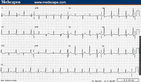
    If you see an ECG with alternating normal complexes and low-voltage complexes, you are looking at electrical alternans which tends to be a sign of pericardial effusion/cardiac tamponade.  Click here to see an image of it.


    So today's battles are over.  If you want to undertake the war to see how much of yesterday's battles you recall, please click here 

    MRCP questions: War 9

    As with previous 'wars' after 'battles' these are just a few quick questions to see if your brain cells have retained the information provided in battles 9.1 to 9.5.


    Grab a piece of paper, jot down your answers then compare them to my answers here


    Question 1:
    Name the classical rash associated with Lyme disease


    Question 2:
    What is the treatment for uncomplicated Lyme disease?


    Question 3:
    A joint aspiration shows negatively birefrigent crystals.  What condition does this suggest?


    Question 4:
    What XR appearance may you see in gout?


    Question 5:
    Which joint is classically affected by pseudogout?


    Question 6:
    What is a craniopharyngioma?


    Question 7:
    What is a catamenial pneumothorax?

    .


    Question 8:
    What is Hammans sign?  What does it signify?


    Question 9:
    A 19 yr old boy presents with shortness of breath.  A CXR shows a 3cm rim of air around his left lung.  How would you manage him?


    Question 10:
    You have a patient with a painful joint and the XR shows chondocalcinosis.  What condition does this suggest?


    answers here

    Sunday, 12 September 2010

    MRCP revision battle 9.1: Lyme Disease

    Working and revising really does feel rather relentless.  I keep thinking of the pop song "and the beat goes on..." - somehow it seems appropriate for the continuous dedication work plus MRCP revision requires!  Anyway, what revision battles does today bring?

    MRCP revision battle 9.1: Lyme disease
    MRCP revision battle 9.2: Gout
    MRCP revision battle 9.3: Pseudogout
    MRCP revision battle 9.4: Craniopharyngioma
    MRCP revision battle9.5: Pneumothorax


    MRCP revision battle 9.1: Lyme disease

    Lyme disease, named after the place in America where the condition was first noted, is a condition caused by the spirochaete borrelia burdorferi.  


    It is spread by tic bites, with certain areas (such as northern America) being higher risk than, for example, Surrey.  However, it is found in the UK and it is important to note the majority of patients will not remember the bite itself.


    Signs, symptoms and complications associated with Lyme disease are multitude and vague, including:
    • malaise
    • fever
    • muscle pain
    • joint swelling
    • decreased cognition
    • encephalitis
    • meningitis
    • lymphadenopathy
    • cranial nerve palsies (MRCP likes bilateral facial nerve palsy for some reason)
    • neuropathy
    • skin conditions


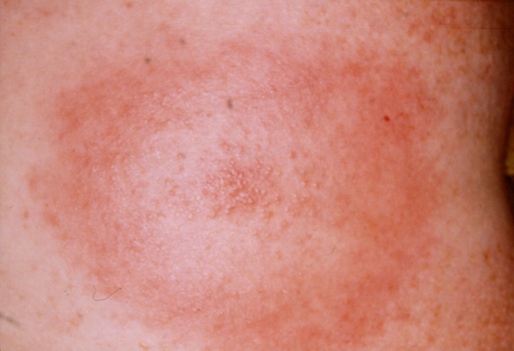
    The classical skin condition associated with Lyme disease is erythema chronicum migrans, which is a spreading erythematous rash which then clears centrally but leaves a spot in the middle (click here for a picture).


    A rarer skin condition is acrodermatitis chronica atrophicans, which occurs late in infection and eventually results in atrophic skin that is thin like cigarette paper.


    Another rarer skin manifestation is borrelial lymphocytoma, which is a blue-red discoloration of the ear lobe.


    Investigation is usually ELISA/PCR.
    If the question throws in a CSF sample look for slightly raised protein and lymphocytosis.


    Treatment of the rash only is doxycycline.  More serious disease may require IV ben pen/a 3rd generation cephalosporin. 


    There is of course far more to learn about Lyme disease should you be so inclined, but if you're content with the barer MRCP-bones of it lets move on to battle 9.2, gout.

    MRCP revision battle 9.2: Gout

    Ah Gout... that classical disease of red-faced old men...

    In terms of MRCP, you need to expand the above knowledge to include the following...


    Gout usually presents as a single, swollen, hot, painful joint.  If the joint affected is the big toe, it is called a podagra.


    Microscopy of joint fluid would reveal negatively birefrigent needle-shaped monosodium urate crystals.

    Uric acid may be >450micromols - but equally may be normal in an acute attack.

    CRP is usually raised, but if the question shows a raised WCC too start thinking about different diagnoses, for example septic arthritis.


    Gout may be either primary or secondary.

    Primary gout may be:
    • idiopathic - usually due to decreased excretion of uric acid 
    • associated with Lesch-Nyhan Syndrome (to be covered in a very brief battle tomorrow)


    Secondary gout can broadly be divided into things which cause increased production/intake of urate, and things which decrease its excretion.


    Causes of increased production/intake include:
    • myeloproliferative/lymphoproliferative disorders
    • psoriasis
    • cytotoxic drugs
    • food - beer, yeast, seafood, liver, kidney
    • exercise
    • fits
    • acidosis

    Causes of decreased excretion include:
    • renal failure
    • diuretics
    • low dose aspirin
    • alcohol
    • lead poisoning

    XR should show a normal joint space (may be decreased in late disease) and large punched-out erosions distant from the joint margins.


    Treatment is NSAIDs - if these are contraindicated, colchine can be used.


    Prophylaxis is with allopurinol (a xanthine oxidase inhibitor).  It should be started 2 to 3 weeks after an acute attack if:
    • >1 attack
    • tophi present
    • renal disease
    • urate stones
    • Lesch-Nyhan syndrome
    • cytotoxic drugs used (in which case it should be given before any episodes of gout)

    So after that quick whizz through gout, lets embrace is near-cousin pseudo-gout...

    MRCP revision battle 9.3: Pseudogout

    Pseudogout involves the deposition of positively birefrigent calcium pyrophosphate dihydrate crystals in joints.


    Pseudogout is associated with a variety of conditions, including:
    • hyperparathyroidism
    • hypothyroidism
    • haemochromatosis
    • Wilsons disease
    • acromegaly
    • old age
    • OA

    It tends to cause a monoarthritis of a larger joint, classically a knee.


    An XR may show chondrocalcinosis = soft tissue calcium deposition.


    Treatment is with NSAIDs.  If this is unsuccessful steroids or hydroxychloriquine can be considered.


    Now on to revision battle 9.4, a treat left over from yesterday, craniopharyngioma...

    MRCP revision battle 9.4: Craniopharyngioma

    Craniopharyngiomas are benign tumours that arise from the remnants of Rathke's pouch (= area between pituitary and 3rd ventricle floor).

    Technically it is not a pituitary tumour as it is derived from the embryonic remnants of the pituitary gland rather than the gland itself.   However, in terms of the mass effects/how it presents it is very similar.  Amenorrhoea and decreased libido are often sited as symptoms.


    Craniopharyngiomas frequently calcify (50%)


    Investigation of choice is MRI, treatment is surgery.


    After that brief squirmish its on to the last battle of the day, the pneumothorax

    MRCP revision battle 9.5: Pneumothorax

    A pneumothorax, as I'm sure you all know, is a collection of gas in the pleural cavity of the chest between the lung and chest wall.


    I'm not about to insult you by going through the basic signs/symptoms of pneumothorax, but I will just briefly mention a rare sign that occasionally pops up in MRCP exams: a clicking sound at the sternal edge synchronised with the heart beat.  This rare sign of a pneumothorax is most commonly assocaited with small left sided pneumothoraces.


    As a quick recap, its important to establish if the pneumothorax is primary (ie no underlying lung disease), secondary (=underlying lung disease) or traumatic (in which case as a medic you shouldn't be treating it!)


    BTS guidelines state that in primary pneumothorax, if the patient is not breathless and the rim of air is <2cm you do not need to necessarily do anything and may consider sending the patient home.  If the rim of air is >2cm or the patient is breathless you should aspirate; try x2, if still unsuccessful insert a drain.


    The guidelines for secondary pneumothorax are slightly more complex:
    • if >50yrs old AND breathless AND rim of air >2cm, insert a drain first line
    • if the above 3 criteria do not apply, try aspirating first
    • regardless of which category your patient falls into, patients with secondary pneumothorax should be admitted for 24 hours observation.


    A random type of pneumothorax to be aware of is catamenial pneumothorax - this is a pneumothorax associated with menstruation, which is due to endometriosis of the lung.  90% of these occur on the right side.


    As a complete aside, as well as recognising the 'click synchronised with heartbeat' as a possible pneumothorax it is worth learning that a 'crunching sound' in the chest can be due to a pneumomediastinum  and is called Hamman's sign.


    Well those are todays battles over, but for those with residual enthusiasm the beat goes on with a war to test your memory of yesterday's topics...

    MRCP questions: War 8

    As with previous 'wars' after 'battles' these are just a few quick questions to see if your brain cells have retained the information provided in battles 8.1 to 8.6.

    Grab a piece of paper, jot down your answers then compare them to my answers here


    Question 1:
    What is the treatment for cluster headache?


    Question 2:
    List 3 symptoms of cluster headache.

    Question 3:
    What odour is associated with acute arsenic poisoning?

    Question 4:
    List 3 causes of Mees lines

    Question 5:
    State the 4 H's and 4 T's which are known as the reversible causes of cardiac arrest.


    Question 6: 
    What kind of visual disturbance is classically associated with a pituitary tumour

    Question 7:
    What histological type of pituitary tumour is most likely to cause a pressure effect?

    Question 8:
    What is Nelson's syndrome?


    Question 9:
    What are the similarities and differences between Romano Ward and Jervell Lange Nielson syndromes?

    Question 10:
    What is the commonest hormone produced by a pituitary tumour?


    answers here

    Saturday, 11 September 2010

    MRCP revision battle 8.1: Cluster headache

    Its Saturday and sunny so I'm not particularly feeling the MRCP revision love.  But the battle must continue, and today's topics are:

    Revision battle 8.1: Cluster headache
    Revision battle 8.2: Arsenic poisoning
    Revision battle 8.3: Mee's lines
    Revision battle 8.4: Reversible causes of cardiac arrest
    Revision battle 8.5 Pituitary tumours
    Revision battle 8.6: Romano-Ward and Jervell-Lange-Nielson


    Revision battle 8.1: Cluster headache


    Cluster headaches appear in the MRCP exam (and occasionally real life) as:
    • severe unilateral pain focused around one eye with
    • redness of the eye and possibly
    • lid swelling with
    • lacrimation and
    • nasal congestion
    It is worth noting that up to 20% will also get ptosis, which in a minority of cases can be permanent.


    Cluster headaches are more common in males (5:1).
    Risk of suffering from cluster headache is increased if you are a smoker.


    Classically patients will suffer from severe 15 minute to 2 hour bouts of pain once or twice a day over a period of 4 to 12 weeks.


    Treatment acutely is 100% oxygen.  S/C sumitriptan and nasal lidocaine are also proposed as treatments.

    Prophylactic options include verapamil, prednisolone or lithium.


    So onwards to a slightly curveball topic - arsenic poisoning...

    MRCP revision battle 8.2: Arsenic poisoning

    Arsenic poisoning used to be both a favourite method of murder (allegedly involved in the demise of Napoleon) and a substance commonly found in paint and water pipes.  It is now thankfully carefully controlled so it tends to be found mainly in MRCP exam questions...


    Arsenic is still used in the glass, smelter and microelectronic industries, and is found in wood preservatives.


    Acute poisoning tends to cause features in 30 mins to 2 hours.

    Features of acute arsenic poisoning include:
    • severe gastroenteritis
    • garlic ordour
    • hypersalivation
    Treatment for acute poisoning is DMPS.


    Symptoms of chronic arsenic poisoning present at different times post-exposure; for example, skin symptoms tend to show clinically around 10 years afterwards.  There are multiple, multiple possible complications of chronic arsenic poisoning but a few which may pop up are:
    • keratoses
    • Mees lines (if you don't know what these are you'll find out in the next battle!)
    • hyperpigmentation
    • neuropathy
    • muscle fasciculation and wasting

    So on to explore Mees lines...

    MRCP revision battle 8.3: Mees lines

    Mees lines are white transverse lines that go all the way across the nail.

    An image of Mees lines can be found here


    Mees lines are found in:
    • arsenic poisoning
    • thalium poisoning
    • heavy metal poisoning
    • renal failure

     Next up: battle 8.4!

    MRCP revision battle 8.4: Reversible causes of cardiac arrest

    A little bit of a random one to throw into MRCP revision but a quick recap of the reversible causes of cardiac arrest, famously recalled as the 4 Hs and 4 Ts, can never go amiss...


    4 H's:
    • hypoxia
    • hypovolaemia
    • hypothermia
    • hypoglycaemia/hyper/hypokalaemia/ other electrolyte imbalance

    4 T's:
    • Tension pneumothorax
    • cardiac Tamponade
    • Thombus
    • Toxins


    I remember the 4 H's in the format of a little picture of a man standing on top of a snow-covered mountain.  He is struggling to breath as there's not much oxygen up there (hypoxia) and  is shivering as its so cold (hypothermia).  He is looking sad and in one hand is an empty water bottle (hypovolaemia) and in the other an empty lunchbox (hypoglycaemia... then remember the hypo/hyper kalaemia from that).

    For the 4 T's I think of the less happy phrase of 'Thomas has Ten Toxic Tampons' (= Thombus, tension pneumothorax, toxins, tamponade)


    So after a couple of nice straightforward battles let face a juicier one: pituitary tumours...

    MRCP revision battle 8.5: Pituitary tumours

    Most pituitary tumours are benign adenomas.


    Symptoms from pituitary tumours are either due to:
    • local pressure
    • hormone secretion
    • hypopituitism

    Local pressure symptoms include:
    • bitemporal hemianopia (pressure on CN III)
    • palsies of CN III/IV/VI (remember the cavernous sinus anatomy at the end of battle 1.1?)

    The commonest hormone secreted by pituitary tumours is prolactin (35%) followed by GH (20%).

    30% of pituitary tumours secrete no obvious hormone


    Potentially a pituitary tumour can disturb hypothalamic control of temperature, sleep or appetite.

    If the tumour erodes the floor of the sella the patient may get CSF rhinorrhoea


    Histologically, pituitary tumours fall into the following catagories:
    • 70% - chromophobes
      • 30% cause a pressure effect
      • can produce prolactin, ACTH, GH
    • 15% - acidophils
      • 10% cause a pressure effect
      • can produce GH, prolactin
    • 15%  - basophils
      • rarely cause a pressure effect
      • can produce ACTH

    The investigation of choice for suspected pituitary tumours  is MRI.


    Finally, in this section we need to cover Nelsons Syndrome.  Nelsons syndrome is a condition that occurs after adrenalectomy for pituitary-dependent cushings.  The adrenalectomy removes the negative feedback on the pituitary which causes ACTH levels to rise massively, leading to hyperpigmentation +/- an invasive pituitary tumour.


    Tomorrow we will look at craniopharyngioma, but to finish today on a lighter note onwards to Romano Ward and Jervell-Lange-Nielson Syndromes.

    MRCP questions: War 7

    As with previous 'wars' after 'battles' these are just a few quick questions to see if your brain cells have retained the information provided in battles 7.1 to 7.5.


    Grab a piece of paper, jot down your answers then compare them to my answers here


    Question 1:
    An efflux of which ion terminates the cardiac action potential?

    Question 2:
    What is the mechanism of action of class III antiarrhythmics?


    Question 3: 
    Name 2 class IV antiarrhythmic drugs

    Question 4:
    What is the relationship between heart rate and QT?

    Question 5:
    How do you calculate QT using the Bazzett formula?

    Question 6:
    State the upper limit of a normal QT

    Question 7: 
    Name 5 drugs that prolong QT

    Question 8:
    State 5 metabolic conditions that can prolong QT

    Question 9:
    Name 2 drugs that increase the effect of adenosine and one that decreases it.

    Question 10:
    Does positive concordance in the chest leads make a broad complex tachycardia more or less likely to be VT?


    answers here


    MRCP revision battle 8.6: Romano Ward and Jervell-Lange-Nielson Syndomes

    Both Romano-Ward and Jervell-Lange-Nielson Syndrome are congenitial causes of a long QT.

    Both are believed to be caused by mutations in the potassium subunit.

    The differences are:
    1. Romano-Ward is autosomal dominant while Jervell-Lange-Nielson is autosomal recessive
    2. Jervell-Lange-Nielson is associated with bilateral deafness; Romano Ward is not.

    Thats all for today; if you want to test your recall of topics covered yesterday click here

    Friday, 10 September 2010

    MRCP revision battle 7.1: cardiac action potentials

    I've got that Friday feeling so I've decided to indulge in a cardiology fest.  Today's revision battles will therefore be:

    MRCP revision battle 7.1: cardiac action potentials
    MRCP revision battle 7.2: the Vaugham-Williams classification system
    MRCP revision battle 7.3: long QT
    MRCP revision battle 7.4: adenosine
    MRCP revision battle 7.5: cause of the broad complex tachycardia

    Now I know that may look like a lot but quite a few of the battles are short and almost sweet, so lets get stuck in!


    MRCP revision battle 7.1: cardiac action potentials


    Understanding the basics of the cardiac action potential is essential to grasp how antiarrhythmic drugs work.  The basic cardiac action potential looks like this:

                                                                                                                thanks to http://commons.wikimedia.org for the image

    In stage 0, there is a rapid influx of sodium
    Stage 1 is then an efflux of potassium, followed by
    Stage 2, which is an influx of calcium.  Finally in
    Stage 3 the efflux of pottasium continues, terminating the action potential.


    Try and keep this image in mind as we move on to the fabulous world of Vaughan-Williams classification...

    MRCP revision battle 7.2: The Vaughan Williams Classification

    The Vaughan-Williams classification is a way of dividing anti-arrhythmic drugs into groups based on their mechanism of action.  It was devised in the 1970s, and like some other inventions of that era (such as flares) it has a few flaws.  The main flaw is that many anti-arrhythmics actually work in several different ways, so although they can be put into a class based on their main mechanism of action this grouping only tells you part of how they work.

    However, understanding it a) makes you seem clever (most consultants would have forgotten it within hours of passing MRCP) and b) helps you answer MRCP questions correctly.  So lets ignore its faults and embrace it whole-heartedly...


    The original classification had groups I, II, III and IV.  A 'catch all' group of V has now been added for awkward drugs who just didn't fit in anywhere originally.  Lets look at each class in turn:


    Class Ia:
    • works by blocking sodium channels - called a 'membrane stabiliser'
    • distinguishes itself from the other class Is by having intermediate association/dissociation with the sodium channels --> end result longer action potential
    • members include disopyramide, quinidine and procainamide

    Class Ib:
    • works by blocking sodium channels - called a 'membrane stabiliser'
    • distinguishes itself from the other class Is by having fast association/dissociation with the sodium channels --> end result shorter action potential
    • members include lidocaine and phenytoin

    Class Ic:
    • works by blocking sodium channels - called a 'membrane stabiliser'
    • distinguises itself from the other class Is by having slow association/dissociation with the sodium channels --> end result same action potential
    • members include flecanide and propafenone

    Class II:
    • are beta blockers 
    • I'm sure you don't really need examples but think atenolol, bisoprolol, misoprolol...

    Class III:
    • works by blocking potassium channels
    • this results in prolonged repolarisation
    • members include amiodarone and sotolol

    Class IV:
    • works by blocking calcium channels
    • members include verapamil and diltiazem

    (Class V: = last minute 'catch all', includes digoxin and adenosine)


    Thats rather a lot to try and remember; understanding and linking it to the cardiac action potential makes it a bit easier to understand, then a bit of word-play might clarify things further:


    To remember the broad classes of drugs, try recalling 'Sodium Blocks Potassium Channels' (just substitute calcium for channels and you have the 4 mechanisms of action)

    To remember the members of each class, think "Double quarter pounder, lemonade please, fries please... and salt uh oh vomiting and diarrhoea..."


    Got it?  Good, cos you'll need in for battle 7.3, long QT.

    MRCP revision battle 7.3: long QT

    The QT interval is that sneaky part of the ECG measured from the start of the QRS complex to the end of the T way.  I call it sneaky as as clinicians we rarely pay it much attention, then it just happens to get a bit on the long side and oh whoopsie we have a ventricular arrhythmia on our hands.


    The first thing to learn about the QT interval is that it decreases as heart rate increases.  It is therefore necessary to correct it, which is achieved by using either Bazetts or Fridericas formula.

    Bazetts = corrected QT = QT/square root of heart rate
    Fridericas = corrected QT = QT/cube root of heart rate

    Upper limit of normal is generally taken to be 450ms in a man and 470ms in a woman (for some bizarre reason I remember 450 by singing the song 'brimful of asha on the 45' then just remember that females are 20 more)


    There are multiple causes of long QT:

    1. familial
      • Romano-Ward Syndrome (to be covered tomorrow)
      • Jervell-Lange-Nielson Syndrome (also to be covered tomorrow)
    2. metabolic
      • low Mg/Ca/K
      • low temp
      • low thyroid (also known as hypothyroidism.. :) )
    3. drugs
      • class I and III antiarrhythmics (which after the last revision battle you should be able to name)
      • erythromycin
      • TCA/haloperidol/risperidone/SSRIs
      • cocaine
      • organophosphates
      • antihistamines
    4. other
      • MI
      • myocarditis
      • SAH

    Treatment is possibly beta blockers, possibly ICD.


    Now on to revision battle 7.4, to meet a drug that fell into class V...

    MRCP revision battle 7.4: Adenosine

    Adenosine is a purine nucleoside which clinically is used to try and terminate SVTs/expose the underlying rhythm.  While doing so it has the potential to cause cardiac standstill, so it can have a unique dual action of increasing the adminstering doctor's heart rate in proportion to how much it decreases the receiving patients heart rate.

    It's half life is 8 to 15 seconds, and it works by activating potassium channels which decreases AVN conduction.


    The effect of adenosine is increased by dipridamol and carbamezepine.
    The effect of adenosine is decreased by aminophylline.


    A positive of adenosine is that it has no significant negatively inotropic effects.


    Risks associated with its use include:
    • enhancing conduction through the accessory pathway in Wolff-Parkinson-White
    • causing a dangerously fast ventricular response in pre-excited AF/flutter

    The side effects include:
    • anxiety
    • chest tightness
    • bronchospasm (avoid in asthma)
    • facial flushing
    • nausea

    The usual dosing regime is 6mg, then if unsuccessful 12mg 2 mins later and if still unsuccessful another 12mg 2 mins after that.  Ensure cardiac monitoring is in place when it is given (it can cause cardiac standstill) and remember that given its short halflife it must be given as a rapid bolus, flushed instantly with saline.


    So to the final revision battle of the day, with that most feared ECG pattern: the broad complex tachycardia.

    MRCP revision battle 7.5: broad complex tachycardia

    At some point an ECG will be shoved under your nose which shows an unnerving broad complex pattern going at a fast rate.  If in doubt, these must always be regarded as VT.  However, there are a few less scary possibilities, such as:
    • AF with LBBB
    • orthodromic tachycardic WPW
    • ventricular paced rhythm


    Essentially the main call is whether the broad complex tachycardia is superventricular or ventricular in origin, and the features the MRCP exam expects you to pick up on to differentiate these two are:

    • AV dissociation - suggests VT
    • fusion/capture beats - suggest VT
    • positive QRS concordance in chest leads - suggests VT
    • marked left axis deviation - suggests VT
    • history of IHD - more likely VT
    • QRS>140ms - suggests VT
    • no response to adenosine/massage - suggests VT


    Well thats if for today.  A quick test of yesterdays topics is available here if you still have the energy.

    MRCP questions: War 6

    As with previous 'wars' after 'battles' these are just a few quick questions to see if your brain cells have retained the information provided in battles 6.1 to 6.5.

    Jot your answers down on a piece of paper then compare them to the answers here

    Question 1:
    Which HLA is ankylosing spondylitis associated with?

    Question 2:
    List 5 symptoms/signs associated with acute iritis

    Question 3:
    What is the correct term for sterile pus in the anterior chamber of the eye?

    Question 4:
    List 7 infections associated with reactive arthritis


    Question 5:
    Name and describe a dermatological condition associated with reactive arthritis

    Question 6:
    What is reiters syndrome?

    Question 7:
    Define Light's criteria
      Question 8:
      List 5 possible causes of transudates and 5 possible causes of exudates

      Question 9:
      Which 5 causes of pleural effusion would cause a low pH (<7.3)?

      Question 10:
      List 3 associations of yellow nail syndrome


      answers here

      Thursday, 9 September 2010

      MRCP revision battle 6.1: ankylosing spondylitis

      Today's revision threw up a mixed bag of topics... a splash of rheumatology with a dash of respiratory and a hint of ophthalmology.

      So...

      Battle 6.1: ankylosing spondylitis
      Battle 6.2: acute iritis
      Battle 6.3: Reiters syndrome
      Battle 6.4: pleural effusion
      Battle 6.5 yellow nail syndrome

      Lets get started!


      6.1: Ankylosing spondylitis


      Ankylosing spondylitis (AS) is a chronic inflammatory disease of the spine and sacroiliac joints.


      It affects males more than females, intially around 6:1 but altering to around 2:1 by the age of 30.

       95% of patients are + for HLA B27


      Patients often present with low back pain which is worse at night and improves on moving.

      Clinically, the things to observe on regarding a patient with AS are:
      • a loss of lumbar lordosis
      • a fixed kyphosis which is compensated by extension of the cervical spine leading to the classical 
      • 'question mark' posture
      • if the patient turns their head to the side, the whole body may turn
      • there may be decreased chest expansion forcing increased diaphragmatic excursion and hence a
      • prominent abdomen
      A possible test to undertake is Schobers test - find L5 (roughly level with the sacral dimples) then mark a level 5cm below and 10cm above.  Get the patient to bend over; this 15cm distance should increase to at least 20cm, if it doesn't is suggests AS.

        Associations of AS include:
        • iritis
        • aortic regurgitation
        • fibrosis (rare)
        • cardiac conduction abnormalities (around 10%, mostly long PR)
        • secondary amyloidosis

        Management is keeping mobile, NSAIDs and in severe cases tumour necrosis factor alpha blockers such as infliximab.

        As an aside, if considering a TNF alpha blocker its a good idea to check TB status as it is likely to reactivate latent TB.


        Onwards.... 6.2!

        MRCP revision battle 6.2: acute iritis

        If you're anything like me eyes are a bit a vague part of the body... as an A&E SHO I hated dealing with 'the red eye'.  The good news is that MRCP doesn't have too many ophthalmology questions so there aren't too many topics you have to be familiar with.  One not to miss however is acute iritis.

        As an aside, acute irits and anterior uveitis are often used by doctors almost interchangeably, but technically anterior uveitis encompasses both iritis (inflammation of the iris) and iridocyclitis (inflammation of the iris and ciliary body).  Fortunately management is pretty much the same.


        Acute iritis presents as:
        • red eye ('circumcorneal redness'
        • pain
        • photophobia
        • blurred vision
        • lacrimation
        • small pupil (in reccurent cases the pupil may be irregular due to adhesions)

        You may be able to illicit a positive Talbot's test, which is an increase in the patient's pain when you get their eyes to converge (due to the pupils constricting)


        On slit lamp examination you may spot:
        • cells in the anterior chamber
        • white precipitates on back of cornea
        • sometimes a hypopyon (= sterile anterior chamber pus)

        It is associated with ankylosing spondylitis and also Bechets disease.
        It may relapse.


        Treatment requires ophthalmic input due to the need for steroids - and if steroids are given inappropriately to an infected eye the patient may become blind....


        On that happy note, lets move on to Reiters Syndrome

        MRCP revision battle 6.3: Reiters syndrome/Reactive Arthritis

        Reactive arthritis is one of the spondyloarthropathies (alongside ankylosing spondylitis, psoriatic arthritis and enteropathic arthritis).  It is characterised by a sterile arthritis which usually affects the lower limb days to weeks following an infection.  It is associated with being HLA B27 positive.


        Infections commonly associated with reactive arthritis fall into 2 groups:
        • GI infections 
          • campylobacter
          • salmonella
          • yersinia
          • shigella
        • GU infections
          • chlamydia
          • gonnorhora
          • ureaplasma

        The classical triad associated with reactive arthritis is Reiters Syndrome of uveitis/conjunctivitis, urethritis and arthritis ("can't see, can't pee, can't climb a tree")  Apparently in some parts this is becoming a politically incorrect term as Dr Reiter was a member of the Nazi party with a less than perfect record by the 1940s...

        Reactive arthritis can also be associated with keratoderma blenorrhagica, which is brown raised plaques on the hands and feet.  Rarely it can cause aortic incompetance.

        Treatment is generally resting/splinting the affected joint and NSAIDS.


        Now time for some diversification to the liquid world of the pleural effusion....

        MRCP revision battle 6.4: Pleural effusions

        Pleural effusions for me are one of the most interesting clinical signs.  A dull day can be made just that tad more interesting by the characteristic stoney dull percussion note, reminding you there is a reason you percuss everyone's chest.  A chest xray can then almost instantly confirm your clinical judgment (unlike with a murmur, which you may never know if you were just imagining or not) but then theres the 'oh bugger' moment as you realise you need to work out why this patient has an effusion... and most of the options aren't entirely positive....


        The main thing to establish with an effusion is if it is an exudate or a transudate.

        Traditionally a transudate was defined as containing <3g/dl of protein.  However, some doctors prefer to use Light's criteria for determining which label to apply (a criteria which will misclassify 25% of transudates as exudates...)

        Lights criteria is that for a fluid to be an exudate it must either have:
        • pleural:serum protein > 0.5 or
        • pleural:serum LDH > 0.6 or
        • pleural LDH >2/3 upper limit of serum LDH

        So, obviously to make your diagnosis you need a pleural fluid sample and a blood sample.  You should send your pleural sample off for:
        • cell count
        • cytology
        • glucose
        • protein
        • LDH
        • amylase
        • pH
        • Ziel-nielson staining
        and remember to send bloods for glucose, LDH and amylase at the same time (plus any other bloods, like FBC, you might need)


        Armed with the knowledge of transudate or exudate, you can spin forth a list of diffentials:

        Causes of transudates:
        • heart failure
        • renal failure
        • liver failure
        • peritoneal dialysis
        • hypothyroidism
        • Meigs
        • constrictive pericarditis

        Causes of exudates:
        • infection: TB, pneumonia, subphrenic abscess
        • inflammation: Dresslers, pancreatitis, SLE, RA
        • malignancy: mesothelioma, local cancer, lymphoma
        • other: PE, uraemia, yellow nail syndrome


        As the list of differentials is still quite big, apart from your clinical acumen some of the other pleural fluid results can help narrow it out:


        Glucose <3.3 or pH <7.2 or raised LDH suggests:
        • TB
        • malignancy
        • empyema
        • SLE
        • RA
         Its worth noting that *really* low glucoses are usually due to RA or empyema


        Raised amylase suggests:
        • pancreatitis
        • carcinoma
        • bacterial pneumonia
        • oesophageal rupture

        Cytology results can give more clues:
        • neutrophils: pneumonia, TB
        • lymphocytes: malignancy, TB, RA, SLE, sarcoid
        • mesothial cells ++ : pulmonary infarction
        • multinucleated giant cells: RA
        • lupus cells: SLE

        Hopefully these lists should equip you well for any MRCP pleural effusion questions (or even, shock horror, real life on the wards!)


        On to the final battle of today, yellow nail syndrome

        MRCP revision battle 6.5: yellow nail syndrome

        Yellow Nail Syndrome falls into that subsection of clinical conditions that is rarely relevant in real life but disproportionately prevalent in MRCP questions.  Thankfully its fairly straightforward.

        Clinically:
        • thick nails which are
        • excessively curved from side to side and
        • pale yellow. They are
        • slow growing (showing uncovered bulbous fingertips) and have
        • absent lunulae (the little 'moon-shaped' white bits at the base of your nails)
        • onycholysis may be present

        The importance from an MRCP point of view is that they are associated with:

        Thats all for today.  If you fancy a quick little test on yesterday's topics, click here

        MRCP questions: War 5

        As with previous 'wars' after 'battles' these are just a few quick questions to see if your brain cells have retained the information provided in battles 5.1 to 4.5.

        Jot down your answers on a piece of paper then compare to mine here


        Question 1:
        State the cardinal triad of nephrotic syndrome.

        Question 2:
        Try and list 10 potential complications of nephrotic syndrome

        Question 3:
        In a patient with nephrotic syndrome, what complication might be implied if the question states they develop 'loin pain'?

        Question 4: 
        What is the commonest glomerulonephritis in adults?

        Question 5:
        Does frank haematuria imply a good or bad prognosis in Bergers disease?

        Question 6:
        What HLA types is Bergers disease associated with?

        Question 7:
        What is the treatment for acute epiglottitis?

        Question 8:
        What is the commonest cause of nephrotic syndrome?

        Question 9:
        What tends to be raised in Bergers disease - C3 or C4?

        Question 10:
        Which gender is more affected by Bergers disease?


        Answers here

        Wednesday, 8 September 2010

        MRCP revision battle 5.1: Nephrotic Syndrome

        After yesterdays big push today's revision was less epic, mainly because I started reading "The Lovely Bones" by Alice Sebold and I just got too involved.  My motivation to revise also wasn't helped by the first onexamination question I did being 'renal based', with renal medicine being my personal black hole of knowledge... it doesn't matter how many facts I try to throw into it, it still remains just as black.

        So, today's battles are:
        5.1 nephrotic syndrome
        5.2 renal vein thrombosis
        5.3 IgA glomerulonephritis
        5.4 acute epiglottis


        Battle 5.1: Nephrotic syndrome


        Nephrotic syndrome is a classic triad of:
        • >3g proteinuria
        • hypoalbuminaemia <25 and
        • oedema
        It is also strongly associated with hypercholesterolaemia


        80% of the time it is associated with glomerulonephritis, a fiendishly complex set of conditions.


        The rest of the time its possible causes are many and varied:
        • diabetes mellitus
        • leprosy/malaria/HBV
        • myeloma/lymphoma
        • amyloidosis
        • SLE
        • gold/penicillamine/NSAIDS/captopril/interferon alpha/heroin
        • pre-eclampsia
        • accelerated hypertension
        • sickle cell disease
        • Alports
        • rarely visico-ureteric reflux
        I'm afraid I don't have a clever way/inclination to remember that list, I just hope that by reading it a few times in the exam a faint light of recognition may go on at the appropriate time.


        Happily I do have a way of remembering the potential complications of nephrotic syndrome, which is good since sneaky MRCP examiners may be trying to get you to guess nephrotic syndrome from a description of the complications alongside part of the classic triad.

        My mnemoric is BOB, I HIT HIM:
        • B12 deficiency
        • Osteomalacia
        • Budd-Chiari
        • Infections
        • Hyperlipidaemia
        • Immunosupression
        • Thrombosis (including renal vein thrombosis)
        • Hyponatraemia
        • Iron deficiency
        • Malnutrition

        Treatment for nephrotic syndrome is to restrict salt, treat the cause, prophylactic heparin, treat any hypertension and potentially give furosemide or ACE-i.


        On to 5.2... renal vein thrombosis

        MRCP revision battle 5.2: Renal vein thrombosis

        Renal vein thrombosis can occur due to:
        • nephrotic syndrome inducing a hypercoagulable state
        • invasion of the renal vein by renal cell carcinoma
        • thombophillia

        It is often asymptomatic, but equally may present with haematuria, loin pain, palpable mass or a sudden worsening of renal function.


        Treatment is with warfarin, anticoagulated to INR 2-3 for 3-6 months.

         Follow me to battle 5.3....

        MRCP revision battle 5.3: IgA Glomerulonephritis

        As if the glomerulonephropathies aren't tricky enough (hence why series 5 of the battles only discusses one of them) they can also have a series of pseudonyms/ 'nicknames.'  For example, IgA nephropathy also goes by the labels 'Bergers disease' and 'mesangioproliferative glomerulonephritis'.  For simplicity, I'll just refer to it as IgA nephropathy.


        IgA nephropathy is the commonest GN in adults, and it classically affects young males after an URTI.  So, in the exam look out for the 25 yr old man who has haematuria following a sore throat.

        Symptoms may just be micro/macro haematuria, or match the nephrotic syndrome triad.

        Incidence is increased in those from the far east, and those with HLA DQw7 or HLA B35.

        Associations include cirrhosis, dermatitis herpetiformis, ank spon, Wiskott-Aldrich syndrome and chronic liver or lung disease.


        IgA is positive in 50% of cases and C3 tends to be raised.


        25% of cases progress to end stage renal disease.

        Frank haematuria is associated with a good prognosis.
        Being male, having proteinuria, hypertension, smoking or high lipids are all associated with a poor prognosis.


        Phew, thats one of the GN covered... a few more to face at a later date... now lets diversify for the last battle of the day to acute epiglottis.

        MRCP revision battle 5.4: Acute Epiglottitis

        Since the introduction of the Hib vaccine the potentially life-threatening condition of acute epiglottitis has happily become rare.  Except, of course, in the MRCP exam.

        The commonest cause of acute epiglottis was always h.influenzae, but many other pathogens can also cause it.

        Presentation tends to be a 'toxic' looking patient with fever and stridor, often with a preceding sore throat.


        Treatment is with a 3rd generation cephalosporin.


        That's it for today, but if you fancy testing yourself on yesterday's battles click here to go to war 4...

        MRCP questions: War 4

        As with previous 'wars' after 'battles' these are just a few quick questions to see if your brain cells have retained the information provided in battles 4.1 to 4.5.

        Jot down your answers on a piece of paper then compare them to my answers here


        Question 1: 
        What pattern would COPD produce on spirometry?

        Question 2:
        With what condition is thermoactinomyces vulgaris associated?

        Question 3:
        In which zone of the lungs would fibrosis secondary to EAA classically occur?


        Question 4:
        Can you name 11 causes of lung fibrosis?

        Question 5:
        Can you name at least 10 causes of clubbing?


        answers here

        Tuesday, 7 September 2010

        MRCP questions: War 3

        As with previous 'wars' after 'battles' these are just a few quick questions to see if your brain cells have retained the information provided in battles 3.1 and 3.2.

        Jot down your answers then look at my answers here


        Question 1: 
        Can you now state 10 causes of bilateral hilar lymphadenopathy?


        Question 2: 
        What are the two main causes of hypercalcaemia?


        Question 3:
        Name the 2 drugs mentioned that can cause hypercalcaemia


        Question 4:
        Toxicity of which 2 vitamins can cause hypercalcaemia?


        Question 5:
        Which 5 cancers are most commonly associated with boney mets?


        Question 6:
        What effect does vit D have on calcium and phosphate?


        Question 7:
        What will bloods show in secondary hyperparathyroidism?


        Question 8: 
        What is the commonest cause of primary hyperparathyroidism?


        Question 9:
        What is vitamin D made from?


        Question 10:
        What is the effect of PTH on calcium and phosphate levels?


        answers here

        MRCP revision battle 4.5: Clubbing

        This clinical sign should be on one of those 'you know you're a medic when...' lists, as in "you know you're a medic when clubbing relates to fingers rather than a night out..."

        As an undergraduate you probably spent cumulative hours staring purposefully at fingers to convince the examiner you were looking for clubbing, and now at MRCP you need to think about it again..


        Firstly, for a definition: clubbing is defined as loss of the obtuse angle between the nail and the dorsum, with thickening of the nail bed and increased curvature of the nail bed in both directions.  There may also be increased fluctuation of the nail bed and the finger can sometimes have a 'drumstick like' appearence.

        A picture can be found here


        And now, for some causes, and a cry of 'ACE MILC (milk) MCFAB'!

        Cardiac causes:
        • atrial myxoma
        • cyanotic congenital heart disease
        • endocarditis
        GI causes:
        • malabsorption
        • inflammatory bowel disease
        • lymphoma
        • cirrhosis
        Lung causes:
        • mesothioloma
        • CF and cancer
        • fibrosis
        • abscess
        • bronchiectasis

        Thats all for today, but if you participated in battle 3 yesterday and fancy testing yourself go on for a brief test here...

        MRCP revision battle 4.4: Lung fibrosis

        You may need to be able to come up with a list of differentials for lung fibrosis... my way of remembering it is C BAR TAPS (mainly because at some point in the revision process I always get an urge to visit a drinking establishment and 'see' the bar taps, and possibly even purchase something from them...)

        So, C BAR TAPS =
        • Cryptogenic fibrosing alveolitis
        • Bleomycin
        • Amiodarone
        • Radiation/rheumatological disease (=RA and SLE)
        • TB
        • Asbestosis
        • Paraquat/pigeon breeders lung
        • Sarcoid/siliosis

        And finally for today, battle 4.5...

        MRCP revision battle 4.3: Aspergillus

        Aspergillus is a group of molds/fungi.  Allegedly they got their name from looking like water does when it falls from an aspergillum, which is an instrument for sprinkling holy water (that fact is unlikely to be necessary in MRCP but its nice for the brain to read bits its not obligated to remember every now and then)


        Aspergillus can affect the lungs in 6 main different ways:
        1. It can cause asthma
          • a type 1 hypersensitivity reaction
          • prick test +, precipitans/culture +
        2.   It can cause an aspergilloma
          • = a fungal ball inside a pre-existing cavity
          • usually asymptomatic but may cause haemoptysis, lethargy or weight loss
          • treated by ?surgery ?amphotericin paste under bronchoscopy
          • prick test -, precipitans/culture ++
        3.  It can cause allergic bronchopulmonary aspergillosis
          • type 1 and type 3 hypersensitivity
          • CXR may show bronchietasis, lobar collapse or upper lobe fibrosis, or patchy lesions
          • prick test +, precipitans/culture +
          • also IgE levels are raised
        4. It can cause invasive aspergillosis
          • only occurs in immunocompromised individuals
          • prick test +/-, precipitans/culture +/-
        5. It can cause EAA
          • + serum precipitans to aspergillus clavatus
        6. It can simply colonise.

        After that slightly heavy-going section, lets go on to think about BAR TAPS...

        MRCP revision battle 4.2: Extrinsic Allergic Alveolitis


        This is inflammation of lung tissue due to hypersensitivity to an allergen.  To get more scientific, it is a Gell and Coombs type 3 reaction acutely and a type 4 reaction chronically.


        Clinically, an acute reaction occurs 4-6 hours post exposure and manifests as fever, rigors, dry cough, dyspnoea and myalgia.  Auscultation of the lungs might reveal crackles but no wheeze.

        Chronically EAA causes dyspnoea, weight loss and can eventually lead to type 1 respiratory failure and cor pulmonale.


        There are several different 'flavours' of EAA, each precipitated by something different.  These include:
        • Farmers lung: micropolyspora faeni or thermoactinomyces vulgaris or Saccharopolyspora rectivirgula
        • malt workers lung: aspergillos clavatus
        • mushroom workers lung: thermophilic actinomycetes
        or, more modernly:
        • hot tub lung: mycobacterium avium in poorly maintaned hottubs!

        A CXR acutely might show mid-zone mottling; chronically upper zone fibrosis might be seen.  It can also be a cause of bilateral hilar lymphadenopathy (remember battle 3.1?)

        Spirometry gives a restrictive picture and lavage will show lymphocytes.


        Treatment is oxygen, possibly steroids.


        Allons-y to battle 4.3!

        MRCP revision battle 4.1: lung function tests

        After working an excessive number of hours and recovering thanks to chocolate cake (do click here to visit the ultimate chocolate cake recipe, which really is just amazing) I've managed a bit of a revision spurt, mainly through the world of lungs.

        So today we have:
        battle 4.1: pulmonary function tests
        battle 4.2: extrinsic allergic alveolitis
        battle 4.3: aspergillosis
        battle 4.4: fibrosis
        then bringing up the rear battle 4.5, that classic clinical sign clubbing.


        Battle 4.1: Pulmonary function tests

        Using a spirometer, ask your patient to blow as hard, as fast and as long as possible.  The spirometer will then give you their FEV1 (=forced expiratory volume in 1 second) and FVC (= forced vital capacity.)  These can be plotted, giving graphs that look like this.

        In the exam you are far more likely to just get figures and need to be able to recognise them as representing either an obstructive or a restrictive picture.


        Obstructive pattern

        Obstructive lung function usually gives a very reduced FEV1 and a less severely reduced FVC.  This means that overall the FEV1/FVC ratio is <75%.

        Classical causes of an obstructive pattern are COPD and asthma


        Restrictive pattern

        Restrictive lung function causes a reduced FVC, and FEV1/FVC remains either the same or may increase.

        Causes include LONE K: lung fibrosis, obesity, neuromuscular problems, effusion, kyphoscoliosis.


        That was short and sweet... onwards to battle 4.2...

        Sunday, 5 September 2010

        MRCP questions: War 2

        As with previous 'wars' after 'battles' these are just a few quick questions to see if your brain cells have retained the information provided in battles 2.1 and 2.2.

        Jot down your answers then compare them to mine here


        Question 1: 
        Name 6 drugs associated with drug-induced lupus


        Question 2: 
        Name the 3 best tests for monitoring SLE activity.


        Question 3:
        What is libman-sacks?


        Question 4:
        Name 3 conditions associated with Jaccouds arthropathy


        Question 5:
        List as many side effects of amiodarone as you can manage


        Click here for the answers

        MRCP revision battle 3.2: hypercalcaemia and hyperparathyroidism

        Hypercalcaemia

        Ah, hypercalcaemia, the classic 'stones, bones, abdominal groans and psychic moans'... and a favourite of MRCP questions.

        So what are the causes?
        The top 2 to consider are always MALIGNANCY and PRIMARY HYPERPARATHYROIDISM.
        Once you've thought of those, think toxic COAT MISST paget...

        Malignancy may be hinted at by other blood tests showing low albumin, chloride or an alkalosis.
        Think of myeloma, the possiblity of boney mets and of tumours producing PTH-P.
        For boney mets remember the brilliant 'bronchus, breast, byroid, brostate and bridney'
        For PTH-P the main culprits are squamous cell lung carcinoma, breast and kidney.

        Primary hyperparathyroidism is usually due to a single adenoma (85% of the time) and is covered more in the topic of that name below.

        So on to the slightly more eoseritc possiblities.... TOXIC COAT MIIST PAGET

        TOXIC = vit D or vit A toxicity

        COAT = the endocrine causes; essentially any 'growth' hormone excess will increase calcium (GH/oestrogen/thyrotoxicosis/steroids) and COAT is a way to recall the important ones: cushings, acromegaly/addissons, thyrotoxicosis.  Phaechromocytoma can occasionally be a culprit too.

        MIIST = the random causes
        Milk-alkali syndrome - treat with n.saline and loop diuretics
        Iatrogenic - thiazides, lithium
        Idiopathic infantile - supravalvular aortic stenosis (think of if 'elfin facies' are mentioned)
        Sarcoid
        TB/any granulomatosis disease (histoplasmosis, wegeners granulomatosis)

        PAGETS - pagets disease of the bone does not normally cause raised calcium but will if the patient has been immobile for a long period.


        Having considered the causes, lets take a step back and look at the 2 main 'characters' involved in calcium balance.

        Firstly, theres vitamin D.  Vit D is made from cholesterol, meaning that while meat-fans can get some of theirs from food vegetarians are reliant on their body making some (made in the skin with the aide of sunlight, making the UK's weather not ideal...)

        Vit D affects calcium by:
        - promoting GI absorption of Ca and phos
        - promoting renal reabsorption of Ca and phos.

        Unsuprisingly, its net effect is therefore to increase both calcium and phosphate.

        The second 'character' is PTH.  This affects calcium by:
        - promoting renal reabsorption of calcium (and encouraging renal loss of phosphate)
        - promoting osteoclast activity --> increasing both Ca and phos
        - increasing Vit D hydroxylation - which indirectly increases GI calcium absorption

        So, overall its net effect is to increase calcium and decrease phosphate.

        Keep these bits in mind and when hypocalcaemia pops up they may come in handy again!


        Hyperparathyroidism

        PTH is released in response to decreased ionised calcium.  It works in the way described above, causing a net increase in calcium and decrease in phosphate.

        Primary hyperparathyroidism causes raised calcium, raised PTH, raised alk phos and low phosphate.
        85% of the time it is a solitary adenoma, 15% of the time hyperplasia of all glands and <0.5% of the time a carcinoma.

        Rarely  osteitis fibrosa cystica can develop = bone marrow fibrosis and cyst formation due to severe reabsorption.  This results in brown tumours, subperiostial erosions of distal phalanges and pepper-pot skull.

        Secondary hyperparathyroidism is the 'good' form - this occurs when calcium is low as a compensatory mechanism, so biochemically the bloods will show low Ca, raised phosphate and raised PTH

        Tertiary hyperparathyroidism is back into the 'inappropriate' catagory - it is seen after long term secondary hyperparathyroidism and is due to the parathyroid glands getting 'too big for their boots' and become hyperplasic/acting autonomously.  Biochemically it causes raised calcium and very high PTH.


        Phew well thats enough for one day... time for some chocolate!
        Unless you're feeling up for testing what you remember from battle 2... if so click here

        MRCP revision battle 3.1: Hilar lymphadenopathy

        Currently in a stint of antisocial long shifts so struggling to squeeze in revision, but have managed to squash a quick review of 3 topics in, all prompted by one question about a patient with bilateral hilar lymphadenopathy:

        1) causes of bilateral hilar lymphadenopathy (sure that one wasn't a suprise)
        2) hypercalcaemia (predictable, given topic 1)
        3) hyperparathyroidism (an obvious progression from topic 2)


        Causes of bilateral hilar lymphadenopathy

        I like to try and remember these as a rhyming song:
        "cancer, sarcoid and TB, lymphoma HIV.... and the other 5 are 2CHEAP2 to me... oh the joys of bilateral hilar lumphadenopathy..."
        CHEAP = 2 Cs (CF, Churg Stauss), EAA, pneumoconiosis, phenytoin (2Ps)


        Onwards to battle 3.2...