Sunday, 26 September 2010

MRCP revision battle 21.4: Rapidly progressive glomerulonephritis

Rapidly progressive glomerulonephritis, AKA crescentic glomerulonephritis (don't you just love the way that in renal medicine everything has multiple names just to make it even more complicated than it already is?!) is, as the name suggests, a particularly aggressive form of glomerulonephritis.  It often presents with acute renal failure, and can progress to end stage renal failure in a matter of days.


It causes crescent-shaped scarring of the glomeruli, hence its name.


Causes of rapidly progressive glomerulonephritis include:
  • Wegeners 
  • microscopic polyangiitis
  • Goodpastures (a topic to be tackled tomorrow)
  • transformation from other glomerulonephritis'

Treatment is with high dose steroids.


The prognosis is poor, especially if the initial creatinine is higher than 600.


Since I find anything renal hard to swallow, achalasia seemed an appropriate next battle...

MRCP revision battle 21.5: Achalasia

Achalasia is abnormal peristalsis and failure of relaxation of the lower oesophageal sphincter.


It causes dysphagia, regurgitation, chest pain and weight loss.


Females aged 30-50 are most commonly affected.


Investigation is with oesophageal manometry and barium swallow.
The barium swallow should show a characteristic 'birds beak' appearance (image below is from wiki commons):


Treatment is with balloon dilation or hellers cardiomyotomy.    Nifedipine may be temporarily helpful.


A rare and late complication of achalsia can be squamous carcinoma.


Now on to battle 21.6, which is short but sure to make you itch...

MRCP revision battle 21.6: Scabies

Scabies is an intensely itchy condition caused by sarcoptes scabei, an arachnid which burrows into skin.


In terms of MRCP the follow facts need to be committed to memory:
  • first line treatment is 5% permethrin which must be applied over whole body, face and scalp and washed off after 8 to 12 hrs and repeated after 7 days
  • second line treatment is 0.5% malatrion
  • pruritus persists for up to 6 weeks post erradication.
  • look out for pictures of lines (burrows) between web spaces of hands


Now you're itching nicely, lets attack the penultimate battle of the day, pre-eclampsia and HELLP syndrome.

MRCP revision battle 21.7: Pre eclampsia and HELLP syndrome

Pre-eclampsia is defined as:
  • gestational hypertension
  • proteinuria
  • occurring after 20 weeks gestation
Oedema is often a feature.
Possible symptoms include blurred vision, headache and abdominal pain.


Treatment is to lower blood pressure but ultimately the only way to treat pre-eclampsia which is advancing towards eclampsia (=fit) is to deliver the baby.


HELLP syndrome is a severe form of pre-eclampsia characterised by:
  • haemolysis
  • elevated liver enzymes
  • low platelets

It complicates 10-15% of cases of pre-eclapsia.
Mortality is 20-25%


The treatment for eclampsia is IV magnesium sulphate


Now to the final battle of the day

MRCP revision battle 21.8: Cafe au lait spots

Cafe au lait spots are light brown patches on the skin (see picture below from wiki commons):


Cafe au lait spots are associated with several conditions. 
The top 4 associations I remember for MRCP are:
  • neurofibromatosis types I and II
  • tuberous sclerosis
  • Fanconi anaemia
  • Mc Cure-Albright syndrome

Which serves as a teaser for conditions I will be covering tomorrow!  See you then.

Saturday, 25 September 2010

MRCP revision battle 20.1: MEN

Yet another sunny day to try and tempt me outside (who am I kidding, it worked, I've been outside had some sunshine, beer and am now back inside trying to settle)

Todays battles are going to be:

MRCP revision battle 20.1: MEN
MRCP revision battle 20.2: Folate
MRCP revision battle 20.3: Subacute combined degeneration of the spinal cord
MRCP revision battle 20.4: Vitamin B12
MRCP revision battle 20.5: Pernicious anaemia
MRCP revision battle 20.6: Churg Strauss
MRCP revision battle 20.7: ANCA


MRCP revision battle 20.1: MEN

As any of my friends will tell you MEN have frequently been a problem in my life and the condition MEN (=multiple endocrine neoplasia) has been no less troublesome.  Yes, I get that they are genetic syndromes in which there are functioning hormone-producing tumours in multiple organs.  My problem lies in managing to associate the different patterns to the different classes of MEN, which MRCP seems to require you to do rather a lot.


Firstly, 2 key facts to grasp:
  1. all MEN can be inherited or sporadic; if inherited they are autosomal dominant
  2. all are associated with hypercalcaemia, especially MEN 1


So, to try and learn the subtypes... I've settled on learning "Para pits against the pan men, for which Phaeo gives a medal to the para"  (= parathyroid, pituitary, pancreas (men gene), phaechromocytoma, medullary thyroid, parathyroid)


Which, in more conventional terms:

MEN1
  • parathyroid (95%), pituitary (70%) and pancreas (50%)
  • caused by mutation of Menin gene (a tumour supressor) on chromosome 10

MEN 2a
  • phaechromocytoma (95%), medullary thyroid cancer (70%) and parathyroid (60%)
  • ret gene on chromosome 11

MEN 2b
  • MEN 2a but without the parathyroid tumours and with a Marfarnoid appearence and mucosal neuromas
  • also caused by the ret gene on chromosome 11


Note that the medullary thyroid cancer in MEN is in general less aggressive than the sporadic forms but prophylactic thyroidectomy should still be considered.


As a random aside it might be worth learning that in MEN the mutation in the ret gene is activating, whereas in Hirschsprung's disease, which is also caused by a ret gene mutation, the mutation is inactivating.


So, have you got Para pits against the pan men, for which phaeo gives a medul to the para... not ideal but its the best I've come up with....


Lets move on for a brief encounter with folate....

MRCP revision battle 20.2: Folate

Have you ever been confused by the seemingly random use of either 'folate' or 'folic acid' and wondered what the difference is?  Well, just in case you have, here is the answer: folate is naturally occurring vitamin B9, while folic acid is the artificial form of vitamin B9.   With that cleared up, lets briefly look at folate.


Folate is important in DNA synthesis.  It is also vital in the remethylation of homocysteine, which is a current area of research I'll touch on at the end.


Low folate levels result in a macrocytic anaemia.  In pregnancy they also predispose to neural tube defects.


Good sources of folate include liver, green vegetables and nuts.


Drugs that decrease absorption of folate include phenytoin
Drugs that decrease its metabolism to its active form include trimethoprim, methotrexate and pyrimethamine


If a patient has low folate you should never give folic acid without B12 as doing so may precipitate, or worsen, subacute combined degeneration of the spinal cord (wait for the next battle...)


So just to round up by speaking about folate and homocysteine.  Raised homocysteine levels are associated with increased risk of cardiovascular events, cerebrovascular events and fractures.  Folate lowers homocysteine levels.  Unfortunately, despite this seemingly simple way to decrease risk trials so far have not shown lowering levels to decrease risk.


On that wet blanket of an observation lets progress to subacute combined degeneration of the spinal cord...

MRCP revision battle 20.3: Subacute combined degeneration of the spinal cord

Subacute combined degeneration of the spinal cord is one of those conditions which does exactly what it says on the tin:
  • subacute - it's onset is insidious
  • combined degeneration - both dorsal and lateral columns affected
  • of the spinal cord

The loss of dorsal columns causes sensory and LMN signs, while the lateral (corticospinal) column loss cause motor and UMN signs.


Clinically the classical triad is:
  • extensor plantars (UMN)
  • absent knee jerks (LMN)
  • absent ankle jerks (LMN)

Pain and temperature sensation are preserved as the spinothalamic tracts are preserved.


Subacute combined degeneration of the spinal cord is caused by B12 deficiency.   Treatment is with B12, with varying levels of success.


Now it seems only sensible to have a quick recap of B12....

MRCP revision battle 20.4: Vitamin B12

Vitamin B12 is essential in DNA synthesis.  The body has approximately 4 yrs worth of B12 stored, 'just in case.'

B12 is found in meat and dairy, so other than vegans most people should get enough.


Absorption of B12 is specific; it requires intrinsic factor (which is released from parietal cells in the stomach) to bind to it and it is then absorbed as a complex uniquely in the terminal ileum.


Deficiency of B12 causes a variety of symptoms and signs:
  • anaemia
  • glossitis
  • dementia/depression
  • peripheral neuropathy
  • subacute combined degeneration of the spinal cord

Investigations will show a macrocytic anaemia, and in severe cases WCC and platelets may also be decreased.


Causes of B12 deficiency can be split into:
  • insufficient intake
    • vegans
    • alcoholics
    • anorexics
  • lack of absorption
    • lack of intrinsic factor
      • pernicious anaemia (see next battle)
      • gastrectomy
    • lack of terminal ileum/absorption space
      • crohns
      • resection
      • giardiasis
      • fish tapeworm, Diphyllobothrium 
      • drugs interfering with absorption, eg metformin


Treatment is to treat the cause/give B12.


Lets have a quick look at pernicious anaemia...

MRCP revision battle 20.5: Pernicious anaemia

Pernicious anaemia is an autoimmune atrophic gastritis which leads to achlorhydria and lack of intrinsic factor.


It affects 1:1000 and is commonest in blood group A


Diagnosis may be by:
  • parietal cell antibodies - present in 90% of patients with PA - but also 3-10% of people without
  • intrinsic factor antibodies - less common but more specific
  • the schilling test - click here if it wasn't drummed into you ad nauseum at med school!

Treatment of pernicious anaemia is by B12 injections.


Note gastric cancer is 3x more common in people with PA.


Now for some diversification into the exciting area of small to medium vessel vasculitis...

MRCP revision battle 20.6: Churg Strauss

Churg Strauss is a rare vasculitic disease of unknown aetiology.


The classic triad associated with it is:
  • asthma
  • eosinophilia
  • vasculitis of small and medium vessels
The vasculitis usually affects the lungs, peripheral nerves and skin.


Glomerulonephritis may occur with Churg Strauss but renal failure is rare.


It is pANCA positive.


Treatment is with high-dose steroids.


Now to the final battle of the day, a bit about ANCA...

MRCP revision battle 20.7: ANCA

I had never heard of ANCA until I started my MRCP revision then it seemed to pop up everywhere!


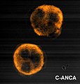
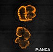
ANCA stands for anti-neutrophil cytoplasmic antibodies.  They are mainly IgG.  They are subdivided into 2 groups based on the patterns produced when they are stained:


1. cANCA
  • cytoplasmic, anti PR3
  • found in Wegeners (90%), mircroscopic polyangiitis (40%)
  • some correlation between level and disease activity


2. pANCA
  • perinuclear, anti MPO
  • found in churg strauss (60%), crescentic glomerulonephritis (80%), microscopic polyangiitis (60%), Wegeners (60%)
  • may also be seen in IBD, RA, SLE, sjogrens, autoimmune hepatitis


Thats it for today, no war at present but I'll try and post one later

Friday, 24 September 2010

MRCP revision battle 19.1: Myelofibrosis

I have more time than usual to revise this week and am actually finding it harder to sit down and get on with it... must have a quiet word with the motivational centres of my brain (which I believe are the mesolimbic areas... anyone care to correct me?)  Anyway, today's battles will be:

MRCP revision battle 19.1: Myelofibrosis
MRCP revision battle 19.2: Hepatitis
MRCP revision battle 19.3: Peutz Jegher Syndrome
MRCP revision battle 19.4: Familial Adenomatous Polyposis
MRCP revision battle 19.5: Hereditary Non-Polyposis Colorectal Cancer
MRCP revision battle 19.6: LTOT
MRCP revision battle 19.7: Exercise tolerance tests
MRCP revision battle 19.8: Lead poisoning


MRCP revision battle 19.1: Myelofibrosis


Myelofibrosis has always been a secret 'sweet-spot' of mine - I think because of its 'teardrop cells' - so its a good battle to start this unmotivated day on...


Myelofibrosis is fundamentally fibrosis of the bone marrow.  There is hyperplasia of megakaryocytes which produce platelet-derived growth factor, leading to:
  1. marrow fibrosis
  2. haemopoesis being forced to move to the spleen and liver.

Features of myelofibrosis include:
  • lethargy
  • weight loss
  • night sweats
  • massive hepato/splenomegaly

Investigations show:
  • raised WCC, low Hb
  • teardrop pokilocytes
  • leucoerythroblastic cells (=nucleated red cells)
  • raised LDH and urate as increased cell turnover
  • bone marrow may produced a 'dry tap'

Image below shows teardrop cells.


    Treatment is ?allogenic stem cell transplant.

    Prognosis is poor with a median survival of 4 to 5 yrs.


    Onwards to hepatitis.... 

    MRCP revision battle 19.2: Hepatitis

    There are 5 'flavours' of viral hepatitis, labeled A to E.  Important general points to learn are:
    • all are caused by RNA virus' except hepatitis B, which is a DNA virus
    • B and C are spread by blood/sexual contact
    • A and E are spread by the faecal-oral route
    • Hepatitis D essentially is only ever found as a co-infection, never alone.

    So to explore the 3 most popular MRCP 'flavours' in more depth....


    Hepatitis A:
    • spread faeco-orally, so look out for questions featuring backpackers returning home
    • incubation period is 2-6 weeks
    • presents as fever, malaise, nausea
    • patient likely to be jaundiced and may have hepato/splenomegaly
    • treatment is supportive
    • patients generally make a full recovery

    Hepatitis B:
    • symptoms are similar to hep A
    • incubation period is longer at 1 - 6 months
    • spread is blood/bodily fluids - questions likely to hint at male business traveller or other innuendo
    • important to fully grasp the various antigens and their meanings:
      • HBsAg: present for 1-6 months after exposure; if present for >6 months patient is a carrier
      • HBeAg: present for 1.5-3 months after exposure and is marker of high infectivity
      • anti HBC IgM - signifies acute infection/carrier
      • anti HBC IgG - may be acute infection, carrier or cleared infection
      • anti HBS - if present with anti HBC suggests recovered from hep B and naturally immune; if present alone suggests hep B vaccination
    • complications of hepatitis B include:
      • 5-10% chronic hepatitis
      • glomerulonephritis
      • increased risk hepatocellular carcinoma
      • cryoglobulinaemia

    Hepatitis C:
    • is spread by blood/sexual contact
    • blood pre 1991 wasn't screened for hep C
    • <20% get an acute hepatitis but 80% get chronic hepatitis
    • breast feeding is NOT contraindicated (a common MRCP fascination for some reason)
    • complications:
      • 80% chronic hepatitis
      • 20% cirrhosis
      • increased risk hepatocellular carcinoma
    • Treatment: IFN alpha and ribavirin


    After that wizz through some high-yield viral hepatitis facts lets move on to Peutz Jegher Syndrome

    MRCP revision battle 19.3: Peutz Jegher Syndrome

    Peutz Jegher Syndrome is a condition characterised by:
    • pigmented 'freckles' (macules) on lips, face, palms and soles
    • hamartomatous polyps in gastrointestinal tract


    It is an autosomal dominant condition caused by a mutation of gene LKB1


    Complications of PJS include:
    • obstruction/intususception
    • GI haemorrhage
    • iron deficiency
    • colicky abdo pain
    • malignant transformation - around 50% of sufferers will have died from a cancer by the age of 60.

    There is no specific treatment.


    So lets move on from a relatively benign cause of multiple colonic polyps to a less benign one....

    MRCP revision battle 19.4: Familial Adenomatous Polyposis

    Familial adenomatous polyposis is an inherited condition in which patients develop literally thousands of polyps in the GI tract.  Virtually all sufferers will get cancer, usually in their 30s or 40s.


    It is caused by a mutation in the tumour supressor gene APC on chromosome 5.


    The appearance of the colon is:


    Treatment tends to be a colectomy before cancer develops.


    As an aside, a variation of FAP called 'Gardners Syndrome' exists.  This is FAP plus osteomas, epidermal cysts, fibromas and retinal pigmentation.


    So after 2 conditions which are defined by polyps, lets look at one which defines itself by not having polyps - Hereditary Non-Polyposis Colorectal Cancer

    MRCP revision battle 19.5: Hereditary Non-Polyposis Colorectal Cancer

    Hereditary Non-Polyposis Colorectal Cancer is an autosomal dominant condition associated with an increased risk of colorectal cancer.


    90% of patients with HNPCC develop cancers, usually in the proximal colon and often poorly differentiated/highly aggressive cancers.


    There is also an increased risk of breast, ovary and endometrial cancers.


    HNPCC is defined by the Amsterdam criteria:
    • at least 3 family members with colon cancer (one a 1st degree relative)
    • at least 2 generations affected
    • at least 1 diagnosed under 50 yrs of age


    Thats quite enough bowels for one day, lets come up for some oxygen...

    MRCP revision battle 19.6: LTOT

    LTOT = long term oxygen therapy = 15 or more hours of oxygen per day.


    This is the only treatment to increase survival in COPD.


    NICE recommends it if:
    • PaO2 <7.3kPa OR
    • PaO2 <8kPa if
      • secondary polycythaemia
      • nocturnal hypoxia (questions may note 'morning headaches;
      • peripheral oedema
      • pulmonary hypertension


    How about now trying our exercise tolerance...

    MRCP revision battle 19.7: Exercise tolerance tests

    Exercise tolerance tests are starting to go out of fashion but are still currently a mainstay of assessment of angina. 

    I'd recommend this BMJ article for a comprehensive discussion; what follows below are just a few salient MRCP points.


    ETTs are carried out according to the Bruce protocol, which is 7 sections each 3 minutes long so a maximum of 21 minutes of exercise.  A modified bruce can be used in higher risk/frailer patients.


    Beta blockers must be stopped the day before an ETT
    Digoxin should be stopped a week before.


    The test is deemed to be positive if:
    • anginal symptoms
    • BP decreases by 15mmHg or fails to increase on exercise
    • arrhythmia
    • ST depression
    • failure to achieve target heartrate (220-age in men, 210-age in women)
    • ST elevation


    Now for the last battle of the day, lead poisoning

    MRCP revision battle 19.8: Lead poisoning

    Lead poisoning is a good topic as it has 2 nice distinctive features (blue lines on nail growth margins and basophillic stippling of red blood cells.)


    A more comprehensive list of lead poisoning features is:
    • abdominal pain
    • constipation
    • peripheral neuropathy
    • fatigue

    Blue lines on the growth margin of nails affect 20% adults with lead poisoning but are rare in children.


    Investigations should show:
    • microcytic anaemia
    • basophillic stippling of red blood cells
    • raised aminolevulinic acid levels (a haem precursor)


     The image below shows basophillic stippling of red blood cells (right and left arrows)


    Treatment is by chelation with either EDTA, d-penicillamine or dimercaprol.


    Thats today wrapped up; questions on yesterday's battles can be found here

    MRCP questions: War 18

    As with previous 'wars' after 'battles' these are just a few quick questions to see if your brain cells have retained the information provided in battles 18.1 to 18.8.

    Grab a piece of paper, jot down your answers then compare them to my answers here


    Question 1:
    Which 4 clotting cascade factors is vitamin K a cofactor for?


    Question 2:
    Your patient has an INR of 7 but no evidence of bleeding.  What would you do?


    Question 3:
    A 20 year old comes to you with a rash across their back of teardrop-shaped lesions topped with some silver scales.  They note they had a sore throat a week or 2 ago.  What is the rash?


    Question 4:
    State the classical triad of symptoms for aortic stenosis.


    Question 5:
    A patient tells you their cardiologist said the pressure gradient across their aortic valve was 52mmHg.  What grade of severity is their stenosis?


    Question 6:
    Your houseofficer has diagnosed PMR and wants to know how to treat it.  What would you prescribe?


    Question 7:
    A mum brings her 16 year old son who has learning difficulties to you, saying he has been itching his bottom a lot recently.  You think he has threadworms.  How would you treat them?


    Question 8:
    Your consultant states he is admitting someone with pseudopseudohypoparathyroidism.  What blood results would you expect?


    The answers are here

    Wednesday, 22 September 2010

    MRCP revision battle 18.1: Warfarin

    Today is another 'octopus' day with 8 battles to embrace (three of which are mercifully brief)

    So the topics are:

    MRCP revision battle 18.1: Warfarin
    MRCP revision battle 18.2: Psoriasis
    MRCP revision battle 18.3: Beau's lines
    MRCP revision battle 18.4: Elliptocytosis
    MRCP revision battle 18.5: Aortic stenosis
    MRCP revision battle 18.6: Polymyalgia rheumatica
    MRCP revision battle 18.7: Threadworm
    MRCP revision battle 18.8: Pseudohypoparathyroidism


    MRCP revision battle 18.1: Warfarin


    Ah, warfarin... everyone's favourite out-of-hours bleep as a houseofficer.  Originally made as a rat poison, it quickly became a favourite in the UK for the treatment of DVTs, PEs and prevention of clots associated with mechanical heart valves.


    Warfarin works by inhibiting vitamin K epoxide reductase, which prevents vitamin K being recycled.  Since vitamin K is a cofactor for factors II, VII, IX and X it decreases coagulation.


    Note however that vitamin K is also a cofactor for protein C and protein S, which help inhibit the clotting cascade.  As protein C is initially affected more than the clotting factors by lack of vitamin K warfarin is initially a procoagulant and when started therefore increases the risk of thrombosis.


    Warfarin is monitored by INR.  The targets are:
    • PE/DVT: INR 2-3
    • metallic valve: INR 3-4

    The duration of anticoagulation depends on the cause of the clot:
    • below knee DVT after surgery: 6 weeks
    • above knee DVT/PE after surgery: 3 months
    • DVT/PE without precipitating factor identified: 6 months

    1-2% of patients on warfarin have a haemorrhage per year.

    Other complications of warfarin treatment include:
    • increased risk of osteoporosis
    • purple toe syndrome - toes turn purple ?secondary to cholesterol deposits 3 to 8 weeks after starting warfarin


    Warfarin levels are notoriously difficult to regulate.

    Things which increase the effect of warfarin include:
    • erythromycine/clarithromycin
    • metronidazole
    • hyperthyroidism
    • cranberry juice
    • alcohol
    • amiodarone
    • propranolol
    • fluconazole
    • isoniazod
    • cimetidine
    • omeprazole

    Things which decrease the effect of warfarin include:
    • rifampacin
    • carbamazepine
    • chlordiazepoxide
    • avocado in excess!

    Management of INR outside the desired range depends both on the level and if there is bleeding:
    • major bleed
      • stop warfarin
      • give vit K 5-10mg IV
      • give prothrombin complex (II, VII, IX, X) or FFP if this is not available
    • INR>8 but no/minimal bleeding
      • stop warfarin
      • give vit K 2.5-5mg PO/0.5-1mg IV
      • restart warfarin when INR<5
    • INR 5-8, no bleeding
      • stop warfarin
      • restart when INR<5
    • INR 5-8 minor bleeding
      • stop warfarin
      • give 1-2.5mg vitamin K PO
      • restart when INR<5


    As an aside, with an eye on the future, a new drug called dabigatran is looking to usurp warfarin as king of the anticoagulants.  Trials such as RELY and RECOVER have shown dabigatran to be non-inferior to warfarin in preventing thrombosis, to cause fewer bleeding adverse effects and even better not require regular blood tests to check levels.  Of course its expensive and so not yet approved...


    On to the second battle of the day, psoriasis...

    MRCP revision battle 18.2: Psoriasis

    Psoriasis is an autoimmune disease that classically causes a silver-scale topped rash on the skin.

    It affects 1-2% of the population.

    The cause is believed to be abnormal activity of the type 1 T helper cells.
    It is associated wtih HLA CW6, B13, B17 and B27.


    Females tend to be affected at a younger age than males.


    The main 'types' of psoriasis are:
    1. chronic plaque
    2. generalised pustular
    3. palmo-plantar pustulosis
      • strong association with smoking
      • tends to affect middle-aged females
    4. guttate
      • usually young adults/teenagers
      • is often preceded by a strep infection 2-4 weeks before
      • resolves spontaneously in 2-3 weeks
    5. nail
    6. flexural


    Athropathy is a feature in 8% and may be in the form of :
    • symmetric arthritis - appears rheumatoid-like; 50%
    • asymmetric arthrtitis - 35% - dactylitis
    • arthritis mutilans - <5%
    • spondylitis
    • distal interphalangeal predominant


    Psoriasis can be worsened by multiple factors, including:
    • trauma
    • infection
    • post-partum
    • beta blockers
    • lithium
    • stress
    • alcohol
    • NSAIDs

    Management for skin psoriasis is by a host of lotions and potions - coal tar, dithranol, topical vitamin D.... if these haven't already been drummed into you a brief visit to the BAD website (british association of dermatologists, trying to sound cool) to recap might be a good idea.


    Now for a brief skirmish with Beau's lines

    MRCP revision battle 18.3: Beau's lines

    Beau's lines are transverse depressions in nails due to temporary arrest in their growth.

    They are associated with:
    • psoriasis
    • diabetes
    • metabolic disturbances


    Now onwards to another very brief battle, elliptocytosis

    MRCP revision battle 18.4: Elliptocytosis

    Elliptocytosis is an autosomal dominant condition which results in cigar-shaped elliptocytes:


    In severe cases they can cause haemolytic anaemia; happily the majority of people with elliptocytosis have no ill effects from it at all.


    Now on to a more substantial battle again: aortic stenosis

    MRCP revision battle 18.5: Aortic stenosis

    Aortic stenosis may be detected incidentally (keen houseofficer noticing the classical ejection systolic murmur radiating to the carotids) or may present with its classical triad of symptoms that can be remembered as 'DAD':
    • exertional dyspnoea
    • etertional angina
    • exertional dizziness


    Signs of aortic stenosis include:
    • ejection systolic murmur radiating to carotids
    • slow rising pulse
    • narrow pulse pressure
    • heaving apex beat


    Things which suggest severe aortic stenosis include:
    • LVF
    • soft S2
    • paradoxically split A2
    • S4

     The formal divisions of severity are as follows:
    • mild: area >1.5cm, gradient <25
    • moderate: area 1-1.5cm, gradient 25-50
    • severe: area <1cm, gradient >50
    • critical: area <0.7cm, gradient >80


    The main causes of aortic stenosis are:
    • calcification of the valve
    • bicuspid aortic valve


    ECG changes which may be associated with aortic stenosis include:
    • p mitrale
    • LVH
    • LAD
    • poor R wave progression
    • complete heart block if calcification involves the conduction tissue.


    Treatment is surgical.  The operative mortality is around 20-25% if there is LVF, 2-8% if not.


    So from the very factual aortic stenosis on to the slightly touchy-feeling PMR...

    MRCP revision battle 18.6: Polymyalgia rheumatica

    Polymyalgia rheumatica (PMR) is a slightly nebulous condition which the Oxford handbook defines as 'symmetrical aching, tenderness and morning stiffness in shoulders and proximal limb muscles, +/- systemic features'.  These systemic features can include anything from fatigue to anorexia...


    It generally affects over 70s and is rare in under 60s.


    Bloods tend to show a raised ESR


    Treatment is with prednisolone (15mg PO OD) which tends to produce a dramatic improvement in days.  This should be decreased by 1mg per month, and of course anti-OP treatment should be given concurrently.

    If symptoms recur on decreasing the dose and you have a lovely little old lady stuck on >10mg OD for over a year, it is worth considering methotrexate or azothioprine to try and allow you to decrease the steroid.


    As a random aside, 10% of patients with PMR will suffer from carpal tunnel syndrome.


    Now get ready to itch as we move on to the penultimate battle of the day, threadworm....

    MRCP revision battle 18.7: threadworm

    Threadworm, AKA enterobius vernicularis, appears frequently in MRCP questions, usually in relation to patients in institutions.

    They tend to present with perianal itching.

    Treatment is mebendazole 100mg (or piperazine if the patient happens to be under 2 yrs of age)


    On that pleasant note, on to the last battle of the day, pseudohypoparathyroidism

    MRCP revision battle 18.8: Pseudohypoparathyroidism

    Pseudohypoparathyroidism is an autosomal dominant condition that occurs when a person is insensitive to PTH.


    Features include:
    • short metacarpals (esp 4th and 5th)
    • short stature
    • round face
    • obese
    • low IQ
    • dental hypoplasia

    It can be diagnosed by a failure of cAMP to increase after injection of parathyroid hormone.  More traditionally, the clinical picture of low calcium, high PTH and normal/raised alk phos is enough.


    Treatment is with alfacalcidol.

    It is worth noting it is associated with slipped femoral epiphysis.


    To end today's battles on an almost comic note (and the fact I find this comic is a reflection that I really should get out more) there also exists a pseudopseudohypoparathyroidism, which is essentially the morphological features of pseudohypoparathyroidism but with normal biochemistry.  Hopefully that'll make you smile if it comes up!


    A few questions on yesterday's battles are now online here, otherwise see you tomorrow!

    MRCP questions: War 17

    As with previous 'wars' after 'battles' these are just a few quick questions to see if your brain cells have retained the information provided in battles 11.1 to 11.4.

    Grab a piece of paper, jot down your answers then compare them to my answers here


    Question 1:
    What haematological picture do you get in DIC?


    Question 2:
    What symptoms are associated with jugular foramen syndrome?


    Question 3:
    Name the commonest form of thyroid cancer.


    Question 4:
    List 3 causes of unilateral pleural calcification


    Question 5:
    What condition is associated with a 'plucked chicken skin' appearence?


    Question 6:
    Legionella is a gram positive bacteria.  True or false?


    Question 7:
    Which biochemical disturbance is classically associated with legionella?


    The answers are here

    Tuesday, 21 September 2010

    MRCP revision battle 17.1: DIC

    Lots of short battles from a variety of specialities today so buckle up for a ride through some haematology, neurology, endocrinology, respiratory and dermatology.  Yeee-hhaaaa!

    MRCP revision battle 17.1: DIC
    MRCP revision battle 17.2: Jugular foramen syndrome
    MRCP revision battle 17.3: Thyroid cancer
    MRCP revision battle 17.4: Legionella infection
    MRCP revision battle 17.5: Pleural calcification
    MRCP revision battle 17.6: Pseudoxathoma elasticum


    MRCP revision battle 17.1: DIC


    Disseminated intravascular coagulation (=DIC) is the pathalogical widespread activation of coagulation.


    Blood tests will show:
    • prolonged PT
    • prolonged aPTT
    • massively raised d-dimer levels
    • low platelets
    • low fibrinogen
    • schistocytes


    Of these, it is the level of fibrinogen which best correlates to the severity.


    Causes of DIC include:
    • obstetric
    • crush injury
    • septicaemia
    • malignancy
    • transfusion reaction

    The treatment is:
    • treat the cause
    • give platelets if platelets <50
    • cryoprecipitate
    • FFP
    • activated protein C if septic.


    Now lets canter onwards to jugular foramen syndrome....

    MRCP revision battle 17.2: Jugular foramen syndrome

    Jugular foramen syndrome, also known as Vermet's Syndrome, is a condition arising from the compression of the cranial nerves that run through the jugular foramen.


    Now just in case they aren't quite on the tip of your tongue, the nerves that run through the jugular foramen are IX, X and XI.


    The syndrome is therefore characterised by:
    • loss of taste to posterior 1/3 of tongue (CN IX)
    • dysphagia (CN X)
    • sternocleidomastoid and trapezius paralysis (CN XI)

    Anything that compresses the jugular foramen can cause this syndrome.  The commonest cause is a paraganglioma.


    Now we've acquired a bit more esoteric MRCP knowledge, lets gallop on to discover some details about thyroid cancer...

    MRCP revision battle 17.3: Thyroid cancer

    Under 5% of thyroid lumps are malignant, and in general those with a malignant mass will be euthyroid.


    There are 4 main types of thyroid cancer:
    1. papillary - 70% - mainly affects young females, excellent prognosis
    2. follicular - 20% 
    3. medullary - 5% - associated with MEN-2 (more on MEN tomorrow)
    4. anaplastic - 1% - very poor prognosis and resistant to treatment.

    Rarely a patient can have lymphoma of the thyroid - this usually follows hashimotos disease.


    The mainstay of treatment for thyroid cancer is surgery.


    Lets break into a canter as we head on to legionella...

    MRCP revision battle 17.4: Legionella

    Legionella is a gram negative bacteria which causes legionella disease/pneumonia and in MRCP questions is most likely found living in a cruise ship/hotel's water tank or airconditioning system.


    The symptoms of legionella disease are:
    • flu-like
    • dry cough
    • dypnoea
    • GI upset


    Complications of legionella which may show up in the blood results include:
    • SIADH - low sodium
    • hepatitis - deranged LFTs


    Legionella is killed above 60c, cannot multiply between 50 and 60c and are happiest between roughly 35 to 45 c


    Males are more affected than females, 3:1


    CXR may show bibasal consolidation.


    Treatment is with antibiotics which achieve a high intracellular concentration, such as clarithromycin, rifampicin or fluroquinolone.


    Legionella has a 10% mortality.


    Lets now trot on to pleural calcification...

    MRCP revision battle 17.5: Pleural calcification

    Pleural calcification pops up both in real life and in MRCP questions intermittently.


    The commonest cause is asbestosis. 
    This tends to be bilateral and spare the costophrenic angles


    So, if the pleural calcification is unilateral consider:
    • previous haemothorax
    • previous TB pleuritis
    • previous empyema

    Other differentials to consider are:
    • previous radiotherapy/radiation exposure
    • post talc pleurodesis, which will mimic calcification.


    Lets return to a full-speed gallop to our last battle of the day, pseudoxanthoma elasticum

    MRCP revision battle 17.6: Pseudoxanthoma elasticum

    Pseudoxanthoma elasticum is fragmentation and calcification of elastic fibres.


    It is an autosomal recessive condition, found on chromosome 16.


    Its various associations are:
    • a characteristic 'plucked chicken' skin (google it, memorise it, get ready to spot it in part 2)
    • angioid streaks in retina/vision problems
    • rarely GI bleeds
    • claudication/cardiovascular problems.


    There is no treatment so management is symptomatic only.


    Thats all for today, proceed to the war if you are up to.

    MRCP questions: War 16

    As with previous 'wars' after 'battles' these are just a few quick questions to see if your brain cells have retained the information provided in battles 16.1 to 16.7.

    Grab a piece of paper, jot down your answers then compare them to my answers here


    Question 1:
    List 8 symptoms/signs of pagets disease


    Question 2:
    Which oral medication would you give to treat pagets disease?


    Question 3:
    Which medication would you use to treat PCP?


    Question 4:
    A patient has muscle weakness that gets worse on exercise.  What medication would you give them?


    Question 5:
    A patient tells you they have weak muscles which get better on exercising.  They state they believe its due to their lung cancer.  What condition do they have?


    Question 6:
    ]What is the commonest cause of hemiballismus?


    Question 7:
    What is the medical treatment for hyperprolactinoma?


    Question 8:
    'Waiters tip' is associated with which palsy?


    The answers are here

    Monday, 20 September 2010

    MRCP revision battle 16.1: Pagets disease of bone

    In between catching up on seemingly endless loads of washing (do socks breed when left alone in a wash basket?) a few hours of MRCP revision has been done and 7 new battles have been written...

    MRCP revision battle 16.1: Pagets disease of bone
    MRCP revision battle 16.2: Pneumocystis carinii
    MRCP revision battle 16.3: Myasthenia gravis
    MRCP revision battle 16.4: Lambert-Eaton syndrome
    MRCP revision battle 16.5: Hemiballismus
    MRCP revision battle 16.6: Prolactin
    MRCP revision battle 16.7: Erbs palsy


    MRCP revision battle 16.1: Pagets disease of bone


    Pagets disease of bone is accelerated, disorganised bone turnover due to increased number and activity of osteoclasts and osteoblasts.


    It is rare in under 40s.


    Predisposing factors include:
    • increasing age
    • northern latitude
    • family history
    • being male


    Features of Pagets include:
    • bone pain
    • bowing of tibia
    • bossing of skull
    • secondary arthritis
    • deafness/other cranial nerve compression
    • high output heart failure
    • pathological fractures
    • bone sarcomas (occur in 1% over 10 yrs)

    My way of remembering the features of pagets is this slightly random rhyming story:
    "Bowing to the Boss, oh what a pain,
    I'm deaf and arthritic and since the fracture I'm lame
    my heart's now a failure, and oh what a shame
    I've got bone sarcoma - thats the end of the game"


    Bloods in Pagets show:
    • raised ALP
    • normal calcium (unless a long period of immobility)
    • normal phosphate


    Treatment is alendronic acid


    That wasn't too bad... onwards to battle 16.2...

    MRCP revision battle 16.2: Pneumocystis carinii

    Pneumocystis carinii, AKA pneumocystis jiroveci, is a unicellular eukaryote.

    It can cause pneumonia in patients with HIV and accounts for 40% of all AIDS-defining illness.
    If CD 4 count is less than 200 PCP prophylaxis (=co-trimexazole) should be started.


    Features of PCP:
    •  dyspnoea
    • dry cough
    • fever
    • few chest signs on examination
    • desaturation on exercise


    CXR typically shows mid-lower bilateral interstitial infiltrates, but it may look normal.

    Sputum cultures are often negative so BAL may be needed.


    Treatment is:
    • co-trimexazole
    • IV pentamidine if severe
    • steroids if hypoxia
      • use of steroids decreases respiratory failure by 50% and death by 1/3)


    On to battle 3 of the day and a classic condition - myasthenia gravis.

    MRCP revision battle 16.3: Myasthenia Gravis

    Myasthenia gravis is an autoimmune condition in which muscles become easily tired and weak.  It affects 1 in 20 000.


    90% of patients have autoantibodies to nicotinic acetylcholine receptors.
    If these are negative consider looking for MUSK antibodies (= muscle specific kinase)


    The key feature of MG is repetitive stimulation leading to decreased evoked response.


    The order in which muscle groups are affected in MG is:
    • extraoccular (--> diplopia)
    • bulbar
    • face
    • neck
    • lumb
    • trunk

    Reflexes are normal in MG.


    Test of choice: tensilon test = edrophonium


    Treatment of MG is:
    • cholinesterase inhibitors eg pyridostigmine
    • prednisolone if pyridostigmine unsuccessful (but be aware that paradoxically steroids can worsen MG)
    • plasmaphoresis/IV IG if respiratory muscle involvement.

    If there is hyperplasia of thymus/thyroma, up to 60% of cases will remit after surgery.


    Up to 10% of patients with MG will have hyperthyroidism.


    Drugs which exacerbate MG include:
    • penicillamine
    • quinidine
    • beta blockers
    • lithium
    • phenytoin
    • gentamycin

    Now on to the 'similar but different' Lambert-Eaton syndrome...

    MRCP revision battle 16.4: Lambert Eaton Syndrome

    Lambert-Eaton syndrome is a rare condition affecting voltage-gated calcium channels which results in:
    • muscular weakness that improves with activity
    • autonomic symptoms (dry mouth, constipation, impotence)
    • hyporeflexia
     

    It is most commonly a paraneoplastic phenomenon, usually due to small cell lung cancer (very rarely due to breast or ovarian cancer).  It can also be autoimmune.


    Treatment is 3,4 diaminopyridine.

    Regular CXR should be performed as Lambert-Eaton may preceed the development of small cell lung cancer by up to 4 years.


    Notice how although both MG and Lambert-Eaton are characterised by muscle weakness there are lots of differences - weakness gets worse on repetition in MG but better in LE, reflexes are normal in MG but reduced in LE and there may be autonomic symptoms in LE but not MG.  Also not involvement of occular muscles is far more common in MG.


    Lets now diversify to something completely different - hemiballismus...

    MRCP revision battle 16.5: Hemiballismus

    Hemiballismus is large amplitude, flinging hemichorea.


    Its cause is damage to the contralateral subthalamic nuclei.


    The commonest cause is stroke.


    Treatment:
    • hemiballismus generally resolves spontaneously in 4 to 8 weeks
    • haloperidol (a dopamine blocker) may be used
    • tetrabenazine (a VMAT inhibitor) may be used

    Bilateral hemiballismus is rare and tends to be associated with HONK.


    After that brief battle onwards to the meatier topic of prolactin

    MRCP revision battle 16.6: Prolactin

    Prolactin is a hormone released from the anterior pituitary.
    It is under the negative control of dopamine (ie dopamine prevents its release)


    Raised prolactin causes a drop in GnRH, LH, oestrogen and testosterone levels.


    Symptoms/signs of raised prolactin include:
    • galactorrhoea
    • decreased libido
    • amenorrhoea
    • infertility

    Note that gynaecomastia is NOT associated with raised prolactin.


    Causes of a raised prolactin include:
    • physiological causes
      • pregnancy
      • breast feeding/nipple stimulation
      • stress
      • post-fit
      • exercise
    • drugs
      • phenothiazides
      • metoclopramide
      • methyl-dopa
      • haloperidol
      • oestrogens
    • metabolic causes
      • hypothyroidism
      • PCOS
      • acromegaly (1/3)
      • chronic renal failure
    • disease
      • prolactinoma
        • microprolactinoma (<10mm) release more prolactin than macroprolactinomas
      • compression of pituitary stalk removing dopamine control

    Treatment is with bromocriptine.
    It is best to give it in the evening as it causes postural hypotension


    Very nearly there.  Last battle of the day will be Erbs palsy....

    MRCP revision battle 16.7: Erbs palsy

    Erbs palsy is the name given to a particular brachial plexus injury, generally caused by shoulder dystocia during birth.


    Any nerve root of the brachial plexus may be affected but the most commonly affected roots are C5 and C6.


    This results in loss of:
    • shoulder abduction
    • elbow flexion
    • suppination

    The net outcome is that the arm hangs medially rotated with the forearm pronated = "waiters tip"


    So thats all for today's battles - on to today's war to check recall of yesterday's battles!

    MRCP questions: War 15

    As with previous 'wars' after 'battles' these are just a few quick questions to see if your brain cells have retained the information provided in battles 15.1 to 15.8.

    The answers are available here


    Question 1:
    List the 3 commonest sites of mets from renal cell carcinomas


    Question 2:
    What abnormalities on blood tests might you see with renal cell carcinoma?


    Question 3:
    What is Von Hippel Lindau disease?


    Question 4:
    A patient complains of severe right shoulder pain followed by weakness of the right arm.  There is no history of trauma.  What is the likely diagnosis?


    Question 5:
    A patient has abdo pain.  The busy nurse just leaves their urine sample in front of you.  Ten minutes later you notice it has turned deep red.  What condition does the patient have?


    Question 6:
    List 3 drugs that can precipitate acute porphyria


    Question 7:
    A medical student excitedly tells you the urine sample they've just taken glows pink under woods light.  What condition does the patient have?


    Question 8:
    Your newest patient has heart block, opthalmoplegia and pigmentary degeneration of the retina.  What test do you want to do to confirm your diagnosis?


    Question 9:
    List 7 symptoms associated with pellegra


    Question 10:
    Which drug is used to treat neuroleptic malignant syndrome?

    Sunday, 19 September 2010

    MRCP revision battle 15.1: Renal cell carcinoma

    I've had a day off in which I've covered literally hundreds of miles and drank excessive amounts of tea and cider and seen far more gingham than any one person ever should.  Net result has been a restoration in my energy levels and so today is going to be a blockbuster of a set of battles, with 8 separate topics to cover including two that I'd never even heard of in my preceding 8 years of studying medicine.  So lets get started!


    MRCP revision battle 15.1: Renal cell carcinoma
    MRCP revision battle 15.2: Von Hippel Lindau
    MRCP revision battle 15.3: Neuralgic amyotrophy
    MRCP revision battle 15.4: Porphyrias
    MRCP revision battle 15.5: Kearns-Sayre
    MRCP revision battle 15.6: Klinefelters
    MRCP revision battle 15.7: Pellegra
    MRCP revision battle 15.8: Neuroleptic malignant syndrome


    MRCP revision battle 15.1: Renal cell carcinoma


    Renal cell carcinoma, also known as hypernephroma or Gravitz Tumour, is a cancer arising in the proximal renal tubular epithelium.

    It accounts for 85-90% of primary renal cancer.


    The classic triad for renal cell carcinoma is:
    1. haematuria
    2. loin pain
    3. abdominal mass
    (NB: classic triads of course rarely exist, but may kindly present themselves during MRCP questions)


    Renal cell carcinoma may also be linked to pyrexias of unknown origin.
    Another clinical sign to look out for is left variocele - caused by invasion of the left renal vein causing compression of the left testicular vein.


    Factors associated with developing renal cell carcinoma include:
    • smoking
    • middle aged males (male:female 2:1)
    • Von-Hippel Lindau syndrome (wait for the next battle if you don't know that that is)
    • tuberous sclerosis
    • acquired cystic kidney disease in renal failure


    25% of patients with renal cell carcinoma have mets at presentation.  The commonest mets are bone, liver and lung.


    Endo effects associated with renal cell carcinoma include:
    • erythropoietin --> polycythaemia
    • PTH --> raised calcium
    • renin --> raised BP
    • ACTH

    Treatment is surgery.  If mets are present IL-2 or interferon alpha may also be considered.


    There is a 45% 5 yr survival.


    Now on to meet Von-Hippel-Lindau syndrome...

    MRCP revision battle 15.2: Von Hippel Lindau disease

    Von Hippel Lindau disease is a rare autosomal dominal condition that results in haemangioblastomas in:
    • cerebellum (may present as ataxia)
    • spinal cord
    • kidney
    • retina (may present as visual loss)

    It is associated with bilateral renal cell carcinomas (develop in approximately 50% of patients with VHL) and pheochromocytomas.


    For those of you who are feeling really geeky the gene responsible is on chromosome 3 and it is a mutation in a tumour supressor gene.


    So after that brief acquaintance, lets move on to a condition I'd never heard of before today - neuralgic amyotrophy...

    MRCP revision battle 15.3: Neuralgic amyotrophy

    Neuralgic amyotrophy.  Never heard of it?  Me neither before today, so here are just a few facts to get you through MRCP...


    Neuralgic amyotrophy is sometimes also known as brachial plexus neuropathy.


    It has a particularly specific presentation:
    • severe pain in shoulder
    • followed by weakness and atrophy of muscles in arms


    This presentation alone in an MRCP question should point you towards neuralgic amyotrophy being the answer.  Other clues which would suggest this diagnosis include:
    • preceeded by a URTI
    • decreased or absent reflexes
    • sensory abnormalities

    Rarely the diaphragm may also be affected.

    There is a risk of shoulder joint subluxation or winging of the scapula.


    This link takes you to an image of what just basic neuralgic amyotrophy can look like.


    In general it resolves spontaneously in a few months, and treatment is conservative.


    After that interesting interlude, lets do battle with the porphyrias...

    MRCP revision battle 15.4: Porphyrias

    Porphyrias are rare genetic disorders causing abnormalities of enzymes responsible for haem synthesis, resulting in accumulation of intermittent compounds (porphobilinogen, aminolaevulinic acid) and porphyrins.


    This battle will focus on 2 acute porphyrias (acute intermittent and variegate) and 1 chronic (porphyria cutanea tarda).


    1. Acute intermittent porphyria


    Acute intermittent porphyria is an autosomal dominant condition in which there is a defect in porphobilinogen deaminase

    Females aged 20-40 are most affected.


    There is always raised urinary porphobilinogen and aminolaevulinic acid, but the rise is greater during acute attacks.  The importance of these substances is that the urine goes deep red on being left.


    Signs and symptoms include:
    • abdominal pain
    • hypertension
    • low sodium
    • low potassium
    • hypotonia
    • psychosis
    • paralysis
    • seizures
    Note there are NO skin symptoms.

    A way to remember this list is to think of a patient saying to you "oh doctor, my abdo pain is giving me high blood pressure and stopping me being able to stand (paralysis)"... to which you may think "ah yes, you are a teaspoon (tsp = low tone, sodium, potassium) of crazy (psychosis) aren't you?"


    Drugs that can precipitate acute porphyria include:
    • alcohol
    • benzos
    • rifampicin
    • tetracyclines
    • phenytoin
    • OCP
    • halothane
    • sulphonamides


    Treatment is:
    • remove precipitating factores
    • IV fluid to correct low sodium/potassium
    • high carb diet
    • IV haematin
    • prochlorperazine for nausea
    • other symptomatic treatment

    Although a rare diagnosis, porphyria should always be a differential in someone presenting with abdominal pain.


    2. Variegate porphyria


    This is an autosomal dominant condition characterised by a deficit in protoporphyrin oxidase.

    It can cause abdominal pain and neuro symptoms, but in contrast to acute intermittent porphyria it also has a photosensitive blistering rash.


    It is commoner in South Africans (afrikaans)


    3. Porphyria Cutanea Tarda


    This is due to uroporphyrinogen decarboxylase deficiency.


    It results in a photosensitive rash with bulla.
    It is also associated with hypertrichosis.
    (these two factors may be the basis of werewolf legends...)


    Urine has raised uroporphyrinogen which glows pink under a Woods light


    Attacks are precipitated by alcohol, iron and oestrogen.


    Treatment is with chloroquine.


    Wow, that was quite long.  The next battle is far shorter, and is on the second condition I'd never heard of before today...

    MRCP revision battle 15.5: Kearns Sayre

    Kearns Sayre is an exceptionally rare condition caused by mitochondrial DNA mutations which results in:
    • progressive external opthalmoplegia
    • pigmentary degeneration of retina
    • heart block

    It may also cause proximal muscle weakness, ptosis and ataxia.
    Symptoms must begin before the age of 20.


    If part 2 asks what investigation you want to do, the correct answer is muscle biopsy looking for ragged red fibres.


    Onwards for another brief eponymous syndrome, Klinefelters..

    MRCP revision battle 15.6: Klinefelters

    Klinefelters is the commonest sex chromosome disorder, affecting roughly 1 in 1000 men.

    Its karyotype is 47 XXY


    The key features are:
    • tall stature
    • small testes
    • azoospermia
    • gynaecomastia


     Bloods show:
    • low testosterone
    • high LH/FSH


    Treatment is with testosterone


    Note there is increased risk of OP and breast cancer.


    So on to the penultimate battle of the day, pellegra...

    MRCP revision battle 15.7: Pellegra

    Pellegra is nicotinic acid deficiency = vitamin B3 deficiency.


    Symptoms include:
    • diarrhoea
    • dementia
    • dermatitis
    • depression
    • ataxia
    • insomnia
    (remember as in a 4d)


    Causes of pellegra include:
    • alcoholism
    • diet
    • isoniazid
    • carcinoid syndrome


    And we're finally about to embark on the last battle of this epic day, neuroleptic malignant syndrome...

    MRCP revision battle 15.8: Neuroleptic malignant syndrome

    Neuroleptic malignant syndrome is a condition that can complicate antipsychotic use. 


    Its features are:
    • pyrexia
    • rigidity
    • tachycardia

    Bloods show:
    • raised CK
    • leucocytosis
    • acidosis

    It is commonest in young males, and onset is usually in the first 10 days of taking the drug or after the dose has been increased.


    It is important not to miss as it has a 10% mortality.


    Treatment is to stop the antipsychotic, give IV fluids, dantrolene and ?bromocriptine.


    That's all for today, I'll catch up on the 'wars' tomorrow after I've caught up on sleep!

    Friday, 17 September 2010

    MRCP revision battle 14.1: Schistosomiasis

    Its a very random bag today, with our first adventure in the world of infectious diseases, some clotting-based battles in the arena of haematology then a tricyclic overdose to round it all off!


    MRCP revision battle 14.1: Schistosomiasis
    MRCP revision battle 14.2: The clotting cascade
    MRCP revision battle 14.3: Factor V Leiden
    MRCP revision battle 14.4: Thrombocytopenia
    MRCP revision battle 14.5: ITP
    MRCP revision battle 14.6: TCA overdose


    MRCP revision battle 14.1: Schistosomiasis

    Warning... this is a revision topic likely to make you itch...


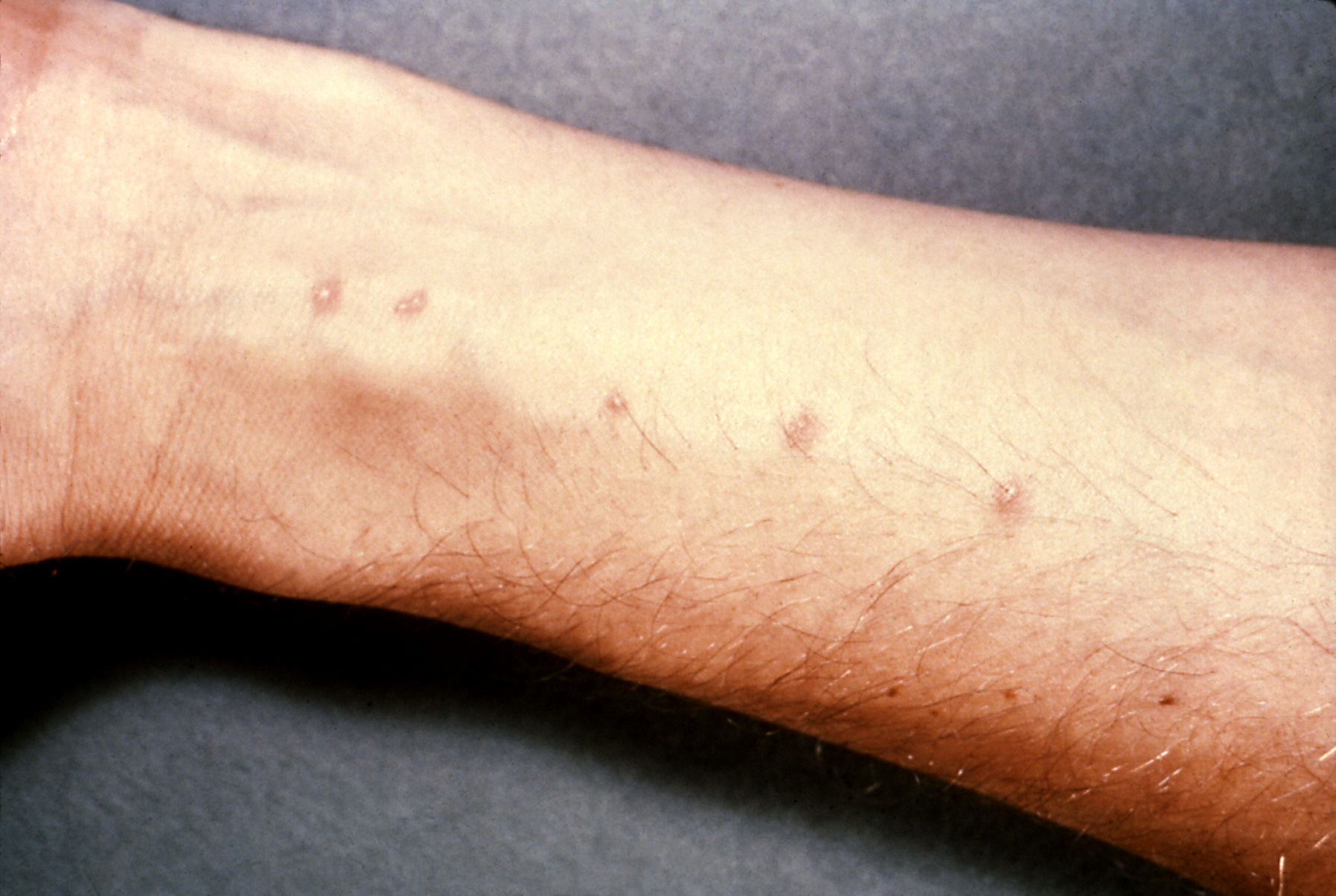
    Schistosomiasis is a parasitic disease caused by flukes (=trematodes.)   It is generally carried by snail vectors that release cercariae (= parasitic lavae)

    The first sign of infection may be 'swimmers itch'.  The MRCP exam may give a history of a patient who has been on holiday to Asia/Africa/South America and has been swimming in a river and is now itchy with a rash.  In part 2 there may also be a picture like the one below:


    The rash tends of appear within hours of infection and disappear within a week.


    The classical presentation of acute schistosomiasis itself occurs roughly 2 weeks after infection with:
    • fever
    • urticaria
    • diarrhoea
    • abdominal pain
    • cough
    • hepatosplenomegaly
    This acute version of schistosomiasis is known as Katayama Fever


    The treatment is praziquantel.


    If schistosomiasis is not recognised and treated it can become a chronic granulomatous disease (granulomas form around the schistosomiasis eggs).  Which parts of the body are affected depends on the type.  For the purposes of MRCP the following associations are worth learning:
    • s.mansoni - liver disease, transverse myelitis
    • s.japonicum - liver disease
    • s.haematobium - urinary tract/bladder disease (haematuria)
      • --> increased risk of squamous cell carcinoma of bladder


    The most practical way of diagnosing is by looking for eggs in stool/urine.


    It goes without saying that any question with an eosinophillia in the blood results of someone who has been travelling should raise the possibility of this as a diagnosis.


    Now on to the less exotic world of haematology and a recap of the clotting cascade!

    MRCP revision battle 14.2: The clotting cascade

    Before we start tackling clotting disorders a recap of the clotting cascade would be useful.


    A schematic of the clotting cascade is:

    Its really important to get this in your head as various questions are likely to be indirectly testing your knowledge of it.  I remember the intrinsic pathway into the common pathway as by a little jingle of     " 12,11,9 then 10 (pause) 2, 1 then back again " with the 'back again' bit referring to factor 13 getting in on the action right at the very end to stablise the clot and hence the numbers returning back to being higher.

    Also note where VIIIa and Va are involved.


    Now look at the same schematic again but with the addition of activated protein C and antithrombin:


    So as well as learning the cascade itself make sure the following are etched in your brain:
    • activated protein C inhitbits VIIIa and Va
    • antithrombin inhibits Xa and IIa 
    • protein C is converted to activated protein C by protein S        and finally
    • factors II, VII, IX, X are vitamin-K dependent (method of remembering: '1972')


    With all that groundwork covered, lets  move on to battle 14.3 and put a bit of it into practice.

    MRCP revision battle 14.3: Factor V Leiden

    A point mutation on the gene for clotting factor V results in a varient known as 'factor V Leiden'.
    It is inherited in an autosomal dominant fashion.


    The result of the mutation is that factor V is insensitive to protein C inactivation, resulting in hypercoagulability.


    This leads to increased risk of PE/DVTs and in females increased miscarriage.


    Merely being factor V leiden positive doesn't necessitate anticoagulation; warfarin etc would only be considered in response to events rather than prophylactically - indeed, up to 7% of all white europeans/north americans have factor V leiden, making it the commonest inherited thrombophilia.


    So having looked at a cause of thrombophillia, lets switch to the opposite and consider low platelets...

    MRCP revision battle 14.4: Thrombocytopenia

    Thrombocytopenia refers to low numbers of platelets.
    Generally the lower limit of normal for platelets is 150 000, with thrombocytopenia often defined as <50 000.


    Low platelets increase the risk of bleeding/bruising, commonly manifesting as bleeding gums or nose bleeds.


    Causes of thrombocytopenia can be broadly divided into causes that increase platelet destruction and causes that decrease production:
    1. increased platelet destruction (= DISC HH)
      • DIC
      • ITP
      • SLE
      • CLL
      • Heparin
      • Hypersplenism (portal hypertension, leukaemia)
    2. decreased platelet production (=CAB M)
      • Chemo/radiotherapy
      • aplastic anaemia
      • B12/folate deficiency
      • marrow infiltration
    Isotretinoin and valproic acid have also been associated with thrombocytopenia.


    Treatment is dependent on the cause/the symptoms.


    Onwards to the last haematological battle of the day, ITP...

    MRCP revision battle 14.5: Idiopathic Thrombocytopenia Purpura

    ITP is a condition which results in low numbers of platelets.  Despite its title of 'idiopathic' it is often autoimmune mediated, with IgG autoantibodies against glycoprotein IIb/IIIa/Ib complex.


    Acute ITP:
    • affects mainly children
    • boys and girls equally affected
    • often after infection/vaccination
    • tends to be self-limiting over 2 weeks

    Chronic ITP:
    • females >males
    • relapsing-remitting


    In ITP the bone marrow shows megakaryocytes


    Treatment is only needed if the patient is symptomatic or the platelets are <20.
    First line = oral prednisolone - 80% respond
    If no response in 3 months --> splenectomy
    Other options include IV IG or immunosupressive drugs.


    One random MRCP type syndrome: Evan's syndrome = ITP +AIHA.


    Nearly there, just a quick tricyclic overdose to go...

    MRCP revision battle 14.6: TCA overdose

    Lets end the day (and the working week) on an overdose.  I've always found it somewhat ironic (and at times in A&E irritating) that a medication which you give to depressed people to treat their depression is so incredibly dangerous in overdose...


    TCA in overdose produce symptoms due to their:
    • anticholinergic effects (dry mouth, dilated pupils, tachycardia, constipation, urinary retention, raised intraocular pressure)
    • alpha blockade (hypotension)
    • sodium channel blockade (my personal favourite, cardiac arrhythmias)


    On the ECG look out for:
    • long PR
    • wide QRS
    • long QT


    In terms of managing these patients you need:
    • ECG and cardiac monitoring
    • consider charcoal if <2 hrs post ingestion
    • bloods for UEs and paracetamol and salicylate (always assume paracetamol taken until proven otherwise)
    • ABG
    • fluids if needed to support BP
    • if acidotic - bicarb
    • dialysis is NOT indicated
    • avoid class Ia/Ic/III antiarrythmics as they all prolong QT.  Also avoid flumazenil (also prolongs QT)
    • observe


    Thats all for today folks, and I'm taking tomorrow off.  And possibly the day after too, depending on my mood.  But battles will definitely recommence on Monday.

    Thursday, 16 September 2010

    MRCP revision battle 13.1: Brugada Syndrome

    I'm afraid I'm too tired to even attempt an insightful intro today so lets just go straight to listing the battles:

    MRCP revision battle 13.1: Brugada Syndrome
    MRCP revision battle 13.2: SVC obstruction
    MRCP revision battle 13.3: Causes long PR
    MRCP revision battle 13.4: Cauda equina syndrome
    MRCP revision battle 13.5: Surviving Sepsis


    MRCP revision battle 13.1: Brugada Syndrome


    Brugada Syndrome is an autosomal dominant condition that causes abnormal sodium channel proteins, leading to a risk of VF/VT and sudden death.


    It is commonest in Asians.

    20-40% of cases are associated with a SCN5A mutation.


    ECG shows partial/complete RBBB with ST elevation in V1 to V2.  The classical ECG is type 1 below, with the other 2 possible variations.

     Treatment is ICD.


    Next up - SVC obstruction...

    MRCP revision battle 13.2: SVC obstruction

    SVC obstruction can be frightening for the doctor as well as the patient - the first lady I saw with it came in with a lovely slim face and as time went on it became more and more puffy and she became increasingly dyspnoeic.  Once seen it will never be forgotten; I can post this photo:
                                                                         Herbert L. Fred, MD and Hendrik A. van Dijk, Wiki Commons

    which demonstrates the type of difference I saw in my patient (normal on right, SVC on left).  My patient however was female and smiled more!


    Anyway, lets refocus on points to learn for the MRCP exam:


    Features of SVC obstruction:
    • dyspnoea
    • orthopnoea
    • headache 
    • cyanosis
    • cough
    • swollen face/arm
    • engorged veins


    There is a rather nice test called Pemberton's Test which involves asking the patient to lift their arms above their head for >1 min and watch for increasing plethora/cyanosis, raised JVP and listen for stridor (that sound every doctor wants to induce in their patient...)  If these things occur it is a positive Pembertons test and suggestive of SVC obstruction.


    Causes of SVC obstruction include:
    • lung cancer
    • lymphoma
    • thymus malignancy
    • thrombus around central line


    Treatment is surgical, with dexamethasone whilst awaiting this.


    Lets return to the small-squared world of the ECG to consider causes of a long PR...

    MRCP revision battle 13.3: Long PR interval

    The PR interval represents the time taken for the electrical impulse to travel from the sino-atrial node and through the atrioventricular node to the ventricles.

    Normal range is 0.12 to 0.2 milliseconds (= 3 to 5 small squares)

    A long PR = first degree heart block.


    Causes of a long PR ( can be remembered as DRIP SAL, easier to remember if you have a drippy friend called Sally):
    • Drugs (beta blockers, calcium channel blockers, digoxin)
    • Rheumatic fever
    • IHD (anterior MIs in particular can prolong the PR interval)
    • Potassium - either high or low levels
    • Sarcoidosis
    • Aortic root pathology
    • Lyme disease


    So that was short and sweet.  Onto another brief battle that should be familiar, cauda equina syndrome...

    MRCP revision battle 13.4: Cauda Equina Syndrome

    Cauda equina syndrome refers to a set of features caused by compression of the cauda equina.

    The cauda equina begins in most people at around L1/L2.


    Features of cauda equina syndrome include:
    • low back pain
    • sciatica
    • urinary incontinence/retention
    • faecal incontinence/retention
    • variable motor/sensory loss
    • classically saddle/perianal paraesthesia


    Causes of cauda equina are logical when you think about it, you just need to consider what could be there to press on it:
    • commonest = large central disc herniation at L4/L5 or L5/S1
    • tumours
    • trauma
    • epidural abscess (consider this one in MRCP questions that mention a 'post op patient')
    • post op haematoma (another reason to look out for that 'post-op patient')
    • IVC thrombus
    • ankylosing spondylitis
    • sarcoidosis

    Treatment is urgent surgery.


    So to the final (and juiciest) battle of the day, surviving sepsis...

    MRCP revision battle 13.5: Surviving sepsis

    There has been a concerted push in recent years to improve the prognosis of patients presenting with sepsis, led by the international 'Surviving Sepsis' campaign.  But what are the key points to pick up for MRCP?


    Firstly, a few definitions.


    SIRS = systemic inflammatory response = 2 or more of:
    • temperature >38 or <36
    • WCC >12 or <4
    • RR>20 or pCO2 <4.3
    • pulse >90 (note this is lower than you'd probably have expected)


    Sepsis is then defined as SIRS plus infection.


    Severe sepsis is then defined as sepsis with evidence of organ dysfunction (hypoxia, anuria, raised lactate... so many possible options for the evidenve)

    And then septic shock is defined as severe sepsis plus hypotension.


    Now we know what sepsis is, what are the key messages from the surviving sepsis campaign?

    1. resuscitate
    • if hypotensive or lactate >4 give fluids
    • aim 
      • CVP 8-12mmHg
      • MAP>65
      • urine output >0.5mls/kg/hr
      • central venous O2 >70% or mixed venous >65%
    • if fluid alone not achieving goals, consider:
      • packed RBC to haematocrit >30% or
      • dobutamine infusion
    1.  =
    2. give antibiotics
    3. keep MAP >65mmHg
      1. norepinephrine 
      2. dopamine
    4. consider hydrocortisone if fluids and vasopressors aren't working
    5. consider recombinant human activated protein c 
      • if apache >25 or multiple organ failure
    6. remember DVT prophylaxis
    7. consider peptic ulceration prophylaxis


    As Bugs Bunny would say, thaatts all folks... unless you fancy participating in today's war to check your recall of yesterday's topics...

    MRCP questions: War 12

    As with previous 'wars' after 'battles' these are just a few quick questions to see if your brain cells have retained the information provided in battles 11.1 to 11.4.

    Grab a piece of paper, jot down your answers then compare them to my answers here


    Question 1:
    Which atria are atrial myxomas found in 75% of the time?


    Question 2:
    What murmur is associated with aortic dissection?


    Question 3:
    What % of patients with aortic dissection will have a pulse deficit?


    Question 4:
    What is the commonest form of aortic dissection?


    Question 5:
    How are type A dissections managed?


    Question 6:
    What is the classic cell associated with CLL on blood film?


    Question 7:
    List 5 features that suggest a poorer prognosis in CLL


    Question 8:
    What condition does tartrate resistant acid phosphatase positive suggest?


    Question 9:
    What is Richters syndrome?


    Question 10:
    How often is the mediastinum wide on a CXR if the patient has aortic dissection?


    The answers are here

    MRCP revision battle 13


    MRCP revision battle 13


    Wednesday, 15 September 2010

    MRCP revision battle 12.1: Atrial myxoma

    Today's battles are a mixed bag; usually they are generated from past questions I've done while today's bunch include some motivated by gaps in clinical knowledge revealed during a recent take.  So the battles are:

    MRCP revision battle 12.1: Atrial Myxoma
    MRCP revision battle 12.2: CLL
    MRCP revision battle 12.3: Hairy cell leukaemia
    MRCP revision battle 12.4: Aortic dissection


    MRCP revision battle 12.1: Atrial Myxoma

    Atrial myxoma are usually benign tumours found in the atria.
    75% are in the left atrium.

    0.3% of the population are found to have an atrial myxoma at post mortem.
    Females >males, 2:1


    Atrial myxoma are usually sporadic but an autosomal dominant inheritance also exists.


    They are usually benign but as they grow rapidly and embolise they require surgery.


    Features of atrial myxoma include:
    • clubbing
    • weight loss
    • fever
    • AF
    • emboli
    • mid diastolic murmur - an 'atrial plop'


    Investigations may show:
    • raised wcc
    • low platelets
    • raised ESR in 60%


    After that brisk whizz through atrial myxoma, onward to CLL!